CSTF2 / Cleavage stimulation factor subunit 2 · IHC design guide

Design Immunohistochemistry for CSTF2

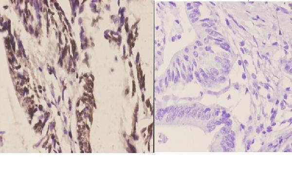
Plan CSTF2 paraffin IHC around general nuclear staining (HPA tissue IHC) and a 1:50–1:200 primary antibody dilution (datasheet). Compare staining by cell type and include a no-primary control, as shown in the catalog antibody’s colon carcinoma IHC image (datasheet).

Evidence assembled Oct 2026 · For research use; verify linked source records and product datasheet before use
Immunohistochemistry protocol sheet for CSTF2 (IHC for CSTF2): expected localisation General nuclear staining (HPA tissue IHC), antibody A07037, validated IHC image, and IHC protocol steps
Printable CSTF2 IHC protocol sheet — expected localisation General nuclear staining (HPA tissue IHC), antibody A07037, controls and protocol steps. Open the full CSTF2 IHC guide →

CSTF2 Immunohistochemistry Experimental Design Guide

Expected localisation, validated protocols, controls and antibodies — the at-a-glance facts below, then the full design guide.

Must know before staining
Expected localisation General nuclear staining (HPA tissue IHC)
Staining pattern Nuclear staining in glandular and respiratory epithelial cells (HPA tissue IHC)
Antigen retrieval Tris-EDTA pH 9.0 HIER, 95–98 °C, 20 min (rule: nuclear antigen)
Positive control ⓘ Adrenal gland+4 more · see all
Negative control ⓘ None in HPA (detected in all 45 tissues); use no-primary + isotype controls
Important caveats
Reasons your staining may differ from the expected pattern.
Fixation Keep fixation consistent across paraffin sections (standard IHC practice; not target-specific)
Caveat Antibody staining and RNA show medium consistency (HPA tissue IHC)
Regulation Low tissue RNA specificity (HPA tissue RNA)
Isoform / epitope 2 isoforms; epitope impact is unknown (UniProt)
Section 1

Recommended CSTF2 IHC & IF Protocols

The catalog antibody’s IHC-P protocol is accompanied by three published CSTF2 IHC protocols (PMC11842344; PMC12751398; PMC9304882).

Recommended immunohistochemistry (IHC-P) protocol parameters
SampleParaffin-embedded human colon carcinoma tissue; fixative not specified (datasheet A07037)
FixationImage fixative and duration unreported (datasheet A07037); verify before use.
Sectioning4–5 µm sections on charged slides (standard)
DeparaffinisationXylene, graded ethanol series to water (standard)
Antigen retrievalHeat-induced epitope retrieval in Tris-EDTA buffer, pH 9.0, 20 min at 95–98 °C (standard rule: nuclear antigen)
Peroxidase block3% H2O2, 10 min, room temperature (standard)
Blocking10% normal serum of the secondary host, 30 min, room temperature (standard)
Primary antibodyRabbit anti-CSTF2, 1:50-1:200 (datasheet A07037)
Primary incubationOvernight at 4 °C (standard)
DetectionHRP-polymer secondary, DAB chromogen 5–10 min (standard)
CounterstainHematoxylin, blue, dehydrate and mount (standard)
Expected resultCSTF2-positive staining in glandular cells of adrenal gland (HPA tissue IHC: High). HPA tissue profile: General nuclear expression. No signal in the no-primary control.
💡Decision noteStart with Tris-EDTA pH 9.0 HIER at 95–98 °C for 20 min (page antigen-retrieval setting). The oral cancer protocol reports microwave retrieval without conditions (PMC11842344).
Section 2

What Is the Expected CSTF2 Staining Pattern?

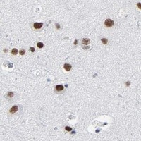
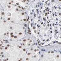
CSTF2 is a nuclear protein with no transmembrane segment (UniProt P33240). In paraffin IHC, expect nuclear staining in several cell populations, including glandular, respiratory epithelial, cerebellar granular-layer and placental decidual cells (HPA tissue IHC). HPA describes general nuclear expression, with “Supported” IHC reliability and medium consistency between antibody staining and RNA expression (HPA tissue IHC).

What am I looking at on my slide?
Distinctly stained nuclei in adrenal or rectal glandular cells, or bronchial respiratory epithelium (HPA: High in each).This matches the reported nuclear pattern (HPA tissue IHC; UniProt P33240). Compare staining within the named cells rather than treating every cell in the section as an equally strong positive.
Strong cytoplasmic staining with little or no nuclear signal in an otherwise well-preserved section.That conflicts with CSTF2’s nuclear location (UniProt P33240; HPA subcellular). Treat it as suspect staining; check the primary-antibody omission control and whether nuclei remain visible before assigning localisation.
Strong staining in liver cholangiocytes or heart cardiomyocytes, especially outside nuclei.HPA reports Low staining in these cells, not absence (HPA tissue IHC). Consider cross-reactivity or endogenous detection activity if the pattern persists; a strong result alone cannot establish either cause.
Diffuse chromogen across cells, tissue edges or blank areas, obscuring nuclear boundaries.This is background that prevents reliable compartment scoring (general IHC practice). Compare a primary-antibody omission control and inspect washing, blocking and detection conditions before interpreting the section.
No nuclear signal in a section containing identifiable bronchial respiratory epithelial cells.Bronchial respiratory epithelial cells are reported High (HPA tissue IHC). A negative run may reflect assay performance; verify a positive-control section and readable counterstained nuclei before interpreting biological absence.
💡Expected CSTF2 appearanceCall a section positive when the relevant cells show clear, predominantly nuclear chromogen, with strong staining plausible in HPA High populations; dominant cytoplasmic colour or diffuse haze is suspect (HPA tissue IHC; UniProt P33240).
How each factor affects the staining
Tissue and cell contextHPA reports High staining in selected glandular, respiratory epithelial, granular-layer and decidual cells, and Low staining in several other populations (HPA tissue IHC). Score the named population; whole-section intensity can hide that distinction.
Strength of the tissue referenceThe tissue profile is “Supported,” with medium staining–RNA consistency and low RNA tissue specificity (HPA tissue IHC). It guides expected localisation and useful positive controls, but does not certify every cell as positive.
Antibody-specific validationHPA lists IHC as Supported for HPA000427 and CAB004680; ICC is Enhanced for HPA000427, with no ICC status listed for CAB004680 (HPA antibodies). Apply each validation label to its own antibody and application.
Isoforms and epitope limitsUniProt lists two CSTF2 isoforms and an RRM at residues 16–94 (UniProt P33240). The supplied evidence gives no antibody epitope or isoform-specific staining result, so neither feature predicts signal from the catalog antibody.
Why is my staining missing, weak or wrong?
SituationLikely causeNext action
No signal in a High reference population (HPA tissue IHC).The result could reflect a failed IHC run or an unsuitable detection setting; the payload gives no CSTF2-specific fixation or retrieval sensitivity.Run a documented High tissue alongside the sample, check counterstain and detection controls, and review the IHC-validated antibody’s supplied protocol (general IHC practice).
Nuclei are faint but the whole section is brown.Background may overwhelm genuine nuclear contrast (general IHC practice); CSTF2 is expected in nuclei (UniProt P33240).Compare the primary-antibody omission control; review blocking, washing and chromogen development, then judge whether nuclear boundaries are distinguishable (general IHC practice).
Strong colour appears mainly in cytoplasm.The compartment conflicts with the reported nuclear localisation (UniProt P33240; HPA subcellular); its source is unresolved.Check controls and repeat with the IHC-validated antibody under its documented conditions; score CSTF2 only where a specific nuclear pattern is supported.
A Low population stains strongly (HPA tissue IHC).Low is not a negative designation (HPA tissue IHC). Cross-reactivity or endogenous detection activity remains possible, especially when staining is extranuclear.Compare a High reference population and primary-antibody omission control; record cell identity and compartment before calling the result unexpected (general IHC practice).
Different tissue regions appear to disagree.HPA levels describe named cell populations, while its tissue IHC profile has medium staining–RNA consistency (HPA tissue IHC). Mixed populations can complicate a regional score.Score identifiable cell populations separately and document the nuclear fraction and intensity; avoid converting one region’s result into a claim about every cell.
IF/ICC: Should CSTF2 appear in nuclear bodies?HPA reports enhanced nucleoplasmic localisation with additional nuclear bodies in ICC-IF images (HPA subcellular); UniProt notes localisation with DDX1 in cleavage bodies (UniProt P33240).Expect nucleoplasm as the main IF/ICC pattern and nuclear bodies as an additional pattern. Use the separate IF/ICC guide for that application; an IHC-P result does not establish ICC performance.

Sample controls for CSTF2 IHC & IF

🧪Run adrenal gland first: glandular cells should show nuclear staining (HPA: High in adrenal gland glandular cells; UniProt P33240: nucleus). HPA detects CSTF2 in all 45 scored tissues, so there is no supported negative tissue; use no-primary and isotype controls, and do not designate any cell type on the positive slide as internally negative (HPA: no negative rows; detected in all 45 scored tissues).
Positive control tissue: Adrenal gland (Glandular cells, HPA High)
Negative control tissue: None in HPA: CSTF2 is detected in all 45 scored tissues. Use a no-primary (secondary-only) and an isotype control instead.
ICC-IF cell lines (HPA subcellular resource): HPA ICC-IF images show CSTF2 in A-431, U-251MG, U2OS, U2OS, siRNA 1 (10x), U2OS, siRNA 1 (40x), U2OS, siRNA 2 (10x), U2OS, siRNA 2 (40x), U2OS, scrambled (10x), U2OS, scrambled (40x), with annotated localisation: Nucleoplasm (enhanced) (HPA subcellular).
Technical controls: Use a no-primary, secondary-only slide and an irrelevant IgG control matched to the primary antibody’s host species and clonality; compare with a matched CSTF2 knockout sample if available (standard IHC controls). For chromogenic adrenal sections, block endogenous peroxidase and check for endogenous biotin if using avidin–biotin detection (standard IHC practice; selected caption: avidin–peroxidase detection).
⚠️Feasibility: No target-specific fixation window or fixation effect is reported, and the A07037 paraffin-section caption does not state a fixative (selected caption: fixative not stated). Retrieval dependence is unreported, so optimize antigen retrieval empirically for paraffin IHC; HPA ICC-IF images support evaluating nucleoplasmic and nuclear-body signal by IF, but do not establish that IF or frozen sections are easier (HPA subcellular: nucleoplasm and nuclear bodies). No adrenal-specific artefact is documented in the supplied evidence (HPA: High in adrenal gland glandular cells).

HPA tissue IHC evidence for CSTF2

Comprehensive Human Protein Atlas IHC scoring per tissue (reliability: Supported — Medium consistency between antibody staining and RNA expression data.). Rows are taken directly from the HPA tissue chart — click any row's HPA link to view the source.

Positive expression · recommended positive controls

TissueCell typeLevelEvidenceSource
Adrenal gland Glandular cells High Protein (IHC) HPA →
Appendix Glandular cells High Protein (IHC) HPA →
Bronchus Respiratory epithelial cells High Protein (IHC) HPA →
Cerebellum Cells in granular layer High Protein (IHC) HPA →
Gallbladder Glandular cells High Protein (IHC) HPA →

Undetected expression · recommended negative controls

TissueCell typeLevelEvidenceSource
None in HPA: CSTF2 is detected in all 45 scored tissues. Use a no-primary (secondary-only) and an isotype control instead.
Section 3

Advanced CSTF2 IHC Tips

Troubleshoot CSTF2 staining in paraffin sections by checking retrieval, nuclear localisation, controls and scoring before interpreting differences between samples.

How should I retrieve CSTF2 in paraffin sections when nuclear staining is weak?
Use Tris-EDTA pH 9.0 HIER at 95–98 °C for 20 min as the starting condition (page retrieval rule: nuclear antigen). Let sections cool in the retrieval buffer, then compare staining with a matched section processed in the same run; the reported paraffin-section image shows nuclear staining at 1:50 (A07037 tissue-IHC caption). If staining remains weak, test a milder citrate pH 6.0 retrieval condition as a fallback while holding detection and exposure to chromogen constant (standard IHC practice). Judge any gain by nuclear signal and preservation of tissue morphology, including comparison with a no-primary control (standard IHC practice).
Could fixation explain variable CSTF2 staining between paraffin blocks?
Target-specific fixation sensitivity is unknown: the selected paraffin-section caption does not report its fixative (A07037 tissue-IHC caption). Record each block’s fixative and fixation duration, and compare blocks with the same retrieval, antibody dilution and chromogen development before attributing a difference to CSTF2 abundance (standard IHC practice). For a controlled trial, process replicate specimens with matched fixation durations and stain them together, including a no-primary section to identify detection background (standard IHC practice). Preserve nuclear detail when assessing the result, because the expected tissue staining is generally nuclear (HPA tissue IHC: general nuclear expression; UniProt P33240: nucleus).
What compartment should contain convincing CSTF2 staining?
Score nuclear staining as the expected pattern: CSTF2 is annotated in the nucleus, including cleavage bodies with DDX1 (UniProt P33240: subcellular location). Nucleoplasmic signal with possible nuclear bodies is also consistent with cellular imaging (HPA subcellular: enhanced nucleoplasm and nuclear bodies). Its sequence has no transmembrane segment, so a crisp membrane-only pattern needs independent validation before it is called CSTF2 (UniProt P33240: topology). Compare nuclear signal with the counterstain in intact cells and inspect a no-primary section; diffuse cytoplasmic chromogen that also appears in the control supports a detection artefact (standard IHC practice).
Could isoforms or epitope accessibility change the CSTF2 IHC pattern?
CSTF2 has 2 annotated isoforms, but the supplied evidence does not map the catalog antibody’s epitope to either one (UniProt P33240: isoforms; A07037 tissue-IHC caption). Its RNA recognition motif spans residues 16–94, and annotated modifications include phosphoserine and methylarginine sites; neither observation establishes antibody sensitivity to those features (UniProt P33240: domains and modified residues). If samples differ, compare them in one staining run and evaluate nuclear signal against matched morphology and controls (standard IHC practice). Treat an isoform-specific or modification-specific interpretation as provisional until the epitope and its recognition in sections are established.
How can I check CSTF2 by IF alongside my chromogenic IHC results?
For exploratory IF, compare nuclear CSTF2 signal with the chromogenic pattern; cellular imaging places CSTF2 mainly in the nucleoplasm and additionally in nuclear bodies (HPA subcellular). Multiplex with a marker for the cell type being evaluated, choosing spectrally separated fluorophores and a far-red channel when tissue autofluorescence interferes with shorter wavelengths (standard IF practice). Because CSTF2 is nuclear and has no transmembrane segment, permeabilise fixed cells or sections to give antibody access to the nuclear epitope, then optimise detergent exposure for morphology (UniProt P33240: location and topology; standard IF practice). Include single-stain and no-primary controls before interpreting overlap or puncta (standard IF practice).
How do I separate CSTF2 signal from chromogenic background?
Begin with the no-primary control used in the reported paraffin-section example, which substituted PBS for primary antibody (A07037 tissue-IHC caption). If both sections stain, check endogenous peroxidase blocking, secondary-reagent binding and DAB development time before changing the CSTF2 antibody concentration (standard IHC practice). If background appears only with primary antibody, compare dilution and incubation settings across matched sections while preserving the same retrieval and detection conditions; 1:50 is reported for one image, not an established optimum for every specimen (A07037 tissue-IHC caption). Accept improvement only when intact nuclei retain distinct signal above adjacent tissue background (UniProt P33240: nucleus; standard IHC practice).
How should I quantify CSTF2 staining across specimens? ⚠ ANSWER MARKED FOR VERIFICATION
Define the tissue compartment and cell population before scoring, then record the percentage of positive nuclei and nuclear intensity on a consistent scale (standard IHC practice; HPA tissue IHC: general nuclear expression). An H-score can combine intensity categories 0–3 with their corresponding percentages, yielding 0–300; report the rule used to classify a nucleus as positive (standard IHC practice). Normalise counts to the number of evaluable nuclei in the selected compartment, or report positive nuclei per mm² when cell density itself matters (standard IHC practice). Apply the same imaging, counterstain and scoring thresholds across batches, and exclude damaged or necrotic areas (standard IHC practice).
When is an apparent CSTF2-positive area likely to be an artefact?
Convincing staining should track intact nuclei, consistent with the reported paraffin-section image and the broader nuclear tissue pattern (A07037 tissue-IHC caption; HPA tissue IHC: general nuclear expression). Treat membrane-only staining or chromogen concentrated at cut edges, folds and necrotic areas cautiously, and compare those regions with the no-primary control (UniProt P33240: no transmembrane segment; standard IHC practice). Signal in an unexpected cell population warrants checking morphology and the corresponding control rather than assigning a new cell-specific pattern; HPA reports low tissue specificity and no negative tissue list here (HPA tissue IHC). If staining persists without primary antibody, investigate endogenous enzyme activity and detection reagents before interpreting CSTF2 expression (standard IHC practice).
Boster reagents

Best CSTF2 / Cleavage stimulation factor subunit 2 IHC Antibodies

A07037 has a paraffin-section IHC image from human colon carcinoma (IHC image caption). IF is listed for human, mouse, and rat, with no IF image supplied (catalog applications/reactivity; IF image list).

Real IHC data Immunohistochemistry (IHC) analyzes of CstF-64 (R46) pAb in paraffin-embedded human colon carcinoma tissue at 1:50.showing nucleus staining. Negative control (the right)Using PBS instead of primary antibody, secondary antibody is Goat Anti-Rabbit IgG-biotin followed by avidin-peroxidase.
Anti-CstF-64 (R46) CSTF2 Antibody
Cat # A07037

A07037 shows nuclear staining in paraffin-embedded human colon carcinoma at 1:50, alongside a PBS-for-primary negative control (IHC image caption). Its listed applications include IHC and IF, and its stated reactivity covers human, mouse, and rat (catalog applications/reactivity).

Which to pick: For tissue IHC, choose A07037: its own image documents paraffin-embedded human colon carcinoma, although the fixative is unreported (A07037 IHC image caption). For IF, A07037 is listed at 1:50–1:200; no IF image or ICC validation is supplied (A07037 catalog applications/IF dilution; IF image list). For mouse or rat work, A07037 is the listed rabbit polyclonal option based on stated reactivity; its supplied tissue image is human only (A07037 catalog reactivity/dilution_raw; IHC image caption).

Each figure is that product's own IHC / IF validation image from its datasheet.

References

  1. UniProt Consortium. UniProt entry P33240 (CSTF2_HUMAN, Cleavage stimulation factor subunit 2).
  2. Human Protein Atlas. CSTF2 tissue IHC expression (reliability: Supported).
  3. Human Protein Atlas. CSTF2 subcellular location (ICC-IF): Mainly localized to the nucleoplasm. In addition localized to the nuclear bodies..
  4. Human Protein Atlas. CSTF2 antibody validation summary (2 antibodies).
  5. Expression of CSTF2 in oral squamous cell carcinoma and its relationship with immune infiltration and poor prognosis. Frontiers in oral health 2025 — PMC11842344.
  6. The role of CSTF2 in gastric cancer: implications for therapy. European journal of medical research 2025 — PMC12751398.
  7. CSTF2 Promotes Hepatocarcinogenesis and Hepatocellular Carcinoma Progression via Aerobic Glycolysis. Frontiers in oncology 2022 — PMC9304882.
  8. CSTF2 Acts as a Prognostic Marker Correlated with Immune Infiltration in Hepatocellular Carcinoma. Cancer management and research 2022 — PMC9481280.
  9. PubMed PMID:1741396 — UniProt-cited evidence.
  10. PubMed PMID:15772651 — UniProt-cited evidence.
  11. PubMed PMID:15489334 — UniProt-cited evidence.