This website uses cookies to ensure you get the best experience on our website.


- Table of Contents
Plan chromogenic IHC for CTRC in paraffin sections using pancreatic exocrine staining as the reference pattern (HPA tissue IHC). The catalog antibody’s human pancreas example uses 2 μg/ml primary antibody and DAB detection (datasheet A03540-1).

Expected localisation, validated protocols, controls and antibodies — the at-a-glance facts below, then the full design guide.
| Expected localisation | Cytoplasmic and extracellular in pancreas (HPA tissue IHC) | |
| Staining pattern | Exocrine cells show cytoplasmic and extracellular staining (HPA tissue IHC) | |
| Antigen retrieval | EDTA pH 8.0 HIER, heat-mediated (datasheet A03540-1) | |
| Positive control | Pancreas | |
| Negative control | Adipose tissue+4 more · see all |
| Fixation | Keep fixation consistent across sections (standard IHC practice; not target-specific) | |
| Caveat | Secretion may extend staining beyond producing cells (HPA tissue IHC) | |
| Regulation | Pancreas enriched; regulation unreported (UniProt) | |
| Isoform / epitope | No isoforms; processing may affect cellular and secreted epitopes (UniProt; HPA tissue IHC) |
The catalog antibody’s IHC-P protocol (datasheet A03540-1) and a published protocol for CTRC staining in tumor and peritumoral tissues (PMC6154863) provide starting conditions.
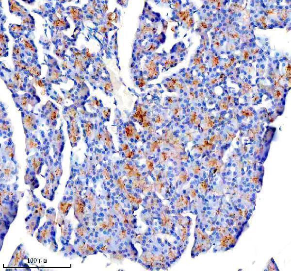
| Sample | Paraffin-embedded rat pancreas tissue; fixative not specified (datasheet A03540-1) |
| Fixation | Image fixative and duration unreported (datasheet A03540-1); verify before use. |
| Sectioning | 4–5 µm sections on charged slides (standard) |
| Deparaffinisation | Xylene, graded ethanol series to water (standard) |
| Antigen retrieval | Heat retrieval: EDTA pH 8.0 (datasheet A03540-1); 20 min, 95–100 °C (standard) |
| Peroxidase block | 3% H2O2, 10 min, room temperature (standard) |
| Blocking | 10% goat serum (datasheet A03540-1) |
| Primary antibody | Rabbit anti-CTRC, 2-5 μg/ml (datasheet A03540-1) |
| Primary incubation | Overnight at 4 °C (datasheet A03540-1) |
| Detection | HRP-conjugated secondary, DAB chromogen (datasheet A03540-1) |
| Counterstain | Hematoxylin, blue, dehydrate and mount (standard) |
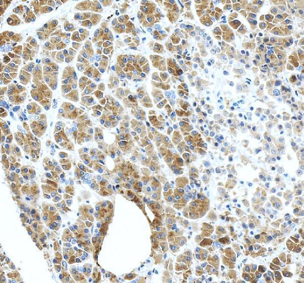
| Expected result | CTRC-positive staining in exocrine glandular cells of pancreas (HPA tissue IHC: Medium). HPA tissue profile: Distinct cytoplasmic expression as well as extracellular positivity in pancreas. No signal in the no-primary control. |
CTRC should show distinct cytoplasmic staining in pancreatic exocrine glandular cells, with extracellular positivity also expected (HPA: pancreas IHC, Enhanced reliability). Its signal peptide and lack of a transmembrane segment are consistent with secretion (UniProt Q99895: signal 1–16; topology). HPA reports that the tissue locations of RNA and secreted protein may differ (HPA: tissue IHC reliability description).
| Distinct cytoplasmic staining in pancreatic exocrine glandular cells, with adjacent extracellular positivity. | This matches the reported tissue pattern. The exocrine glandular cell staining level is Medium; extracellular signal can accompany a secreted protein (HPA: pancreas IHC; HPA: Secreted). |
| Predominantly nuclear staining or a crisp cell-surface outline, without the expected cytoplasmic pattern. | Treat this as a localisation mismatch requiring review, rather than a confirmed CTRC pattern. HPA reports cytoplasmic and extracellular positivity; UniProt lists no transmembrane segment and does not assign a subcellular location (HPA: pancreas IHC; UniProt Q99895: topology, subcellular annotation). |
| Strong staining in an unexpected cell population while pancreatic exocrine glandular cells are weak or negative. | Consider cross-reactivity or detection background. Cell identity matters: HPA reports CTRC staining in pancreatic exocrine glandular cells, while its listed adipocytes, respiratory epithelial cells and glial cells are Not detected in their respective tissues (HPA: tissue IHC). |
| A uniform haze covers cells and extracellular spaces, including a control section processed without primary antibody. | A signal that persists without primary antibody suggests background from the detection workflow, such as endogenous enzyme activity in chromogenic IHC. This is a general IHC interpretation, not an HPA finding about CTRC; it obscures the distinct pancreatic pattern reported by HPA (HPA: pancreas IHC). |
| No convincing signal in pancreatic exocrine glandular cells on an otherwise readable section. | This conflicts with the reported positive reference tissue. First check that exocrine glandular cells are present and assess the assay controls; absent signal alone cannot distinguish technical failure from a difference between samples or antibodies (HPA: pancreas IHC, Medium, Enhanced reliability; general IHC practice). |
| Tissue and cell context | Use pancreatic exocrine glandular cells as the positive reference: HPA reports Medium staining there and Not detected staining in its listed negative cell populations. Those findings describe the sampled tissues, not every cell type or specimen (HPA: tissue IHC). |
| Secretion and precursor processing | CTRC has a signal peptide at residues 1–16, a propeptide at 17–29, and a listed chain at 30–268 (UniProt Q99895: processing). Its reported extracellular positivity is consistent with secretion (HPA: pancreas IHC; HPA: Secreted). An antibody epitope in a removed segment could affect what it detects; no epitope is supplied here, so this remains conditional. |
| Antibody evidence | HPA lists antibody HPA046920 as rabbit polyclonal with Enhanced IHC validation; its tissue reliability description reports high agreement between staining and RNA data (HPA: antibody record; HPA: tissue IHC). This supports the reported pattern but supplies no dilution, retrieval condition or epitope for another antibody. |
| IF/ICC Q&A: Is the IHC pattern an IF/ICC localisation reference? | Use it as a tissue level expectation only. HPA calls CTRC Secreted, but lists no main ICC-IF location or cell line images, and the listed antibody has no ICC validation status (HPA: subcellular record; HPA: antibody record). An IF/ICC result therefore needs its own controls and interpretation. |
| Situation | Likely cause | Next action |
|---|---|---|
| Pancreatic exocrine glandular cells show no staining. | The assay may have failed, the relevant cells may be absent, or this specimen may differ from HPA's reference; the slide alone cannot distinguish these possibilities (HPA: pancreas IHC; general IHC practice). | Confirm exocrine glandular cells on the counterstain, review positive and negative assay controls, and check the antibody's documented IHC-P conditions before changing one workflow variable at a time (general IHC practice). |
| The whole section develops brown signal, including a no-primary control. | The pattern suggests detection-system background or endogenous chromogenic activity, not a cell-specific CTRC call (general IHC practice). | Review the no-primary control, blocking and endogenous enzyme suppression appropriate to the detection system; rescore only after the background clears (general IHC practice). |
| Staining is mainly nuclear or outlines cell membranes. | This differs from HPA's cytoplasmic and extracellular pancreatic pattern; UniProt reports no transmembrane segment, but gives no subcellular annotation (HPA: pancreas IHC; UniProt Q99895: topology, subcellular annotation). | Check morphology and compartment assignment, then compare with the pancreatic positive reference and detection controls before accepting the signal as CTRC (HPA: pancreas IHC; general IHC practice). |
| Unexpected cells stain strongly while the expected exocrine cells do not. | Cross-reactivity or local background is plausible; HPA's positive call is for pancreatic exocrine glandular cells, and its listed negative populations are Not detected (HPA: tissue IHC). | Identify the stained cells on the counterstain and compare a reported negative tissue under the same run conditions; treat discordant staining as unconfirmed (HPA: tissue IHC; general IHC practice). |
| Extracellular signal is present but cytoplasmic exocrine staining is hard to see. | Extracellular positivity is part of the reported pattern for this secreted protein; it does not by itself establish which nearby cells produced the signal (HPA: pancreas IHC; HPA: Secreted). | Inspect intact exocrine glandular cells at suitable magnification and score cellular and extracellular staining separately, using the HPA pattern as the reference (HPA: pancreas IHC; general IHC practice). |
| An IF/ICC image is being judged against the IHC section. | HPA provides no main ICC-IF location or cell line images, and its listed antibody has no ICC validation status (HPA: subcellular record; HPA: antibody record). | Interpret the IF/ICC result with its own controls and avoid assigning a precise intracellular location from the tissue IHC pattern alone (HPA: pancreas IHC; HPA: subcellular record; general IF practice). |


Comprehensive Human Protein Atlas IHC scoring per tissue (reliability: Enhanced — High consistency between antibody staining and RNA expression data. Secreted protein, tissue location of RNA and protein is expected to differ.). Rows are taken directly from the HPA tissue chart — click any row's HPA link to view the source.
| Tissue | Cell type | Level | Evidence | Source |
|---|---|---|---|---|
| Pancreas | Exocrine glandular cells | Medium | Protein (IHC) | HPA → |
| Tissue | Cell type | Level | Evidence | Source |
|---|---|---|---|---|
| Adipose tissue | Adipocytes | Not detected | Protein (IHC) | HPA → |
| Adrenal gland | Glandular cells | Not detected | Protein (IHC) | HPA → |
| Appendix | Glandular cells | Not detected | Protein (IHC) | HPA → |
| Bone marrow | Hematopoietic cells | Not detected | Protein (IHC) | HPA → |
| Breast | Adipocytes | Not detected | Protein (IHC) | HPA → |
Troubleshoot CTRC staining in paraffin sections by checking retrieval, tissue pattern, and controls against the reported pancreas IHC evidence (datasheet A03540-1; HPA tissue IHC).
A03540-1 has real IHC images from paraffin-embedded rat and human pancreas sections (catalog IHC image captions). IF/ICC validation is not reported (catalog applications; IF image alts absent).
A03540-1 is the only SKU and lists IHC with human and rat reactivity (catalog applications/reactivity). Its captions document staining in paraffin-embedded rat and human pancreas sections; the rendered card shows the rat section (catalog IHC image captions; card figure tag).
Which to pick: Choose A03540-1 for tissue IHC: this rabbit polyclonal antibody has paraffin-section pancreas captions for rat and human, with EDTA pH 8.0 retrieval and 2 μg/ml primary antibody documented in the rat caption (catalog host/dilution_raw; IHC image captions). For cross-species IHC, the same SKU lists human and rat reactivity and has an image caption for each (catalog reactivity; IHC image captions). No IF/ICC protocol option is supported here, and the IHC captions do not report the fixative (catalog applications; IF image alts absent; IHC image captions).