CTSE / Cathepsin E · IHC design guide

Design Immunohistochemistry for CTSE

Plan CTSE IHC in paraffin sections using stomach glandular cells as a high-staining reference (HPA tissue IHC). Use consistent fixation and score staining by cell type and compartment.

Evidence assembled Oct 2026 · For research use; verify linked source records and product datasheet before use
Immunohistochemistry protocol sheet for CTSE (IHC for CTSE): expected localisation Cytoplasmic and membranous tissue staining (HPA tissue IHC), antibody A04874-1, validated IHC image, and IHC protocol steps
Printable CTSE IHC protocol sheet — expected localisation Cytoplasmic and membranous tissue staining (HPA tissue IHC), antibody A04874-1, controls and protocol steps. Open the full CTSE IHC guide →

CTSE Immunohistochemistry Experimental Design Guide

Expected localisation, validated protocols, controls and antibodies — the at-a-glance facts below, then the full design guide.

Must know before staining
Expected localisation Cytoplasmic and membranous tissue staining (HPA tissue IHC)
Staining pattern Stomach glandular cells stain in cytoplasm and membrane (HPA tissue IHC)
Antigen retrieval Citrate pH 6 HIER, heat-mediated (datasheet A04874-1)
Positive control ⓘ Appendix+4 more · see all
Negative control ⓘ Adipose tissue+4 more · see all
Important caveats
Reasons your staining may differ from the expected pattern.
Fixation Keep paraffin-section fixation consistent across samples. (standard IHC practice; not target-specific)
Caveat Signal varies with glandular or endocrine cell content (HPA tissue IHC)
Regulation Present in activated, absent in resting B cells (UniProt)
Isoform / epitope 3 isoforms; processed forms warrant epitope checks (UniProt)
Section 1

Recommended CTSE IHC & IF Protocols

The catalog antibody protocol uses citrate pH 6 heat retrieval (datasheet A04874-1). The published IHC protocols below provide tissue preparation and staining details (PMC4153675; PMC5460450; PMC3922809).

Recommended immunohistochemistry (IHC-P) protocol parameters
SampleParaffin-embedded human Lung cancer tissue; fixative not specified (datasheet A04874-1)
FixationImage fixative and duration unreported (datasheet A04874-1); verify before use.
Sectioning4–5 µm sections on charged slides (standard)
DeparaffinisationXylene, graded ethanol series to water (standard)
Antigen retrievalHeat retrieval: Citrate pH 6, 20 min (datasheet A04874-1)
Peroxidase block3% H2O2, 10 min, room temperature (standard)
Blocking10% goat serum (datasheet A04874-1)
Primary antibodyRabbit anti-CTSE, 2μg/ml (datasheet A04874-1)
Primary incubationOvernight at 4 °C (datasheet A04874-1)
DetectionStreptavidin-biotin complex (SABC), DAB chromogen (datasheet A04874-1)
CounterstainHematoxylin, blue, dehydrate and mount (standard)
Expected resultCTSE-positive staining in endocrine cells of appendix (HPA tissue IHC: High). HPA tissue profile: Cytoplasmic and membranous expression most abundant in stomach but also in intestine, gallbladder, urinary bladder and squamous epithelia of tonsil. No signal in the no-primary control.
💡Decision noteStart with citrate pH 6 heat retrieval for the catalog antibody (datasheet A04874-1). The colon study reports citrate or Tris-EDTA retrieval without specifying which was used for CTSE (PMC3922809).
Section 2

What Is the Expected CTSE Staining Pattern?

CTSE is expected in stomach glandular cells and intestinal endocrine cells, with cytoplasmic and membranous IHC staining (HPA tissue IHC). Mature CTSE localizes to endosomes; its proenzyme localizes to the endoplasmic reticulum and Golgi, and CTSE has no transmembrane segment (UniProt P14091). Treat the tissue pattern as a guide to interpretation: HPA rates its IHC reliability Enhanced while reporting medium consistency between staining and RNA data (HPA tissue IHC).

What am I looking at on my slide?
Strong cytoplasmic staining in stomach glandular cells, possibly with membranous staining (HPA tissue IHC).This matches a high-staining cell population and the reported cytoplasmic and membranous tissue profile (HPA tissue IHC). Assess the named cells, since staining intensity elsewhere in the section cannot establish that the expected population is positive.
Staining in duodenal, rectal or appendix endocrine cells (HPA: High in each population).These are additional observed high-staining populations (HPA tissue IHC). Colon and small-intestinal endocrine cells are listed at Medium, so weaker staining there can still agree with the tissue record (HPA tissue IHC).
Predominantly nuclear signal, with little staining in the expected cytoplasmic compartment.A nuclear pattern is discordant with the reported cytoplasmic and membranous IHC profile (HPA tissue IHC) and endosomal localization (UniProt P14091). Consider nonspecific staining or a slide artefact; compartment alone does not identify its cause.
Strong staining mainly in adipocytes while stomach glandular cells are unstained.HPA reports CTSE as not detected in adipose-tissue adipocytes and High in stomach glandular cells (HPA tissue IHC). Investigate antibody cross-reactivity or endogenous chromogen activity before calling the adipocyte signal CTSE; check controls and tissue identity.
Diffuse color across cells and tissue spaces, or no signal in stomach glandular cells.Diffuse color lacks the reported cell-restricted profile; an absent stomach signal conflicts with a High reference population (HPA tissue IHC). General IHC practice calls for checking background controls, detection performance and section quality before assigning a biological negative.
💡Expected CTSE appearanceCall a section positive when stomach glandular cells show strong cytoplasmic, sometimes membranous staining (HPA: High; HPA tissue IHC profile); broad uniform or chiefly nuclear color is discordant with that pattern (HPA tissue IHC; UniProt P14091).
How each factor affects the staining
Cell population and tissue (HPA tissue IHC).Reference intensity varies by named cell population: stomach glandular and duodenal, rectal and appendix endocrine cells are High; gallbladder glandular and pancreatic exocrine glandular cells are Medium (HPA tissue IHC). Compare like cells when judging a result.
CTSE maturation and location (UniProt P14091).The proenzyme is reported in endoplasmic reticulum and Golgi, while mature CTSE is endosomal (UniProt P14091). This supports intracellular staining but does not predict which pool a particular antibody detects; its epitope is unspecified in the supplied record.
IHC evidence and antibody validation (HPA tissue IHC; HPA antibodies).HPA assigns Enhanced IHC status to HPA012940 and CAB032687, and describes medium consistency between antibody staining and RNA data (HPA). Use the observed pattern as a reference, while treating unexpected cells or compartments as findings to verify.
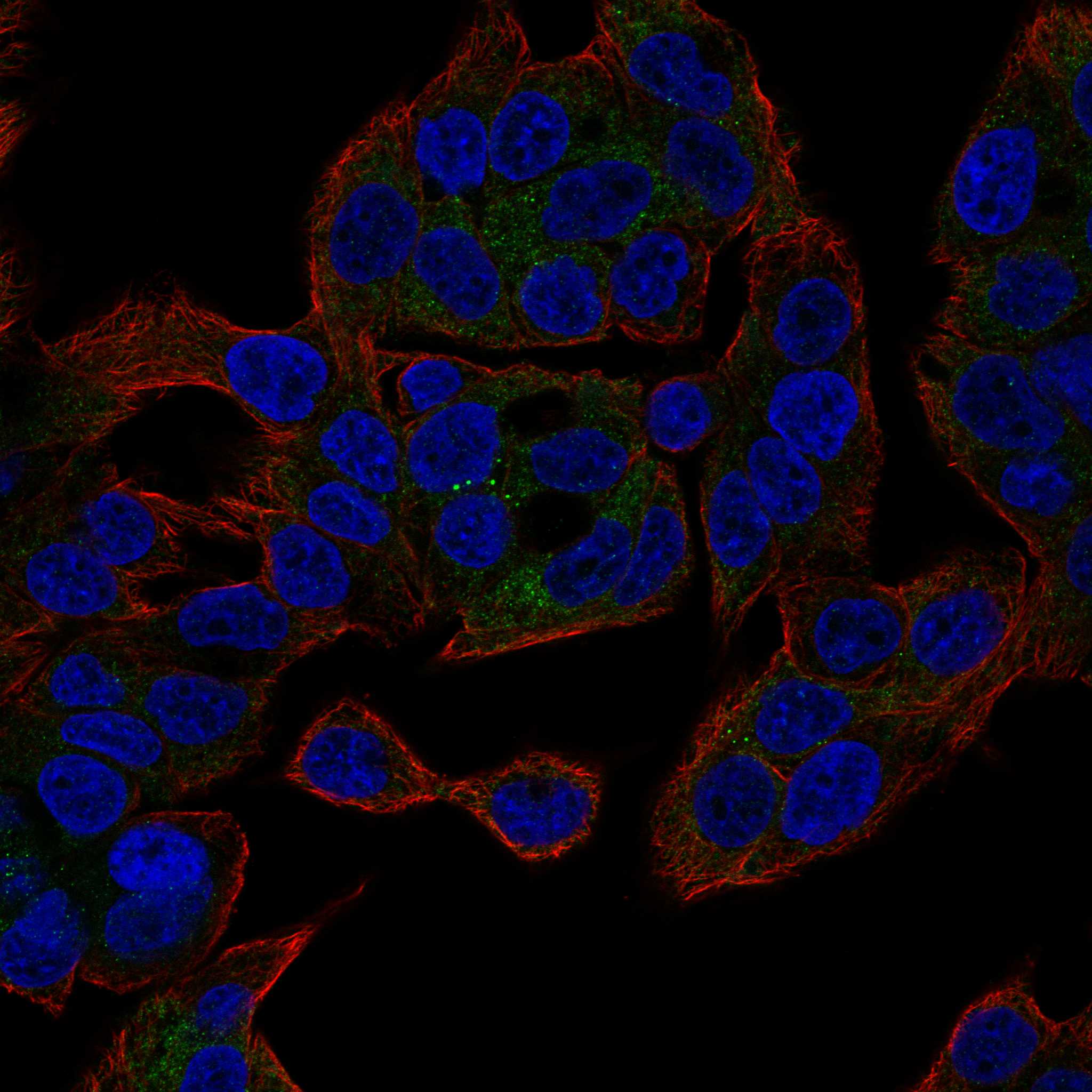
IF localization evidence (HPA subcellular; UniProt P14091).HPA reports approved vesicle localization in ICC-IF, compatible with mature CTSE's endosomal location (HPA subcellular; UniProt P14091). The IF observation helps interpret compartment, but it does not specify an IHC staining intensity or establish an IHC protocol.
Why is my staining missing, weak or wrong?
SituationLikely causeNext action
No color in stomach glandular cells, including the intended positive control.The result misses an HPA High population (HPA tissue IHC); a detection failure is possible, but the slide alone cannot identify the failed step.In general IHC practice, verify section identity and morphology, antibody and detection controls, and that the planned staining steps were completed. Reassess before reporting CTSE absent.
Uniform brown haze obscures glandular-cell boundaries.The reported tissue pattern is cell-associated and cytoplasmic or membranous (HPA tissue IHC); widespread haze can arise from nonspecific binding or endogenous detection activity (general IHC practice).Compare a reagent control and review blocking, washing and detection conditions (general IHC practice). Judge CTSE only where cellular staining can be distinguished from haze.
A predominantly nuclear signal appears in the positive tissue.Nuclear staining does not match the HPA cytoplasmic and membranous profile or UniProt endosomal location (HPA tissue IHC; UniProt P14091).Check counterstain and reagent controls, then confirm whether glandular-cell cytoplasm carries a separate signal (general IHC practice). Do not score nuclear color alone as the expected CTSE pattern.
Unexpected cells stain strongly, especially adipocytes.HPA lists adipose-tissue adipocytes as Not detected (HPA tissue IHC). Cross-reactivity, nonspecific binding or endogenous detection activity are possible explanations (general IHC practice).Confirm the cell type, compare an appropriate reagent control and inspect a known-positive stomach section. An unexpected positive requires verification; HPA's cell-specific entry does not make every cell in adipose tissue negative.
A weak intestinal result seems inconsistent with a strong stomach control.HPA levels differ by cell population: stomach glandular cells are High, while colon and small-intestinal endocrine cells are Medium (HPA tissue IHC).Identify the intestinal cell population before comparing intensity, and score its cellular pattern alongside a control stained in the same run (general IHC practice).
Q (IF/ICC): What intracellular pattern should support CTSE localization?HPA reports approved vesicle localization in ICC-IF, and UniProt places mature CTSE in endosomes (HPA subcellular; UniProt P14091).A (IF/ICC): Look for vesicular signal in the relevant cells (HPA subcellular). Interpret diffuse or nuclear fluorescence cautiously; this localization evidence does not provide an IF/ICC protocol or an IHC-P staining threshold.

Sample controls for CTSE IHC & IF

🧪Run stomach first: glandular cells should stain (HPA: High in stomach glandular cells). Use adipose tissue as the negative, focusing on adipocytes (HPA: Not detected in adipocytes); on the stomach slide, assess cells outside the stained glandular population for nonspecific signal without assuming they are CTSE-negative.
Positive control tissue: Appendix (Endocrine cells, HPA High)
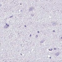
Negative control tissue: Adipose tissue (HPA Not detected)
ICC-IF cell lines (HPA subcellular resource): HPA ICC-IF images show CTSE in OE19, U2OS, with annotated localisation: Vesicles (approved) (HPA subcellular).
Technical controls: Include a no-primary, secondary-only control and a host- and isotype-matched nonimmune IgG control (caption: rabbit primary antibody). Use CTSE knockout material or a validated immunizing-peptide block as a biological specificity control; block endogenous peroxidase and assess endogenous biotin background when using the caption’s streptavidin–biotin/DAB detection (caption: SABC with DAB).
⚠️Feasibility: A target-specific fixation window or fixation effect is unreported, and the selected A04874-1 tissue-IHC caption does not state a fixative (caption: fixative not stated). The caption uses citrate pH 6 heat retrieval for 20 minutes, but does not establish that retrieval is required (caption: citrate retrieval); the supplied evidence does not establish whether frozen sections or IF are easier. In stomach, assess glandular staining against control slides for endogenous peroxidase or biotin background from the chromogenic detection method (caption: SABC with DAB).

HPA tissue IHC evidence for CTSE

Comprehensive Human Protein Atlas IHC scoring per tissue (reliability: Enhanced — Medium consistency between antibody staining and RNA expression data.). Rows are taken directly from the HPA tissue chart — click any row's HPA link to view the source.

Positive expression · recommended positive controls

TissueCell typeLevelEvidenceSource
Appendix Endocrine cells High Protein (IHC) HPA →
Duodenum Endocrine cells High Protein (IHC) HPA →
Rectum Endocrine cells High Protein (IHC) HPA →
Stomach Glandular cells High Protein (IHC) HPA →
Colon Endocrine cells Medium Protein (IHC) HPA →

Undetected expression · recommended negative controls

TissueCell typeLevelEvidenceSource
Adipose tissue Adipocytes Not detected Protein (IHC) HPA →
Adrenal gland Glandular cells Not detected Protein (IHC) HPA →
Breast Adipocytes Not detected Protein (IHC) HPA →
Bronchus Respiratory epithelial cells Not detected Protein (IHC) HPA →
Caudate Glial cells Not detected Protein (IHC) HPA →
Section 3

Advanced CTSE IHC Tips

Troubleshoot CTSE staining in paraffin sections by checking retrieval, cellular distribution, controls and scoring before interpreting chromogenic signal.

How should I optimize retrieval when CTSE staining is weak?
Use heat-mediated citrate retrieval at pH 6 for 20 minutes before chromogenic CTSE staining in paraffin sections (datasheet A04874-1). The demonstrated section used 10% goat serum, 2 µg/mL catalog antibody overnight at 4 °C, then a biotinylated secondary and DAB detection (datasheet A04874-1). If signal is weak, compare heating intervals on adjacent sections while holding antibody concentration and detection constant; inspect morphology and background after each condition (standard IHC practice). Include stomach glandular cells as a positive reference and assess the expected cell type, since staining varies across tissues (HPA tissue IHC: high in stomach glandular cells).
Could fixation explain variable CTSE staining between paraffin blocks?
Target-specific fixation sensitivity is unknown: the selected paraffin-section caption does not state its fixative (datasheet A04874-1: fixative not stated). Record the fixative, fixation duration, processing history and section age for each block before comparing CTSE staining (standard IHC practice). Run blocks together using citrate retrieval at pH 6 for 20 minutes and the same detection conditions, then assess both signal and tissue preservation (datasheet A04874-1; standard IHC practice). If staining differs, test matched material under controlled fixation conditions before assigning causation; neither the expression pattern nor protein topology establishes a fixation effect (HPA tissue IHC; UniProt P14091 topology).
Where should CTSE chromogenic staining appear within positive cells?
Look for predominantly cytoplasmic, potentially granular staining, consistent with mature CTSE in endosomes and vesicular localization reported separately (UniProt P14091 subcellular; HPA subcellular). The proenzyme is associated with the endoplasmic reticulum and Golgi apparatus, so intracellular staining need not have one identical pattern across cells (UniProt P14091 subcellular). Compare the stained compartment with cell identity on the counterstained section; stomach glandular cells provide a strongly stained tissue reference, while lung alveolar type II cells have low reported staining (HPA tissue IHC). Uniform nuclear staining or staining confined to section edges warrants comparison with controls and neighboring intact tissue before it is scored as CTSE (standard IHC practice).
Could CTSE processing or isoforms change which cells stain?
CTSE has 3 reported isoforms, and its precursor contains a signal sequence at residues 1–19 and a propeptide at 20–53 (UniProt P14091). Two reported mature chains start at residues 54 and 57, so epitope position could affect recognition of precursor versus processed protein (UniProt P14091 processing). CTSE also has a glycosylation site at residue 90; the supplied caption does not identify the catalog antibody's epitope or establish an effect of glycosylation on staining (UniProt P14091 glycosylation; datasheet A04874-1). If compartment patterns differ between samples, obtain epitope information and compare appropriately controlled sections before attributing the difference to an isoform (standard IHC practice).
How can IF help check a puzzling CTSE IHC pattern?
Use IF as a separate localization check, pairing CTSE with a validated marker for the expected cell type, such as stomach glandular cells when that tissue is examined (HPA tissue IHC: high in stomach glandular cells; standard IF practice). Select fluorophores after checking tissue autofluorescence and include single-label controls to assess spectral bleed-through before interpreting overlap (standard IF practice). Because CTSE has a signal sequence, lacks a transmembrane segment and occupies intracellular compartments, assess whether permeabilisation gives the antibody access to its mapped epitope (UniProt P14091 topology and subcellular; standard IF practice). Vesicular IF localization can support a compartment assignment, but use the IHC section's cell identity and controls when interpreting its DAB signal (HPA subcellular: vesicles; standard IHC practice).
How do I reduce diffuse brown background without losing CTSE signal?
Inspect a no-primary control and adjacent intact tissue to distinguish reagent background from cellular CTSE staining (standard IHC practice). For DAB detection, verify the peroxidase block, adjust antibody exposure if needed, and check washing and serum blocking; the demonstrated section used 10% goat serum and 2 µg/mL primary antibody (standard IHC practice; datasheet A04874-1). Because that demonstration used a biotinylated secondary and streptavidin–biotin detection, assess background attributable to the detection system with suitable controls (datasheet A04874-1; standard IHC practice). Preserve a positive reference during optimization: stomach glandular cells show high CTSE staining, whereas adipocytes in adipose tissue are reported as undetected (HPA tissue IHC).
How should I score CTSE across heterogeneous paraffin sections? ⚠ ANSWER MARKED FOR VERIFICATION
Define the cell population and compartment before scoring; CTSE is reported as most abundant in stomach glandular cells, while other listed cell populations show different levels (HPA tissue IHC). For a chromogenic section, record the percentage of positive target cells and an intensity score, then calculate an H-score from 0–300 if the study uses that convention (standard IHC practice). Alternatively, count positive cells per mm², normalizing to evaluable tissue area, and report the number of target cells examined when comparing cell populations (standard IHC practice). Exclude necrotic or damaged regions consistently, apply the same threshold and retrieval conditions across sections, and report cell-type-specific results (standard IHC practice).
What distinguishes credible CTSE staining from an IHC artefact?
A credible result has cellular staining in an expected population and an intracellular pattern compatible with CTSE trafficking from endoplasmic reticulum and Golgi to endosomes (UniProt P14091 subcellular; standard IHC practice). Stomach glandular cells are a useful positive reference, while bronchial respiratory epithelial cells were reported as undetected in the supplied tissue profile (HPA tissue IHC). Treat isolated edge staining, necrotic areas, diffuse extracellular DAB or a dominant nuclear pattern cautiously; compare intact neighboring tissue and a no-primary control (standard IHC practice). Check for endogenous peroxidase signal with the detection controls before calling weak brown staining positive, and interpret discordance in light of the tissue profile's medium staining–RNA consistency (standard IHC practice; HPA tissue IHC: Enhanced reliability, medium consistency).
Boster reagents

Best CTSE / Cathepsin E IHC Antibodies

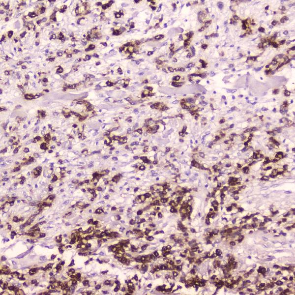
The anti-CTSE catalog antibody has IHC images from paraffin sections of human lung and gastric cancer tissue (A04874-1 IHC captions); listed reactivity covers human, mouse and rat (catalog reactivity).

Real IHC data IHC analysis of Cathepsin E using anti-Cathepsin E antibody (A04874-1). Cathepsin E was detected in paraffin-embedded section of human Lung cancer tissue. Heat mediated antigen retrieval was performed in citrate buffer (pH6, epitope retrieval solution) for 20 mins. The tissue section was blocked with 10% goat serum. The tissue section was then incubated with 2μg/ml rabbit anti-Cathepsin E Antibody (A04874-1) overnight at 4℃. Biotinylated goat anti-rabbit IgG was used as secondary antibody and incubated for 30 minutes at 37℃. The tissue section was developed using Strepavidin-Biotin-Complex (SABC)(Catalog # SA1022) with DAB as the chromogen.
Anti-Cathepsin E/CTSE Antibody ®
Cat # A04874-1

A04874-1 is listed for IHC and WB in human, mouse and rat (catalog applications/reactivity). Its own IHC images show paraffin sections of human lung cancer and gastric cancer tissue (A04874-1 IHC captions).

Which to pick: Choose A04874-1 for tissue IHC: this rabbit polyclonal is listed for IHC, with images from human paraffin sections (catalog dilution_raw; A04874-1 IHC captions). There is no IF/ICC-validated pick in this payload because A04874-1 has no listed IF/ICC application or IF image (catalog applications; catalog IF image alts). For cross-species planning, A04874-1 lists human, mouse and rat reactivity, but its supplied IHC images show human tissue only; the fixative is unreported (catalog reactivity; A04874-1 IHC captions).

Each figure is that product's own IHC / IF validation image from its datasheet.

References

  1. UniProt Consortium. UniProt entry P14091 (CATE_HUMAN, Cathepsin E).
  2. Human Protein Atlas. CTSE tissue IHC expression (reliability: Enhanced).
  3. Human Protein Atlas. CTSE subcellular location (ICC-IF): Localized to vesicles..
  4. Human Protein Atlas. CTSE antibody validation summary (3 antibodies).
  5. Monitoring pancreatic carcinogenesis by the molecular imaging of cathepsin E in vivo using confocal laser endomicroscopy. PloS one 2014 — PMC4153675.
  6. CTSE Overexpression Is an Adverse Prognostic Factor for Survival among Rectal Cancer Patients Receiving CCRT. Life (Basel, Switzerland) 2021 — PMC8304221.
  7. Differential expression of Cathepsin E in transthyretin amyloidosis: from neuropathology to the immune system. Journal of neuroinflammation 2017 — PMC5460450.
  8. RNA sequencing of sessile serrated colon polyps identifies differentially expressed genes and immunohistochemical markers. PloS one 2014 — PMC3922809.
  9. PubMed PMID:2674141 — UniProt-cited evidence.
  10. PubMed PMID:1370478 — UniProt-cited evidence.
  11. PubMed PMID:7983070 — UniProt-cited evidence.