This website uses cookies to ensure you get the best experience on our website.


- Table of Contents
Plan CTSF chromogenic IHC in paraffin sections using hepatocytes or cortical neurons as positive controls (HPA tissue IHC). This guide covers granular cytoplasmic staining (HPA tissue IHC), lysosomal context (UniProt), and an antibody starting range of 2–5 μg/ml (datasheet A06600-1).

Expected localisation, validated protocols, controls and antibodies — the at-a-glance facts below, then the full design guide.
| Expected localisation | Granular cytoplasm in tissue; lysosomal by annotation (HPA tissue IHC; UniProt) | |
| Staining pattern | Granular cytoplasm in most tissues; neurons and hepatocytes high (HPA tissue IHC) | |
| Antigen retrieval | EDTA pH 8.0 HIER, heat-mediated (datasheet A06600-1) | |
| Positive control | Cerebellum+4 more · see all | |
| Negative control | Adipose tissue+4 more · see all |
| Fixation | Keep paraffin-section fixation consistent. (standard IHC practice; not target-specific) | |
| Caveat | Colon glands lack staining despite moderate colon expression (HPA tissue IHC; UniProt) | |
| Regulation | Tissue-dependent; high in brain and testis (UniProt) | |
| Isoform / epitope | No annotated isoforms; processing requires epitope mapping (UniProt) |
The catalog antibody protocol is accompanied by published CTSF IHC methods for lung tumour and brain metastasis tissue and human skin (PMC9174239; PMC9886782).
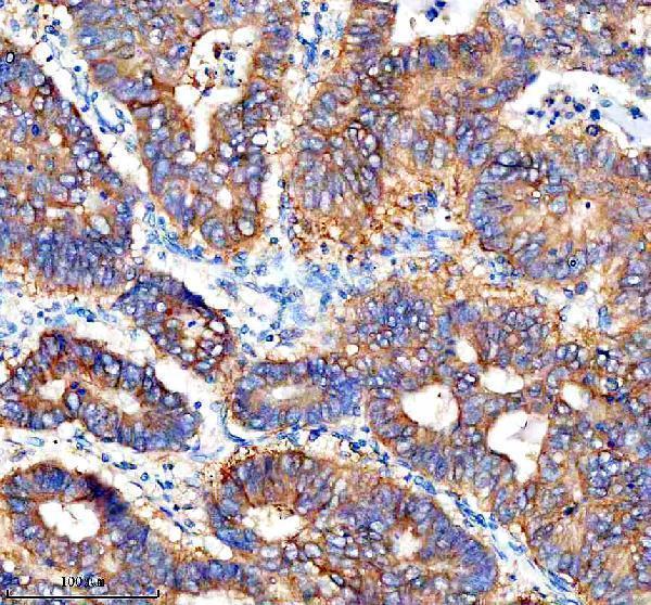
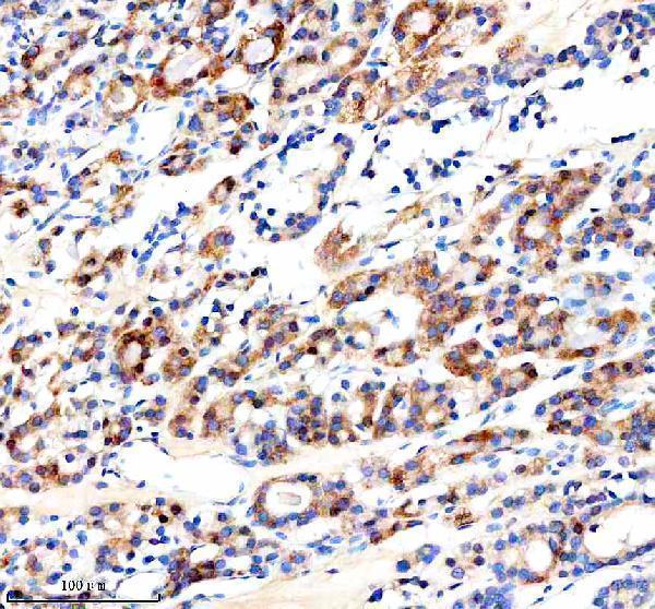
| Sample | Paraffin-embedded human stomach cancer tissue; fixative not specified (datasheet A06600-1) |
| Fixation | Image fixative and duration unreported (datasheet A06600-1); verify before use. |
| Sectioning | 4–5 µm sections on charged slides (standard) |
| Deparaffinisation | Xylene, graded ethanol series to water (standard) |
| Antigen retrieval | Heat retrieval: EDTA pH 8.0 (datasheet A06600-1); 20 min, 95–100 °C (standard) |
| Peroxidase block | 3% H2O2, 10 min, room temperature (standard) |
| Blocking | 10% goat serum (datasheet A06600-1) |
| Primary antibody | Rabbit anti-CTSF, 2-5 μg/ml (datasheet A06600-1) |
| Primary incubation | Overnight at 4 °C (datasheet A06600-1) |
| Detection | HRP-conjugated secondary, DAB chromogen (datasheet A06600-1) |
| Counterstain | Hematoxylin, blue, dehydrate and mount (standard) |
| Expected result | CTSF-positive staining in cells in molecular layer of cerebellum (HPA tissue IHC: High). HPA tissue profile: Cytoplasmic expression with a granular pattern in most tissues. No signal in the no-primary control. |
CTSF localizes to lysosomes and has no transmembrane segment (UniProt Q9UBX1 localization/topology). In paraffin IHC, expect granular cytoplasmic staining, including strong signal in hepatocytes, cortical and hippocampal neurons, and testicular Leydig cells (HPA: tissue IHC). HPA rates the tissue IHC profile Enhanced while describing only medium consistency between staining and RNA data (HPA: reliability).
| Discrete cytoplasmic granules in hepatocytes or cortical neurons, with stronger staining than adjacent negative cells (HPA: High in hepatocytes and cortical neurons). | This matches the reported tissue pattern and lysosomal location (HPA: granular cytoplasm; UniProt Q9UBX1: lysosome). Granularity alone cannot prove lysosomal identity or antibody specificity; assess the cell type and controls together (general IHC interpretation). |
| Predominantly nuclear staining or a continuous cell-edge outline replaces cytoplasmic granules (HPA: granular cytoplasmic tissue pattern). | Treat this as discordant with the expected IHC pattern and check controls and staining conditions (HPA: tissue IHC; general IHC practice). HPA also reports plasma-membrane IF signal with uncertain support, so an isolated edge signal is not decisive evidence of CTSF localization (HPA: subcellular IF). |
| Strong signal appears in adipocytes or bone-marrow hematopoietic cells (HPA: Not detected in these cell types). | Consider nonspecific antibody binding or detection-system activity, especially when the expected positive cells also stain poorly (general IHC interpretation). HPA's cell-specific observations are comparison points, not proof that every cell in those tissues lacks CTSF (HPA: tissue IHC). |
| Uniform haze covers cells and extracellular spaces, obscuring granules (HPA: expected granular cytoplasmic pattern). | This prevents a confident positive call (general IHC interpretation). Review primary-antibody concentration, blocking, washing, and chromogen development with a no-primary control; these are general IHC checks, not documented CTSF-specific sensitivities (general IHC practice). |
| No granular signal is visible in adequately sampled hepatocytes or cortical neurons (HPA: High in both cell types). | An absent expected positive weakens the run's interpretability (general IHC interpretation). Check tissue preservation, retrieval, primary-antibody conditions, and detection with controls; the supplied sources do not identify a CTSF-specific fixation or retrieval failure mode (general IHC practice). |
| Cellular location and topology (UniProt Q9UBX1: lysosome; no transmembrane segment). | The expected tissue readout is intracellular granularity, consistent with HPA's cytoplasmic pattern (HPA: tissue IHC). A chromogenic granule cannot identify its organelle by itself; assess morphology and controls (general IHC interpretation). |
| Proteolytic processing and antibody epitope (UniProt Q9UBX1: signal peptide 1–19, propeptide 20–270, mature chain 271–484). | Different epitopes could recognize different processing states, but the supplied record gives no epitope for the catalog antibody (UniProt Q9UBX1: processing; supplied antibody data). Do not predict which form produces tissue staining from these boundaries alone. |
| Tissue and cell-type differences (HPA: tissue IHC; UniProt Q9UBX1: tissue specificity). | HPA reports High hepatocyte staining but Not detected in colon glandular cells and ovarian stroma (HPA: tissue IHC). UniProt reports moderate colon and high ovary expression (UniProt Q9UBX1: tissue specificity); tissue-level expression and selected-cell IHC observations should be interpreted at their respective scales. |
| Antibody-specific validation (HPA: antibody records and tissue IHC reliability). | CAB002141 has Enhanced IHC status, whereas HPA031431 has Supported ICC status without an IHC rating in the supplied list (HPA: antibodies). HPA's Enhanced tissue profile includes medium staining–RNA consistency; neither label transfers automatically to a different catalog antibody (HPA: reliability; general assay interpretation). |
| IF/ICC Q: Should vesicles appear? (HPA: subcellular IF). | A: Vesicles are the supported main IF location; endoplasmic-reticulum and plasma-membrane locations are uncertain (HPA: subcellular IF). This helps interpret images from an independently validated IF assay but does not establish an IHC compartment beyond the reported granular cytoplasm (HPA: tissue IHC). |
| Situation | Likely cause | Next action |
|---|---|---|
| Expected positive tissue has no staining (HPA: High in hepatocytes or cortical neurons). | The run may have failed at retrieval, primary incubation, or detection, but no CTSF-specific failure step is identified (general IHC practice; supplied sources). | Check a known-positive section in the same run, then review retrieval and reagent performance; avoid declaring the sample negative until its positive control works (general IHC practice). |
| Granules are weak in a reported High cell type (HPA: tissue IHC). | Weak detection or tissue condition is possible; HPA's High category does not set a required intensity for every section (general IHC interpretation; HPA: tissue IHC). | Compare matched positive and no-primary controls, and optimize antibody dilution or detection within the validated IHC workflow (general IHC practice). |
| Signal fills nuclei or traces cell borders more than cytoplasmic granules (HPA: tissue IHC). | The pattern conflicts with the reported lysosomal and granular distribution; its precise cause cannot be assigned from appearance alone (UniProt Q9UBX1: lysosome; HPA: tissue IHC). | Recheck morphology, positive tissue, and no-primary control; repeat with the IHC-validated antibody if the discordance persists (general IHC practice). |
| Cells reported as Not detected stain as strongly as expected positives (HPA: adipocytes or bone-marrow hematopoietic cells). | Nonspecific binding or endogenous detection activity is possible; an HPA negative call remains cell-type specific (general IHC interpretation; HPA: tissue IHC). | Compare a no-primary control; if the detection chemistry uses an enzyme, check its endogenous-activity block, then reassess antibody conditions (general IHC practice). |
| Diffuse brown background obscures cell boundaries and granules (HPA: granular cytoplasmic pattern). | Excess primary antibody, incomplete washing, or prolonged chromogen development can raise background (general chromogenic IHC practice). | Inspect no-primary background, then adjust blocking, dilution, washing, and development using the validated IHC workflow (general IHC practice). |
| Colon or ovary appears inconsistent with UniProt tissue expression (UniProt Q9UBX1: tissue specificity). | UniProt's tissue-level expression and HPA's Not detected calls for colon glandular cells and ovarian stroma describe different scopes (UniProt Q9UBX1; HPA: tissue IHC). | Score the identified cell population and report the observed compartment and intensity; use HPA's cell-specific pattern as the comparison, with its medium RNA consistency caveat (HPA: tissue IHC/reliability). |


Comprehensive Human Protein Atlas IHC scoring per tissue (reliability: Enhanced — Medium consistency between antibody staining and RNA expression data.). Rows are taken directly from the HPA tissue chart — click any row's HPA link to view the source.
| Tissue | Cell type | Level | Evidence | Source |
|---|---|---|---|---|
| Cerebellum | Cells in molecular layer | High | Protein (IHC) | HPA → |
| Cerebral cortex | Neuronal cells | High | Protein (IHC) | HPA → |
| Hippocampus | Neuronal cells | High | Protein (IHC) | HPA → |
| Liver | Hepatocytes | High | Protein (IHC) | HPA → |
| Seminal vesicle | Glandular cells | High | Protein (IHC) | HPA → |
| Tissue | Cell type | Level | Evidence | Source |
|---|---|---|---|---|
| Adipose tissue | Adipocytes | Not detected | Protein (IHC) | HPA → |
| Appendix | Glandular cells | Not detected | Protein (IHC) | HPA → |
| Bone marrow | Hematopoietic cells | Not detected | Protein (IHC) | HPA → |
| Colon | Glandular cells | Not detected | Protein (IHC) | HPA → |
| Kidney | Cells in glomeruli | Not detected | Protein (IHC) | HPA → |
Troubleshoot CTSF staining in paraffin sections by checking retrieval, compartment, cell type, controls, and scoring before interpreting chromogenic signal.
A06600-1 has real IHC data from human paraffin sections of stomach and thyroid cancer tissue (catalog IHC captions); no IF/ICC data are listed (catalog applications and images).
A06600-1 is listed for human IHC (catalog applications and reactivity). Its IHC captions show staining in paraffin sections of human stomach and thyroid cancer tissue (catalog IHC captions).
Which to pick: Choose A06600-1 for human paraffin-section IHC; its listed IHC concentration is 2–5 μg/ml (datasheet), and its captions document staining at 2 μg/ml after EDTA pH 8.0 retrieval (catalog IHC captions). The captions do not report the fixative (catalog IHC captions). No listed SKU supports an IF/ICC or cross-species recommendation: A06600-1 lists human reactivity and IHC, but no IF/ICC application or image (catalog applications, reactivity and images).