CUEDC1 / CUE domain-containing protein 1 · IHC design guide

Design Immunohistochemistry for CUEDC1

Plan chromogenic CUEDC1 IHC-P around cytoplasmic staining, with high staining in cerebellar Purkinje cells and stomach glandular cells (HPA tissue IHC). Start the catalog antibody at 2 μg/mL (datasheet: 2 μg/mL) and score cytoplasmic staining against controls (standard IHC practice).

Evidence assembled Oct 2026 · For research use; verify linked source records and product datasheet before use
Immunohistochemistry protocol sheet for CUEDC1 (IHC for CUEDC1): expected localisation Cytoplasmic tissue staining (HPA tissue IHC), antibody A17855, validated IHC image, and IHC protocol steps
Printable CUEDC1 IHC protocol sheet — expected localisation Cytoplasmic tissue staining (HPA tissue IHC), antibody A17855, controls and protocol steps. Open the full CUEDC1 IHC guide →

CUEDC1 Immunohistochemistry Experimental Design Guide

Expected localisation, validated protocols, controls and antibodies — the at-a-glance facts below, then the full design guide.

Must know before staining
Expected localisation Cytoplasmic tissue staining (HPA tissue IHC)
Staining pattern General cytoplasmic staining in tissue cells (HPA tissue IHC)
Antigen retrieval Citrate pH 6.0 HIER, 95–98 °C, 20 min (rule: cytoplasmic / membrane antigen)
Positive control ⓘ Cerebellum+4 more · see all
Negative control ⓘ None in HPA (detected in all 45 tissues); use no-primary + isotype controls
Important caveats
Reasons your staining may differ from the expected pattern.
Fixation Keep fixation consistent across sections (standard IHC practice; not target-specific)
Caveat Staining–RNA consistency is medium; verify specificity (HPA tissue IHC)
Regulation No target-specific regulation annotated (UniProt)
Isoform / epitope 2 isoforms; epitope differences unknown (UniProt)
Section 1

Recommended CUEDC1 IHC & IF Protocols

The catalog antibody has a datasheet IHC-P protocol (datasheet: IHC-P). Two published CUEDC1 IHC studies add tissue-specific examples (PMC7655170; PMC12664147).

Recommended immunohistochemistry (IHC-P) protocol parameters
SampleTissue sections; selected-image fixative not specified (standard IHC workflow)
FixationImage fixative and duration unreported (datasheet A17855); verify before use.
Sectioning4–5 µm sections on charged slides (standard)
DeparaffinisationXylene, graded ethanol series to water (standard)
Antigen retrievalHeat-induced epitope retrieval in citrate buffer, pH 6.0, 20 min at 95–98 °C (standard rule: cytoplasmic / membrane antigen)
Peroxidase block3% H2O2, 10 min, room temperature (standard)
Blocking10% normal serum of the secondary host, 30 min, room temperature (standard)
Primary antibodyRabbit anti-CUEDC1, 2 μg/mL (datasheet A17855)
Primary incubationOvernight at 4 °C (standard)
DetectionHRP-polymer secondary, DAB chromogen 5–10 min (standard)
CounterstainHematoxylin, blue, dehydrate and mount (standard)
Expected resultCUEDC1-positive staining in purkinje cells of cerebellum (HPA tissue IHC: High). HPA tissue profile: General cytoplasmic expression. No signal in the no-primary control.
💡Decision noteStart with citrate pH 6.0 HIER at 95–98 °C for 20 min (page antigen retrieval); neither article specifies retrieval conditions (PMC7655170; PMC12664147).
Section 2

What Is the Expected CUEDC1 Staining Pattern?

CUEDC1 tissue IHC is generally cytoplasmic, with high staining reported in cerebellar Purkinje cells and stomach glandular cells (HPA: tissue IHC). HPA rates the tissue pattern Approved, with medium consistency between antibody staining and RNA data and external verification pending (HPA: reliability). UniProt does not annotate a subcellular location or transmembrane segment (UniProt Q9NWM3: subcellular annotation and topology).

What am I looking at on my slide?
Cytoplasmic staining in Purkinje cells or stomach glandular cells.This matches the reported IHC distribution; these cells show high staining (HPA: tissue IHC). Compare signal with neighboring cells and the negative control before calling a section positive (general IHC practice).
Predominantly nuclear staining, with little cytoplasmic signal.This departs from the general cytoplasmic tissue IHC profile (HPA: tissue IHC). Consider nonspecific staining or a detection artefact, then check controls; compartment alone cannot prove the signal is false (general IHC practice).
Strong staining in an unexpected cell population.Check morphology and the reported cell distribution before assigning specificity (HPA: tissue IHC). Cross-reactivity or endogenous detection activity can produce misleading chromogenic signal; an antibody omission control helps distinguish detection background (general IHC practice).
Diffuse colour across cells and extracellular spaces.A widespread haze is difficult to reconcile with the reported cell-associated cytoplasmic pattern (HPA: tissue IHC). Assess blocking, washing, detection chemistry and counterstain against a negative control before scoring cells (general IHC practice).
No stain in Purkinje cells or stomach glandular cells.Absent signal in these reported high populations weakens interpretation of a negative test section (HPA: tissue IHC). Check the antibody and detection run with a positive control; HPA reliability does not guarantee every specimen will stain (HPA: reliability; general IHC practice).
💡Expected CUEDC1 appearanceCall a section positive when identifiable Purkinje cells or stomach glandular cells show clear, chiefly cytoplasmic high staining (HPA: tissue IHC); diffuse haze or nuclear-only colour warrants a control check (general IHC practice).
How each factor affects the staining
Tissue and cell selectionPurkinje and stomach glandular cells have high reported IHC staining; several other listed populations are Medium, while spleen red-pulp cells are Low (HPA: tissue IHC).
Strength of tissue evidenceThe tissue IHC profile is Approved, but antibody staining and RNA expression show medium consistency and external verification is pending (HPA: reliability). Treat the pattern as a guide, not proof of specificity in each specimen.
Antibody validationBoth listed antibodies, HPA023050 and HPA024836, are Approved for IHC (HPA: antibodies). Approval supports use of the reported pattern but does not by itself establish an Enhanced IHC validation result.
Does ICC-IF predict paraffin IHC?ICC-IF reports approved plasma membrane, cilium, centrosome and basal-body locations, plus cytosol and other structures (HPA: subcellular). Interpret these separately from the general cytoplasmic tissue IHC profile (HPA: tissue IHC).
Protein architecture and isoformsUniProt lists 2 isoforms, no transmembrane segment and no signal peptide or propeptide (UniProt Q9NWM3). The supplied record does not map an antibody epitope, so isoform recognition cannot be inferred.
Antigen retrieval and fixationRetrieval is a routine paraffin IHC workflow variable (general IHC practice). No target-specific CUEDC1 fixation sensitivity or retrieval response is supplied; do not infer one from topology, modifications or tissue staining (UniProt Q9NWM3; HPA: tissue IHC).
Why is my staining missing, weak or wrong?
SituationLikely causeNext action
Positive control and test section both lack signal.A run-level antibody, detection or retrieval problem is possible (general IHC practice); the observation alone does not establish CUEDC1 fixation sensitivity.Check control records, reagent preparation, retrieval settings and detector function; repeat with a reported high tissue population (HPA: tissue IHC; general IHC practice).
Test section is negative but the positive control stains.The tested cell population may express less detectable protein; HPA reports Low staining in spleen red-pulp cells and Medium staining in several listed populations (HPA: tissue IHC).Score the correct cell type and inspect morphology and internal controls. Report a negative finding for the tested specimen without extending it to all tissues (general IHC practice).
Colour appears without primary antibody.Endogenous enzyme activity or nonspecific detection chemistry may contribute to chromogenic signal (general IHC practice).Review the omission control and detector-specific blocking and washing steps. Interpret cell-associated staining only after background is controlled (general IHC practice).
Most of the section has diffuse background.Incomplete blocking or washing, excessive antibody concentration, or detection background are possible workflow causes (general IHC practice).Compare negative controls; check blocking, wash conditions and the catalog antibody's validated IHC-P dilution before changing one workflow variable at a time (general IHC practice).
Signal is strong in nuclei rather than cytoplasm.The distribution conflicts with HPA's general cytoplasmic tissue IHC profile, although that conflict alone does not establish cross-reactivity (HPA: tissue IHC).Recheck tissue morphology, counterstain and controls. If feasible, compare the result with another IHC-approved antibody before assigning a nuclear CUEDC1 pattern (HPA: antibodies; general IHC practice).
A punctate membrane or ciliary pattern is expected in paraffin sections.HPA reports those locations in ICC-IF, while its tissue IHC summary reports general cytoplasmic expression (HPA: subcellular; HPA: tissue IHC).Use the tissue IHC pattern to score chromogenic sections. Consult the separate IF/ICC guide when testing fine subcellular localisation (HPA: tissue IHC; HPA: subcellular).

Sample controls for CUEDC1 IHC & IF

🧪Run cerebellum first and score Purkinje cells for staining (HPA: High in Purkinje cells). HPA detects CUEDC1 in all 45 scored tissues, so there is no HPA-supported negative tissue; use no-primary and isotype controls, and expect cells without specific signal on the positive slide to show only counterstain or background (HPA: no negative tissue listed; standard IHC practice).
Positive control tissue: Cerebellum (Purkinje cells, HPA High)
Negative control tissue: None in HPA: CUEDC1 is detected in all 45 scored tissues. Use a no-primary (secondary-only) and an isotype control instead.
ICC-IF cell lines (HPA subcellular resource): HPA ICC-IF images show CUEDC1 in A-431, U-251MG, U2OS, ASC52telo, RPTEC/TERT1, hTERT-RPE1 (serum starved), with annotated localisation: Plasma membrane (approved), Primary cilium (approved), Centrosome (approved), Basal body (approved) (HPA subcellular).
Technical controls: Include a no-primary, secondary-only control and an isotype control matched to the primary antibody’s host and immunoglobulin class; for a polyclonal primary, use matched nonimmune IgG (standard IHC practice). If available, use a CUEDC1 knockout sample as a biological specificity control, and quench endogenous peroxidase for HRP detection in cerebellum (standard IHC practice).
⚠️Feasibility: A target-specific fixation window and antigen-retrieval dependence are unreported in the supplied evidence; the A17855 liver IHC caption does not state a fixative (A17855 caption: fixative unreported). The evidence does not establish whether frozen sections or IF are easier than paraffin IHC, although HPA provides ICC-IF images for CUEDC1 (HPA: ICC-IF image cell lines). Cerebellar neuronal lipofuscin can cause autofluorescent background if this tissue is examined by IF (standard IF practice).

HPA tissue IHC evidence for CUEDC1

Comprehensive Human Protein Atlas IHC scoring per tissue (reliability: Approved — Medium consistency between antibody staining and RNA expression data. Pending external verification.). Rows are taken directly from the HPA tissue chart — click any row's HPA link to view the source.

Positive expression · recommended positive controls

TissueCell typeLevelEvidenceSource
Cerebellum Purkinje cells High Protein (IHC) HPA →
Stomach Glandular cells High Protein (IHC) HPA →
Adipose tissue Adipocytes Medium Protein (IHC) HPA →
Adrenal gland Glandular cells Medium Protein (IHC) HPA →
Appendix Glandular cells Medium Protein (IHC) HPA →

Undetected expression · recommended negative controls

TissueCell typeLevelEvidenceSource
None in HPA: CUEDC1 is detected in all 45 scored tissues. Use a no-primary (secondary-only) and an isotype control instead.
Section 3

Advanced CUEDC1 IHC Tips

Use the IHC-validated antibody with tissue controls, then assess staining by cell type, compartment and reproducibility (HPA tissue IHC; standard IHC practice).

How should I optimize retrieval when CUEDC1 staining is weak in paraffin sections?
Start CUEDC1 paraffin IHC with citrate pH 6.0 heat-induced epitope retrieval at 95–98 °C for 20 min (page retrieval rule: cytoplasmic / membrane antigen). Run a known positive control alongside the test section, such as cerebellum containing Purkinje cells or stomach containing glandular cells (HPA: High in both cell types; standard IHC practice). If staining remains weak, compare a modestly longer retrieval time on matched sections while holding antibody concentration and detection constant (standard IHC practice). If tissue damage or diffuse background increases, return to the starting conditions and check section adhesion and blocking before changing retrieval chemistry (standard IHC practice).
Can fixation account for weak or uneven CUEDC1 staining?
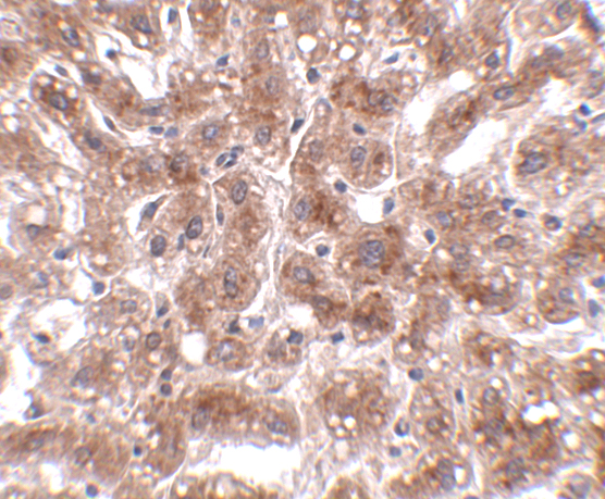
CUEDC1-specific fixation sensitivity is unknown: the selected A17855 tissue-IHC caption does not state a fixative (A17855 tissue-IHC caption). Record the specimen’s fixative, fixation duration and processing history, then compare sections with similar handling before attributing a staining difference to CUEDC1 abundance (standard IHC practice). Uneven fixation can complicate morphology and antigen detection, so examine section centers and edges separately and include a concurrently processed control (standard IHC practice). The reported liver staining at 2 µg/mL establishes a captioned IHC concentration, but provides no basis for choosing or ranking fixation conditions (A17855 tissue-IHC caption).
Which CUEDC1 staining pattern should count as plausible in tissue IHC?
Assess a broadly cytoplasmic pattern first in tissue sections, because that is the reported tissue-IHC profile (HPA: general cytoplasmic expression). High staining in cerebellar Purkinje cells and stomach glandular cells provides cell-type context for controls, while spleen red-pulp cells are listed as low (HPA: tissue IHC). Approved cell-imaging locations also include plasma membrane, primary cilium, centrosome and basal body, with additional cytosolic and microtubule-associated locations (HPA: subcellular). At chromogenic IHC resolution, score a fine punctate or rim pattern cautiously and compare it with morphology and controls before assigning an organelle (standard IHC practice; HPA: subcellular).
How can isoforms and an unknown epitope affect CUEDC1 IHC interpretation?
CUEDC1 has 2 annotated isoforms, while the supplied evidence does not map this antibody’s epitope to either isoform (UniProt Q9NWM3: isoforms 1 and 2; supplied A17855 caption). The annotated CUE domain spans residues 46–89, so an epitope claim involving that region requires antibody-specific mapping (UniProt Q9NWM3: CUE domain; standard IHC practice). Compare staining across validated controls and, if available, an independently validated antibody recognizing a known distinct epitope (standard IHC practice). Do not interpret a tissue staining difference as isoform-specific expression without epitope and isoform evidence, even when the cellular pattern appears plausible (standard IHC practice; UniProt Q9NWM3: alternative splicing).
How should IF help investigate an ambiguous CUEDC1 IHC pattern?
Use IF as a separate follow-up and multiplex CUEDC1 with a validated marker for the expected cell type, such as Purkinje cells or stomach glandular cells (HPA: High tissue-IHC staining; standard IF practice). Choose a fluorophore and imaging channel with low specimen autofluorescence, and include single-label and no-primary controls to assess bleed-through and background (standard IF practice). HPA cell imaging reports plasma membrane, cilium, centrosome and basal-body locations, alongside additional cytosolic locations (HPA: subcellular). Select permeabilisation according to the antibody epitope’s accessibility on either side of the membrane; its side is not supplied here, so verify antibody-specific guidance before interpreting absent signal (standard IF practice; supplied A17855 caption).
What should I check when CUEDC1 IHC has diffuse brown background?
First compare the tissue with a no-primary control and inspect whether brown signal follows cells, section edges or damaged areas (standard IHC practice). For peroxidase-based chromogenic detection, check the hydrogen-peroxide block and wash steps; endogenous peroxidase and residual detection reagent can produce misleading color (standard IHC practice). Titrate the primary antibody around the captioned 2 µg/mL condition on matched sections, while keeping retrieval and development time constant (A17855 tissue-IHC caption; standard IHC practice). A credible result should preserve cell morphology and a reproducible cellular distribution consistent with the reported general cytoplasmic tissue profile (HPA: tissue IHC; standard IHC practice).
How should I score CUEDC1 chromogenic staining across specimens? ⚠ ANSWER MARKED FOR VERIFICATION
Define the scored compartment and cell population before analysis, using the reported general cytoplasmic tissue pattern as a starting point (HPA: tissue IHC; standard IHC practice). Record either an H-score from intensity and percentage of positive cells, the percentage of positive cells alone, or positive-cell density per mm² for a clearly defined region (standard IHC practice). Normalize cell counts to the number of eligible cells, or area measurements to analyzable tissue area, and exclude folds, necrosis and edge artefacts consistently (standard IHC practice). Keep retrieval, antibody concentration, chromogen development, imaging and thresholds aligned across the compared sections, with blinded scoring where feasible (standard IHC practice).
How can I distinguish genuine CUEDC1 staining from an IHC artefact?
Prioritize reproducible cellular staining with intact morphology and a plausible broadly cytoplasmic pattern, especially in Purkinje cells or stomach glandular cells used as positive references (HPA: tissue IHC; standard IHC practice). Treat signal confined to necrosis, section edges or damaged tissue as suspect, and compare it with a no-primary control (standard IHC practice). Check endogenous peroxidase blocking when brown deposit appears independent of the primary antibody (standard IHC practice). Strong isolated nuclear staining needs further validation because the supplied tissue profile is cytoplasmic, while the approved cell-imaging locations emphasize membrane, cilium, centrosome and related structures (HPA: tissue IHC; HPA: subcellular).
Boster reagents

Best CUEDC1 / CUE domain-containing protein 1 IHC Antibodies

A17855 lists IHC-P and IF applications, with images of human liver tissue for both; listed reactivity covers human, mouse, and rat (catalog: applications, reactivity and image captions).

Real IHC data Immunohistochemistry of CUEDC1 in human liver tissue with CUEDC1 antibody at 2 μg/mL.
Anti-CUEDC1 Antibody
Cat # A17855

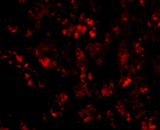
A17855 will render with its human liver IHC image at 2 μg/mL (catalog: IHC image caption). Its IF image also shows human liver tissue at 20 μg/mL; human, mouse, and rat are listed as reactive species (catalog: IF image caption and reactivity).

Which to pick: For paraffin-section IHC, choose A17855: IHC-P is listed, and its own IHC image shows human liver tissue at 2 μg/mL; the fixative is unreported (catalog: applications and IHC image caption). For IF, choose A17855 based on its human liver IF image at 20 μg/mL; ICC validation is unreported (catalog: applications and IF image caption). For work across species, A17855 lists human, mouse, and rat reactivity, though its IHC and IF images show human tissue only; clonality is unreported (catalog: reactivity, image captions and clone field).

Each figure is that product's own IHC / IF validation image from its datasheet.

References

  1. UniProt Consortium. UniProt entry Q9NWM3 (CUED1_HUMAN, CUE domain-containing protein 1).
  2. Human Protein Atlas. CUEDC1 tissue IHC expression (reliability: Approved).
  3. Human Protein Atlas. CUEDC1 subcellular location (ICC-IF): Mainly localized to the plasma membrane, centrosome, primary cilium and basal body. In addition localized to the cytosol, microtubules, cytokinetic bridge, mitotic spindle and primary cilium tip..
  4. Human Protein Atlas. CUEDC1 antibody validation summary (2 antibodies).
  5. CUEDC1 inhibits epithelial-mesenchymal transition via the TβRI/Smad signaling pathway and suppresses tumor progression in non-small cell lung cancer. Aging 2020 — PMC7655170.
  6. CUEDC1 promotes glycolytic metabolism reprogramming through the CUEDC1/CACNG4/PI3K axis to promote ER-positive breast cancer growth. Cellular & molecular biology letters 2025 — PMC12664147.
  7. PubMed PMID:14702039 — UniProt-cited evidence.
  8. PubMed PMID:15489334 — UniProt-cited evidence.