This website uses cookies to ensure you get the best experience on our website.


- Table of Contents
Plan chromogenic CX3CR1 IHC in paraffin sections using the catalog antibody at 2–5 μg/ml (datasheet A00280-2). Compare the expected membrane location (UniProt) with reported cytoplasmic tissue staining (HPA tissue IHC), using tissue controls to assess the result.

Expected localisation, validated protocols, controls and antibodies — the at-a-glance facts below, then the full design guide.
| Expected localisation | Membrane expected; cytoplasm seen in tissue IHC (UniProt) (HPA tissue IHC) | |
| Staining pattern | Cytoplasmic signal in several tissues; caudate glia stain (HPA tissue IHC) | |
| Antigen retrieval | EDTA pH 8.0 HIER, heat-mediated (datasheet A00280-2) | |
| Positive control | Adrenal gland+4 more · see all | |
| Negative control | Adipose tissue+4 more · see all |
| Fixation | Keep fixation consistent across paraffin sections. (standard IHC practice; not target-specific) | |
| Caveat | IHC staining has low consistency with RNA expression (HPA tissue IHC) | |
| Regulation | Inflammation recruits CX3CR1+ cells (UniProt) | |
| Isoform / epitope | 4 isoforms; extracellular vs cytoplasmic epitope coverage is unknown (UniProt) |
The catalog antibody protocol uses heat-mediated EDTA retrieval (datasheet A00280-2). The four published IHC protocols below provide tissue-specific staining conditions (PMC3921448; PMC3399807; PMC5471565; PMC3730155).
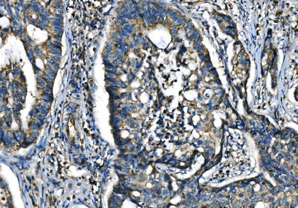
| Sample | Paraffin-embedded human adenocarcinoma of the right colon tissue; fixative not specified (datasheet A00280-2) |
| Fixation | Image fixative and duration unreported (datasheet A00280-2); verify before use. |
| Sectioning | 4–5 µm sections on charged slides (standard) |
| Deparaffinisation | Xylene, graded ethanol series to water (standard) |
| Antigen retrieval | Heat retrieval: EDTA pH 8.0 (datasheet A00280-2); 20 min, 95–100 °C (standard) |
| Peroxidase block | 3% H2O2, 10 min, room temperature (standard) |
| Blocking | 10% goat serum (datasheet A00280-2) |
| Primary antibody | Rabbit anti-CX3CR1, 2-5 μg/ml (datasheet A00280-2) |
| Primary incubation | Overnight at 4 °C (datasheet A00280-2) |
| Detection | HRP-conjugated secondary, DAB chromogen (datasheet A00280-2) |
| Counterstain | Hematoxylin, blue, dehydrate and mount (standard) |
| Expected result | CX3CR1-positive staining in glandular cells of adrenal gland (HPA tissue IHC: Medium). HPA tissue profile: Cytoplasmic expression in several tissues, including cerebral cortex. No signal in the no-primary control. |
CX3CR1 is a seven-pass cell-membrane receptor (UniProt P49238 topology). In paraffin-section IHC, assess staining in lymphocyte subsets and neural tissue, including microglia (UniProt P49238 tissue specificity and function). HPA reports medium glial-cell staining in caudate and cytoplasmic expression across several tissues, but rates its tissue IHC data Approved with low consistency between antibody staining and RNA expression (HPA tissue IHC).
| Cell outlines stain in an anatomically plausible population, with limited signal in adjacent cells. | Membrane-associated staining fits receptor topology (UniProt P49238 topology). Interpret the cell population alongside tissue context: HPA reports medium caudate glial-cell staining, while its tissue IHC profile also describes cytoplasmic staining (HPA tissue IHC). |
| Signal is confined to nuclei, with no convincing cell-outline staining. | An exclusively nuclear pattern conflicts with the reported cell-membrane location (UniProt P49238 subcellular location; HPA subcellular ICC-IF). Check the negative control and antibody dilution before scoring it as CX3CR1 (standard IHC practice). |
| Strong staining appears in a cell type listed as not detected, such as bronchial respiratory epithelium. | This conflicts with the sampled HPA tissue pattern, but one negative entry is not proof of universal absence (HPA tissue IHC). Consider cross-reactivity or endogenous detection activity; compare a negative control and tissue morphology (standard IHC practice). |
| Diffuse colour covers stroma and multiple cell types without clear cellular boundaries. | This is difficult to assign to a CX3CR1-positive population (UniProt P49238 tissue specificity; HPA tissue IHC). Background from detection or blocking is possible; inspect the negative control and reassess detection conditions (standard IHC practice). |
| No signal is visible in a section expected to contain HPA-positive cells. | HPA reports medium staining in caudate glial cells and several specified glandular-cell populations, not guaranteed positivity in every section (HPA tissue IHC). Confirm that the relevant cells are present, then review the IHC controls and assay conditions (standard IHC practice). |
| Receptor topology | Seven transmembrane segments and extracellular and cytoplasmic regions support a membrane-localised interpretation (UniProt P49238 topology). Apparent intracellular colour in tissue IHC should be judged in context because HPA also describes cytoplasmic expression (HPA tissue IHC). |
| Tissue and cell context | HPA lists medium staining in caudate glia and cells of several glandular tissues, but not-detected staining in selected other cell types (HPA tissue IHC). Its Approved tissue profile has low staining–RNA consistency, so use these entries as comparison points rather than definitive identity tests (HPA tissue IHC). |
| Antibody validation | HPA lists HPA046587 and CAB032478 as Approved for IHC; HPA046587 is Supported for ICC (HPA antibodies). Those ratings do not make every stained cell CX3CR1-positive, particularly given the tissue-profile consistency caveat (HPA tissue IHC; standard IHC practice). |
| Isoforms | Four isoforms are listed (UniProt P49238 isoforms). This record supplies no isoform-specific tissue pattern or antibody epitope, so do not assign different staining patterns to particular isoforms (UniProt P49238 isoforms; HPA antibodies). |
| What should IF/ICC show? | HPA reports supported plasma-membrane localisation in ICC-IF and lists images from HEL, THP-1 and U2OS (HPA subcellular ICC-IF). Use that localisation as a comparison; IF/ICC assay design belongs in its own guide (HPA subcellular ICC-IF). |
| Situation | Likely cause | Next action |
|---|---|---|
| No colour in the chosen positive-control tissue | The sampled section may lack the relevant cells, or the IHC run may have failed (HPA tissue IHC; standard IHC practice). | Confirm the reported cell population is present; check a run control and review the validated IHC procedure, retrieval and antibody dilution before interpreting absence (standard IHC practice). |
| Uniform haze or stromal colour obscures cells | Background detection or incomplete blocking can obscure cellular staining (standard IHC practice). | Compare the negative control; optimise blocking, washes and detection conditions, then judge whether cell boundaries become discernible (standard IHC practice). |
| Only nuclei are strongly coloured | This disagrees with cell-membrane localisation (UniProt P49238 subcellular location; HPA subcellular ICC-IF). | Inspect the negative control and morphology; review antibody dilution and detection before assigning nuclear colour to CX3CR1 (standard IHC practice). |
| Unexpected epithelial cells stain strongly | Some glandular cells show medium HPA staining, while specified bronchial respiratory epithelial cells are not detected; tissue and cell identity matter (HPA tissue IHC). | Identify the exact cell population, compare its HPA tissue entry and check negative-control staining before concluding cross-reactivity (HPA tissue IHC; standard IHC practice). |
| Signal is mainly cytoplasmic rather than outlining cells | HPA tissue IHC describes cytoplasmic expression, whereas UniProt and HPA ICC-IF place CX3CR1 at the membrane (HPA tissue IHC; UniProt P49238 subcellular location; HPA subcellular ICC-IF). | Record the compartment and cell type separately; compare controls and treat compartment agreement across methods as supportive rather than required (standard IHC practice). |
| An HPA-listed negative cell type shows faint colour | Faint signal may reflect background; an HPA not-detected result describes its sampled tissue IHC observation (HPA tissue IHC; standard IHC practice). | Compare nearby cells and the negative control, then report intensity and distribution without treating faint colour alone as confirmed CX3CR1 (standard IHC practice). |


Comprehensive Human Protein Atlas IHC scoring per tissue (reliability: Approved — Low consistency between antibody staining and RNA expression data.). Rows are taken directly from the HPA tissue chart — click any row's HPA link to view the source.
| Tissue | Cell type | Level | Evidence | Source |
|---|---|---|---|---|
| Adrenal gland | Glandular cells | Medium | Protein (IHC) | HPA → |
| Appendix | Glandular cells | Medium | Protein (IHC) | HPA → |
| Breast | Glandular cells | Medium | Protein (IHC) | HPA → |
| Caudate | Glial cells | Medium | Protein (IHC) | HPA → |
| Colon | Glandular cells | Medium | Protein (IHC) | HPA → |
| Tissue | Cell type | Level | Evidence | Source |
|---|---|---|---|---|
| Adipose tissue | Adipocytes | Not detected | Protein (IHC) | HPA → |
| Bronchus | Respiratory epithelial cells | Not detected | Protein (IHC) | HPA → |
| Cerebellum | Cells in granular layer | Not detected | Protein (IHC) | HPA → |
| Cervix | Glandular cells | Not detected | Protein (IHC) | HPA → |
| Endometrium | Cells in endometrial stroma | Not detected | Protein (IHC) | HPA → |
Troubleshoot CX3CR1 staining in paraffin sections by checking retrieval, compartment, cell identity and controls before assigning biological meaning.
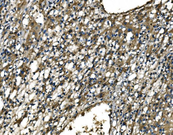
A00280-2 has real IHC data from human paraffin sections of right-colon adenocarcinoma, liver cancer, and lymphoma; no IF data are supplied (catalog image captions; catalog applications).
A00280-2 is listed for human IHC, with a recommended concentration of 2–5 μg/ml (catalog applications and reactivity; datasheet: 2–5 μg/ml). Its IHC captions show staining in paraffin sections of human right-colon adenocarcinoma, liver cancer, and lymphoma (catalog image captions).
Which to pick: For human tissue IHC, choose polyclonal A00280-2: its paraffin-section caption reports EDTA retrieval at pH 8.0 and primary antibody at 2 μg/ml (catalog antibody description; A00280-2 IHC caption). The fixative is unreported, and A00280-2 has no listed IF/ICC application or IF image, so there is no supported IF/ICC choice here (A00280-2 IHC caption; catalog applications and images). For cross-species work, no SKU qualifies because A00280-2 lists human reactivity only (catalog reactivity).