CXCR5 / C-X-C chemokine receptor type 5 · IHC design guide

Design Immunohistochemistry for CXCR5

Plan CXCR5 paraffin-section IHC around cytoplasmic staining in a subset of immune cells (HPA tissue IHC), while accounting for its membrane receptor topology (UniProt). Use tonsil germinal center cells as a positive staining reference (HPA tissue IHC) and keep fixation consistent across sections (standard IHC practice).

Evidence assembled Oct 2026 · For research use; verify linked source records and product datasheet before use
Immunohistochemistry protocol sheet for CXCR5 (IHC for CXCR5): expected localisation Cytoplasmic in tissue (HPA tissue IHC); membrane protein (UniProt), antibody M00663-1, validated IHC image, and IHC protocol steps
Printable CXCR5 IHC protocol sheet — expected localisation Cytoplasmic in tissue (HPA tissue IHC); membrane protein (UniProt), antibody M00663-1, controls and protocol steps. Open the full CXCR5 IHC guide →

CXCR5 Immunohistochemistry Experimental Design Guide

Expected localisation, validated protocols, controls and antibodies — the at-a-glance facts below, then the full design guide.

Must know before staining
Expected localisation Cytoplasmic in tissue (HPA tissue IHC); membrane protein (UniProt)
Staining pattern Cytoplasmic staining in a subset of immune cells (HPA tissue IHC)
Antigen retrieval EDTA pH 8.0 HIER, heat-mediated (datasheet M00663-1)
Positive control ⓘ Appendix+4 more · see all
Negative control ⓘ Adipose tissue+4 more · see all
Important caveats
Reasons your staining may differ from the expected pattern.
Fixation Keep fixation consistent across sections (standard IHC practice; not target-specific)
Caveat Presumed off-target binding may mimic staining (HPA tissue IHC)
Regulation Expression in mature B cells (UniProt)
Isoform / epitope 2 isoforms; verify whether the epitope is extracellular or cytoplasmic (UniProt)
Section 1

Recommended CXCR5 IHC & IF Protocols

The catalog antibody’s IHC-P protocol is accompanied by three published CXCR5 staining protocols for synovium, lymphoma tissue, and liver tissue (PMC1065316; PMC4437674; PMC4792554).

Recommended immunohistochemistry (IHC-P) protocol parameters
SampleParaffin-embedded rat spleen tissue; fixative not specified (datasheet M00663-1)
FixationImage fixative and duration unreported (datasheet M00663-1); verify before use.
Sectioning4–5 µm sections on charged slides (standard)
DeparaffinisationXylene, graded ethanol series to water (standard)
Antigen retrievalHeat retrieval: EDTA pH 8.0 (datasheet M00663-1); 20 min, 95–100 °C (standard)
Peroxidase block3% H2O2, 10 min, room temperature (standard)
Blocking10% goat serum (datasheet M00663-1)
Primary antibodyRabbit monoclonal (clone 20C58) anti-CXCR5, 1:50 (datasheet M00663-1)
Primary incubationOvernight at 4 °C (datasheet M00663-1)
DetectionHRP-conjugated secondary, DAB chromogen (datasheet M00663-1)
CounterstainHematoxylin, blue, dehydrate and mount (standard)
Expected resultCXCR5-positive staining in non-germinal center cells of appendix (HPA tissue IHC: High). HPA tissue profile: Cytoplasmic expression in a subset of immune cells. No signal in the no-primary control.
💡Decision noteStart with heat-mediated EDTA at pH 8.0 for the catalog antibody (datasheet: M00663-1). The published citrate retrieval applies to double-label staining (PMC1065316).
Section 2

What Is the Expected CXCR5 Staining Pattern?

CXCR5 is a seven-pass cell-membrane receptor expected in mature B cells (UniProt P32302: topology and tissue specificity). In paraffin IHC, look for staining in a subset of immune cells; HPA describes the observed pattern as cytoplasmic, so cytoplasmic signal alone is not grounds for rejection (HPA: tissue IHC). HPA rates the tissue profile Enhanced but reports medium staining–RNA consistency and presumed off-target binding (HPA: reliability description).

What am I looking at on my slide?
Selected immune cells stain in a lymphoid section, with membrane-associated or cytoplasmic signal.This fits the receptor’s membrane location and the subset-of-immune-cells pattern observed in tissue IHC (UniProt P32302: subcellular location; HPA: tissue IHC). Score the stained cells and their compartment separately; do not require a sharp membrane rim before calling a cellular signal compatible with the HPA pattern.
Strong nuclear staining dominates cells that otherwise lack a convincing membrane or cytoplasmic pattern.Nuclear localization is unsupported by the supplied CXCR5 localization records (UniProt P32302: cell membrane; HPA: cytoplasmic tissue profile). Treat the result as suspect, then compare it with positive tissue and detection controls before attributing it to CXCR5. The supplied records do not identify a cause for nuclear signal.
Staining appears broadly in cells outside the expected subset, including cells scored as undetected in HPA.Consider nonspecific antibody binding or detection activity, especially if the pattern recurs in a control lacking primary antibody (HPA: presumed off-target binding; standard chromogenic IHC practice). Cell identity matters: HPA also reports endocrine-cell staining in duodenum, colon and rectum, so epithelial-region signal cannot automatically be called artefactual (HPA: tissue IHC).
Diffuse colour covers tissue, empty spaces or most cells without a discernible cellular pattern.This cannot establish CXCR5-positive cells; review the primary-free control and the detection and blocking steps (standard chromogenic IHC practice). HPA’s reported pattern is restricted to a subset of immune cells, although its tissue record also lists particular endocrine populations (HPA: tissue IHC). Record background separately from cell-specific intensity.
No signal appears in a section expected to contain positive cells.First verify that the relevant cell population is present: HPA reports High staining in tonsil germinal center cells, spleen white-pulp cells, and specified appendix and lymph-node cells (HPA: tissue IHC). A blank slide despite those cells prompts a check of the IHC-validated antibody, retrieval and detection workflow (standard IHC practice); it does not by itself establish CXCR5 absence.
💡Expected CXCR5 appearanceCall a result compatible when selected cells show discernible membrane-associated or cytoplasmic staining in an HPA-supported population, potentially High in tonsil germinal center or spleen white-pulp cells; widespread nuclear or cell-independent colour is suspect (UniProt P32302: membrane location; HPA: tissue IHC; standard IHC practice).
How each factor affects the staining
Membrane topology and epitope locationCXCR5 has seven transmembrane segments with extracellular and cytoplasmic regions (UniProt P32302: topology). An antibody’s epitope would determine which region it recognizes, but no epitope is supplied here; topology alone cannot set antigen retrieval, permeabilisation or dilution conditions.
Tissue and cell selectionHPA reports High staining in specified cells of appendix, duodenum, lymph node, spleen and tonsil, and Medium staining in bone marrow, colon and rectum (HPA: tissue IHC). Use the named cells when interpreting a section; an organ-wide positive or negative label would overstate those observations.
Antibody validation and specificityTwo listed antibodies, HPA042432 and CAB026149, each have Enhanced IHC status (HPA: antibody validation). The tissue profile also notes medium staining–RNA consistency and presumed off-target binding (HPA: reliability description). Read validation alongside morphology and controls rather than treating every coloured cell as confirmed CXCR5.
Isoforms and processingUniProt lists Long and Short isoforms, one chain spanning residues 1–372, and no annotated signal peptide or propeptide (UniProt P32302: isoforms and processing). The supplied evidence does not show isoform-specific IHC patterns or establish shedding; do not assign a staining difference to either mechanism.
IF/ICC: what should be expected?A membrane pattern is the available localization expectation (UniProt P32302: cell membrane; HPA: ICC-IF summary). HPA provides no ICC-IF image cell lines or main-location assignment in this payload, so it offers no image-based IF confirmation here (HPA: subcellular record). Assess IF/ICC with its own guide.
Why is my staining missing, weak or wrong?
SituationLikely causeNext action
Known-positive lymphoid section is blank.The expected HPA-positive cells may be absent from the field, or an IHC step may have failed (HPA: tissue IHC; standard IHC practice).Confirm the named cell population on the section, then review the IHC-validated antibody, retrieval and chromogenic detection controls (HPA: tissue IHC; standard IHC practice). No target-specific retrieval setting is supplied.
Only diffuse cytoplasmic staining is seen.Cytoplasmic staining can match HPA’s tissue profile, but haze without cell boundaries may be background (HPA: tissue IHC; standard IHC practice).Check whether signal resolves to a subset of cells and compare a primary-free control; document compartment and background independently (HPA: tissue IHC; standard IHC practice).
Many unexpected cells stain, including a population HPA scores Not detected.Presumed off-target binding is noted for the tissue profile; endogenous chromogenic activity is another general IHC possibility (HPA: reliability description; standard IHC practice).Compare the same cell type in an HPA-supported positive section and a primary-free control; review detection and blocking controls before assigning CXCR5 expression (HPA: tissue IHC; standard IHC practice).
A strong nuclear pattern overwhelms other signal.Nuclear localization lacks support in the supplied localization records; the precise source of the colour is undetermined (UniProt P32302: cell membrane; HPA: tissue IHC).Inspect positive and primary-free controls and record the nuclear result as unconfirmed rather than scoring it as receptor localization (standard IHC practice; UniProt P32302: cell membrane).
A negative-control tissue contains isolated coloured cells.An HPA Not detected call applies to its named cell type, not every cell in that tissue; background or an unlisted cell population remains possible (HPA: tissue IHC; standard IHC practice).Identify the stained cell type first. For example, compare adipocytes with HPA’s adipocyte call, then check the primary-free control before judging specificity (HPA: adipose tissue, adipocytes Not detected; standard IHC practice).
Membrane rims are weak while some relevant cells show cytoplasmic colour.The membrane location predicted from receptor topology and HPA’s observed cytoplasmic tissue profile describe different evidence and need not produce identical visual patterns (UniProt P32302: topology; HPA: tissue IHC).Score the observed compartment and cell identity, compare an HPA-supported positive population, and avoid declaring failure solely because membrane outlines are faint (HPA: tissue IHC; standard IHC practice).

Sample controls for CXCR5 IHC & IF

🧪Run spleen first and assess staining in white-pulp cells, where CXCR5 is High (HPA: spleen, cells in white pulp, High). Use adipose tissue adipocytes as the negative comparator (HPA: adipose tissue, adipocytes, Not detected); on the spleen slide, compare white-pulp staining with cells outside the stained region, without assuming every other cell type is CXCR5-negative (HPA: spleen, cells in white pulp, High).
Positive control tissue: Appendix (Non-germinal center cells, HPA High)
Negative control tissue: Adipose tissue (HPA Not detected)
ICC-IF cell lines (HPA subcellular resource): HPA carries no ICC-IF cell line for CXCR5; derive a cell-line control from the positive tissue's cell type (Non-germinal center cells) and confirm it by RNA or western blot first.
Technical controls: Include a no-primary, secondary-only control; a rabbit isotype control matched to the primary antibody’s isotype and monoclonal or polyclonal format; and, if available, a CXCR5 knockout specimen or antigen-peptide competition control (caption: rabbit anti-CXCR5 antibody; standard IHC practice). For HRP–DAB staining of spleen, block endogenous peroxidase and check background in the no-primary control (caption: rat spleen, HRP–DAB; standard IHC practice).
⚠️Feasibility: The supplied evidence reports no target-specific fixation window or fixation effect, and the selected SKU’s paraffin-section caption does not state a fixative (caption: M00663-1, fixative unreported). Heat retrieval in EDTA at pH 8.0 produced the illustrated spleen IHC result, but the caption does not establish that retrieval is required in every preparation (caption: M00663-1). The evidence does not establish whether frozen sections or IF are easier; for spleen IHC, check endogenous peroxidase background when using HRP–DAB (caption: HRP–DAB detection; standard IHC practice).

HPA tissue IHC evidence for CXCR5

Comprehensive Human Protein Atlas IHC scoring per tissue (reliability: Enhanced — Medium consistency between antibody staining and RNA expression data. Presumed off target binding observed and disregarded.). Rows are taken directly from the HPA tissue chart — click any row's HPA link to view the source.

Positive expression · recommended positive controls

TissueCell typeLevelEvidenceSource
Appendix Non-germinal center cells High Protein (IHC) HPA →
Duodenum Endocrine cells High Protein (IHC) HPA →
Lymph node Non-germinal center cells High Protein (IHC) HPA →
Spleen Cells in white pulp High Protein (IHC) HPA →
Tonsil Germinal center cells High Protein (IHC) HPA →

Undetected expression · recommended negative controls

TissueCell typeLevelEvidenceSource
Adipose tissue Adipocytes Not detected Protein (IHC) HPA →
Adrenal gland Glandular cells Not detected Protein (IHC) HPA →
Breast Adipocytes Not detected Protein (IHC) HPA →
Bronchus Respiratory epithelial cells Not detected Protein (IHC) HPA →
Caudate Glial cells Not detected Protein (IHC) HPA →
Section 3

Advanced CXCR5 IHC Tips

These questions address CXCR5 staining in paraffin sections with chromogenic detection, using the selected antibody’s tissue image and the supplied expression evidence.

Which retrieval condition should I start with for CXCR5 in paraffin sections?
Start with heat-mediated EDTA retrieval at pH 8.0 for paraffin-section CXCR5 IHC (datasheet M00663-1). The selected rat spleen image used this retrieval, a 10% goat-serum block, and 1:50 primary antibody overnight at 4°C (caption M00663-1). If staining is weak, compare another retrieval buffer only after confirming that the EDTA run worked on a positive control (standard IHC practice). Keep section thickness, detection, and DAB development matched across serial sections so a stronger signal can be assessed alongside tissue damage and background (standard IHC practice). Look for membrane-associated signal consistent with this 7-pass receptor (UniProt P32302 topology and subcellular location).
How should I troubleshoot CXCR5 staining differences between paraffin blocks with different fixation histories?
Target-specific fixation sensitivity is unknown, and the selected paraffin-section caption does not report its fixative (caption M00663-1). Record each block’s fixative, delay before fixation, and fixation duration before comparing staining across blocks (standard IHC practice). For prospectively collected material, keep the fixation schedule consistent and document departures from it (standard IHC practice). Test matched sections with EDTA at pH 8.0 and the reported 1:50 overnight primary incubation at 4°C (caption M00663-1). Include a positive rat spleen section and a no-primary control in the same run to assess staining and background under each processing condition (caption M00663-1; standard IHC practice).
Should I score cytoplasmic CXCR5 staining as confidently as membrane staining?
Prioritize convincing membrane-associated staining because CXCR5 is assigned to the cell membrane and has 7 transmembrane segments (UniProt P32302 subcellular location and topology). The tissue IHC profile also reports cytoplasmic staining in a subset of immune cells, so record that pattern separately (HPA tissue IHC profile). Compare membrane and cytoplasmic signal in the same annotated cell population rather than combining them into one positive score (standard IHC practice). Review a no-primary section for diffuse DAB and check whether staining persists in well-preserved areas away from section edges (standard IHC practice). Treat cytoplasmic-only staining cautiously because presumed off-target binding was observed in the tissue survey (HPA tissue IHC reliability description).
Could CXCR5 isoforms or epitope position explain inconsistent IHC staining?
CXCR5 has 2 annotated isoforms, Long and Short, but the supplied evidence does not locate this antibody’s epitope or establish isoform coverage (UniProt P32302 isoforms; caption M00663-1). Its topology includes extracellular residues 1–55 and a cytoplasmic tail at 326–372 (UniProt P32302 topology). Glycosylation is annotated at residues 28 and 196, both in extracellular regions (UniProt P32302 glycosylation and topology). Ask for epitope mapping before attributing a negative section to an isoform or glycosylation effect (standard IHC interpretation). Meanwhile, compare matched positive-control sections under the reported EDTA pH 8.0 retrieval and a no-primary control (caption M00663-1; standard IHC practice).
How can an IF follow-up help assess a CXCR5 chromogenic IHC pattern?
Use IF as a separate validation experiment: the supplied CXCR5 evidence gives a paraffin-section IHC image, but no antibody-specific IF result (caption M00663-1; HPA subcellular record). Multiplex CXCR5 with a mature B-cell marker such as CD20 to assess the expected cell population (UniProt P32302 tissue specificity; standard IF practice). Choose a fluorophore in a channel with low measured tissue autofluorescence, and include single-stain and no-primary controls before interpreting overlap (standard IF practice). If the epitope maps to an extracellular loop, assess staining without permeabilisation; an intracellular epitope requires a controlled permeabilisation comparison (UniProt P32302 topology; standard IF practice). Do not assume the IHC 1:50 dilution transfers to IF (caption M00663-1; standard IF practice).
What should I change when CXCR5 DAB staining is diffuse or widespread?
First compare the stained section with a no-primary control and inspect whether DAB accumulates at folds, damaged edges, or poorly preserved areas (standard IHC practice). Apply an endogenous peroxidase block before HRP detection and keep DAB development time consistent across sections (standard IHC practice). The selected image used 10% goat serum, 1:50 primary antibody overnight at 4°C, and an HRP-based DAB workflow (caption M00663-1). If background persists, titrate the primary antibody while holding retrieval and detection constant (standard IHC practice). Weigh diffuse staining against the reported subset-of-immune-cells profile and the tissue survey’s warning about presumed off-target binding (HPA tissue IHC profile and reliability description).
How should I quantify CXCR5-positive cells in chromogenic tissue sections? ⚠ ANSWER MARKED FOR VERIFICATION
Define the anatomical region and cell population before scoring, then apply the same boundaries and DAB threshold to every section (standard IHC quantification practice). Report the percentage of positive cells and, when cell counts and area are reliable, positive-cell density per mm² (standard IHC quantification practice). An H-score can summarize intensity grades 0–3 weighted by the percentage of cells at each grade, yielding 0–300 (standard IHC quantification practice). Normalize density to the analyzed tissue area and report positivity relative to the eligible cells in that region (standard IHC quantification practice). Keep membrane and cytoplasmic scores separate because membrane localization is annotated while cytoplasmic immune-cell staining is also reported (UniProt P32302 subcellular location; HPA tissue IHC profile).
What distinguishes credible CXCR5 positivity from staining artefact in IHC?
Seek a reproducible cell-associated pattern in well-preserved tissue, with membrane staining consistent with CXCR5 localization (UniProt P32302 subcellular location; standard IHC interpretation). Check whether positive cells occupy plausible immune-cell regions: the tissue survey reports high staining in splenic white-pulp cells and tonsillar germinal-center cells (HPA tissue IHC). Compare suspicious signal with a no-primary control, especially around section edges, folds, necrotic areas, and sites of endogenous peroxidase activity (standard IHC practice). Record cytoplasmic-only staining separately, since the survey reports cytoplasmic immune-cell staining and presumed off-target binding (HPA tissue IHC profile and reliability description). Require agreement across controls and replicate sections before calling a marginal DAB signal positive (standard IHC practice).
Boster reagents

Best CXCR5 / C-X-C chemokine receptor type 5 IHC Antibodies

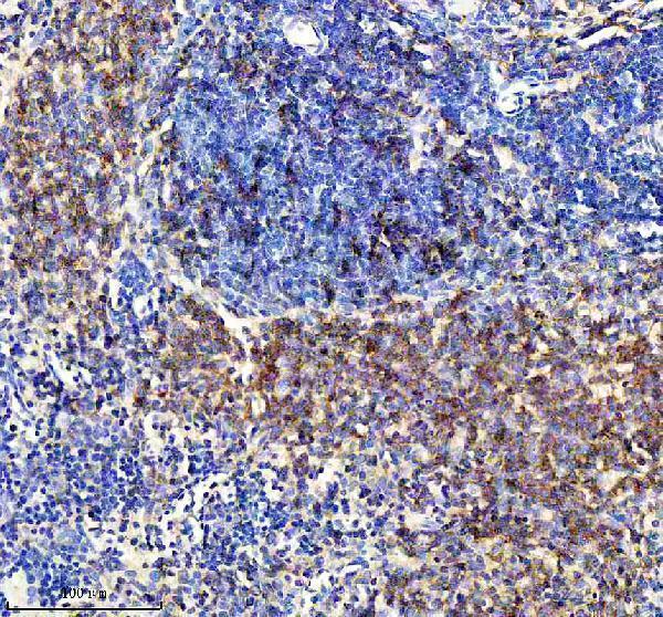
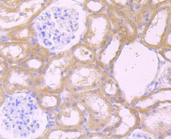
Anti-CXCR5 antibodies have rat spleen and kidney paraffin-section IHC images (IHC image captions). Both list human, mouse and rat reactivity (catalog); M00663-1 also lists IF/ICC applications (catalog).

Real IHC data IHC analysis of CXCR5 using anti-CXCR5 antibody (M00663-1). CXCR5 was detected in a paraffin-embedded section of rat spleen tissue. Heat mediated antigen retrieval was performed in EDTA buffer (pH 8.0, epitope retrieval solution). The tissue section was blocked with 10% goat serum. The tissue section was then incubated with 1:50 rabbit anti-CXCR5 Antibody (M00663-1) overnight at 4°C. Peroxidase Conjugated Goat Anti-rabbit IgG was used as secondary antibody and incubated for 30 minutes at 37°C. The tissue section was developed using HRP Conjugated Rabbit IgG Super Vision Assay Kit (Catalog # SV0002) with DAB as the chromogen.
Anti-CXCR5 Rabbit Monoclonal Antibody
Cat # M00663-1
Real IHC data Immunohistochemical analysis of paraffin-embedded rat kidney tissue using anti-CXCR5 antibody. Counter stained with hematoxylin.
Anti-CXCR5 Antibody
Cat # A00663

M00663-1 has a rat spleen paraffin-section IHC image and lists IHC, IF and ICC applications (M00663-1 IHC image caption; catalog). A00663 has a rat kidney paraffin-section IHC image and lists IHC, but not IF/ICC (A00663 IHC image caption; catalog).

Which to pick: For tissue IHC, both have paraffin-section examples: M00663-1 in rat spleen and A00663 in rat kidney (respective IHC image captions). Choose M00663-1 for IF/ICC because those applications are listed for this rabbit monoclonal; A00663 lists IHC but not IF/ICC (catalog). Both list human, mouse and rat reactivity, but their image captions document rat tissue only; neither caption reports the fixative (catalog; respective IHC image captions).

Each figure is that product's own IHC / IF validation image from its datasheet.

References

  1. UniProt Consortium. UniProt entry P32302 (CXCR5_HUMAN, C-X-C chemokine receptor type 5).
  2. Human Protein Atlas. CXCR5 tissue IHC expression (reliability: Enhanced).
  3. Human Protein Atlas. CXCR5 subcellular location (ICC-IF): Membrane.
  4. Human Protein Atlas. CXCR5 antibody validation summary (2 antibodies).
  5. Chemokine receptors in the rheumatoid synovium: upregulation of CXCR5. Arthritis research & therapy 2005 — PMC1065316.
  6. The serum CXCL13 level is associated with the Glasgow Prognostic Score in extranodal NK/T-cell lymphoma patients. Journal of hematology & oncology 2015 — PMC4437674.
  7. Phenotype and function of CXCR5+CD45RA-CD4+ T cells were altered in HBV-related hepatocellular carcinoma and elevated serum CXCL13 predicted better prognosis. Oncotarget 2015 — PMC4792554.
  8. The chemokine receptor cxcr5 regulates the regenerative neurogenesis response in the adult zebrafish brain. Neural development 2012 — PMC3441421.
  9. PubMed PMID:1425907 — UniProt-cited evidence.
  10. PubMed PMID:7639692 — UniProt-cited evidence.
  11. PubMed PMID:9463416 — UniProt-cited evidence.