CXXC5 / CXXC-type zinc finger protein 5 · IHC design guide

Design Immunohistochemistry for CXXC5

Plan chromogenic CXXC5 IHC in paraffin sections using cytoplasmic staining in cortical neurons and Leydig cells as reference patterns (HPA tissue IHC). Compare staining across consistently fixed sections, and confirm that the chosen antibody recognizes the isoforms relevant to your sample (UniProt).

Evidence assembled Oct 2026 · For research use; verify linked source records and product datasheet before use
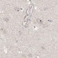
Immunohistochemistry protocol sheet for CXXC5 (IHC for CXXC5): expected localisation Cytoplasmic tissue staining (HPA tissue IHC); nuclear location annotated (UniProt), antibody A06865-1, validated IHC image, and IHC protocol steps
Printable CXXC5 IHC protocol sheet — expected localisation Cytoplasmic tissue staining (HPA tissue IHC); nuclear location annotated (UniProt), antibody A06865-1, controls and protocol steps. Open the full CXXC5 IHC guide →

CXXC5 Immunohistochemistry Experimental Design Guide

Expected localisation, validated protocols, controls and antibodies — the at-a-glance facts below, then the full design guide.

Must know before staining
Expected localisation Cytoplasmic tissue staining (HPA tissue IHC); nuclear location annotated (UniProt)
Staining pattern Cytoplasmic staining in cortical neurons and Leydig cells (HPA tissue IHC)
Antigen retrieval EDTA pH 8.0 HIER, heat-mediated (datasheet A06865-1)
Positive control ⓘ Cerebral cortex+4 more · see all
Negative control ⓘ Adipose tissue+4 more · see all
Important caveats
Reasons your staining may differ from the expected pattern.
Fixation Keep fixation consistent across paraffin sections (standard IHC practice; not target-specific)
Caveat Staining and RNA show only medium consistency (HPA tissue IHC)
Regulation Brain-enhanced RNA expression (HPA tissue RNA)
Isoform / epitope 2 isoforms; epitope coverage is unspecified (UniProt)
Section 1

Recommended CXXC5 IHC & IF Protocols

The catalog antibody protocol is accompanied by two published chromogenic CXXC5 IHC protocols (PMC9712602; PMC6458850).

Recommended immunohistochemistry (IHC-P) protocol parameters
SampleParaffin-embedded mouse brain tissue; fixative not specified (datasheet A06865-1)
FixationImage fixative and duration unreported (datasheet A06865-1); verify before use.
Sectioning4–5 µm sections on charged slides (standard)
DeparaffinisationXylene, graded ethanol series to water (standard)
Antigen retrievalHeat retrieval: EDTA pH 8.0 (datasheet A06865-1); 20 min, 95–100 °C (standard)
Peroxidase block3% H2O2, 10 min, room temperature (standard)
Blocking10% goat serum (datasheet A06865-1)
Primary antibodyRabbit anti-CXXC5, 2-5 μg/ml (datasheet A06865-1)
Primary incubationOvernight at 4 °C (datasheet A06865-1)
DetectionHRP-conjugated secondary, DAB chromogen (datasheet A06865-1)
CounterstainHematoxylin, blue, dehydrate and mount (standard)
Expected resultCXXC5-positive staining in neuronal cells of cerebral cortex (HPA tissue IHC: High). HPA tissue profile: Cytoplasmic expression in several tissues. No signal in the no-primary control.
💡Decision noteStart with heat-mediated EDTA retrieval at pH 8.0 (datasheet A06865-1); citrate at pH 6.0 is a published alternative (PMC9712602; PMC6458850).
Section 2

What Is the Expected CXXC5 Staining Pattern?

CXXC5 is found in the nucleus and cytoplasm and has no transmembrane segment (UniProt Q7LFL8). In tissue IHC, cytoplasmic staining is reported in several tissues, with high staining in cerebral cortex neuronal cells and testis Leydig cells (HPA tissue IHC). The tissue profile is Approved, with medium consistency between staining and RNA data (HPA tissue IHC).

What am I looking at on my slide?
Cytoplasmic signal in cerebral cortex neuronal cells or testis Leydig cells.This matches the reported high tissue IHC pattern (HPA tissue IHC). Score the named cell population and its compartment; signal in nearby cells should be assessed separately. An expected pattern supports interpretation, but the Approved profile has only medium consistency with RNA data (HPA tissue IHC).
Signal appears chiefly at the cell membrane, in extracellular material, or as an isolated chromogenic precipitate.Treat this as a suspected artefact: CXXC5 is nuclear and cytoplasmic, with no transmembrane segment (UniProt Q7LFL8). HPA tissue IHC reports a cytoplasmic profile, while HPA ICC-IF supports nucleoplasmic localisation. Review morphology and controls before assigning such signal to CXXC5.
Strong staining appears in a cell population reported as not detected, such as adipocytes or lung alveolar cells.This conflicts with those specific HPA observations (HPA tissue IHC: adipocytes and alveolar cells Not detected). Consider antibody cross-reactivity or endogenous chromogenic detection activity (general IHC practice). Confirm the cell identity and compare control sections; a tissue-level label does not make every cell in that tissue negative.
Weak, diffuse colour covers cells and surrounding tissue without a clear cellular pattern.This is difficult to score as CXXC5 because the reported tissue profile is cellular and cytoplasmic (HPA tissue IHC). Non-specific antibody binding, endogenous enzyme activity, or detection background can produce diffuse colour (general IHC practice). Compare the negative control and examine whether signal follows recognizable cells.
No signal is seen in cerebral cortex neuronal cells or testis Leydig cells.A blank result in either reported high population raises concern about assay performance (HPA tissue IHC). Verify that the expected cells are present and that the positive control developed; then review the validated IHC-P workflow (general IHC practice). One negative section does not establish absent CXXC5 expression.
💡Expected CXXC5 appearanceCall a section positive when identifiable cerebral cortex neuronal cells or testis Leydig cells show clear cytoplasmic staining consistent with the reported high level (HPA tissue IHC); isolated membrane, extracellular, or diffuse colour is suspect given CXXC5 localisation (UniProt Q7LFL8; HPA tissue IHC).
How each factor affects the staining
Tissue and cell selectionThe strongest listed IHC examples are cerebral cortex neuronal cells and testis Leydig cells (HPA tissue IHC: High). Breast and colon glandular cells are listed at Medium, whereas endometrial glandular cells are Low (HPA tissue IHC). Choose a positive control with the stated cell population present.
Compartment and assayUniProt places CXXC5 in the nucleus and cytoplasm (UniProt Q7LFL8). HPA describes tissue IHC as cytoplasmic, but ICC-IF as mainly nucleoplasmic, with additional cytosolic localisation marked uncertain (HPA tissue IHC; HPA subcellular). Interpret each assay against its own observed pattern.
Validation scopeThe tissue IHC profile is Approved with medium consistency against RNA data (HPA tissue IHC). HPA036508 is Approved for IHC, while HPA058148 is Supported for ICC; these statuses belong to different antibodies and assays (HPA antibodies). Neither status proves that every stained cell or compartment is specific.
Protein form and topologyUniProt lists two isoforms, a full-length chain spanning residues 1–322, no signal peptide or propeptide, and no transmembrane segment (UniProt Q7LFL8). The payload gives no epitope map, so it cannot predict whether either isoform stains differently or whether a particular retrieval step alters recognition.
Why is my staining missing, weak or wrong?
SituationLikely causeNext action
Reported high-positive cells are blank.The target cells may be absent from the section, or the IHC detection workflow may have failed (general IHC practice); the expected cells are High in the HPA profile (HPA tissue IHC).Confirm cell identity on the counterstained section, check a known-positive control, and review the catalog antibody’s IHC-P instructions, including retrieval and detection steps (general IHC practice).
Colour is widespread, including cell-free areas.Diffuse deposition or endogenous detection activity may obscure a cellular result (general IHC practice). This appearance does not match the reported cytoplasmic tissue profile (HPA tissue IHC).Compare a negative control, inspect for precipitate, and review blocking, washes, and chromogen development (general IHC practice). Score only signal that can be assigned to intact cells.
A listed negative cell population stains strongly.Cross-reactivity or endogenous activity is possible (general IHC practice); adipocytes and lung alveolar cells are listed as Not detected (HPA tissue IHC).Confirm the stained cells, check detection controls, and compare with a reported high-positive population (HPA tissue IHC). Do not treat the negative listing as proof that every cell in the tissue must be unstained.
Signal is confined to membrane or extracellular space.That distribution conflicts with nuclear and cytoplasmic localisation and the absence of a transmembrane segment (UniProt Q7LFL8).Inspect section morphology and control staining, then reassess antibody specificity and detection background (general IHC practice). Avoid scoring isolated deposits as CXXC5-positive cells.
Nuclear staining appears in tissue IHC.Nuclear localisation is biologically plausible (UniProt Q7LFL8), although HPA’s tissue IHC summary emphasizes cytoplasmic expression (HPA tissue IHC).Record nuclear and cytoplasmic scores separately, verify cell identity, and compare controls (general IHC practice). Do not assume the ICC-IF nucleoplasmic pattern must reproduce exactly in chromogenic tissue IHC (HPA subcellular).
IF/ICC: should the tissue IHC pattern be expected unchanged?The assays have different reported observations: tissue IHC is cytoplasmic, whereas ICC-IF is mainly nucleoplasmic with uncertain additional cytosolic localisation (HPA tissue IHC; HPA subcellular).Use the separate IF/ICC guide for that assay. For interpretation here, report the compartment actually observed and its controls; HPA’s Supported ICC status applies to HPA058148, while Approved IHC applies to HPA036508 (HPA antibodies).

Sample controls for CXXC5 IHC & IF

🧪Run cerebral cortex first and look for staining in neuronal cells (High; HPA: cerebral cortex). Run adipose tissue as a negative comparator, assessing adipocytes (Not detected; HPA: adipose tissue). On the cortex slide, use non-neuronal cells without specific staining as an internal background reference; HPA does not identify a validated negative cell type within that tissue (HPA: cerebral cortex).
Positive control tissue: Cerebral cortex (Neuronal cells, HPA High)
Negative control tissue: Adipose tissue (HPA Not detected)
ICC-IF cell lines (HPA subcellular resource): HPA ICC-IF images show CXXC5 in A-431, U-251MG, U2OS, with annotated localisation: Nucleoplasm (supported) (HPA subcellular).
Technical controls: Include a no-primary (secondary-only) slide, a matched rabbit IgG isotype control, and CXXC5 knockout tissue or a validated immunizing-peptide block where available (caption: rabbit anti-CXXC5; standard IHC practice). Block endogenous peroxidase and check for brain pigment that could be mistaken for DAB staining (caption: mouse brain, HRP/DAB; standard IHC practice).
⚠️Feasibility: A target-specific fixation window or fixation effect is unreported, and the selected A06865-1 paraffin-section caption does not state a fixative (selected tissue-IHC caption). The caption reports heat retrieval in EDTA at pH 8.0, but does not establish whether CXXC5 staining depends on that retrieval condition (selected tissue-IHC caption). Neither frozen sections nor IF/ICC are established as easier by the supplied evidence; brain autofluorescence warrants checking in IF, while HPA ICC-IF supports mainly nucleoplasmic localization (standard IF practice; HPA: subcellular).

HPA tissue IHC evidence for CXXC5

Comprehensive Human Protein Atlas IHC scoring per tissue (reliability: Approved — Medium consistency between antibody staining and RNA expression data.). Rows are taken directly from the HPA tissue chart — click any row's HPA link to view the source.

Positive expression · recommended positive controls

TissueCell typeLevelEvidenceSource
Cerebral cortex Neuronal cells High Protein (IHC) HPA →
Testis Leydig cells High Protein (IHC) HPA →
Breast Glandular cells Medium Protein (IHC) HPA →
Caudate Neuronal cells Medium Protein (IHC) HPA →
Cervix Squamous epithelial cells Medium Protein (IHC) HPA →

Undetected expression · recommended negative controls

TissueCell typeLevelEvidenceSource
Adipose tissue Adipocytes Not detected Protein (IHC) HPA →
Lung Alveolar cells Not detected Protein (IHC) HPA →
Lymph node Germinal center cells Not detected Protein (IHC) HPA →
Ovary Ovarian stroma cells Not detected Protein (IHC) HPA →
Parathyroid gland Glandular cells Not detected Protein (IHC) HPA →
Section 3

Advanced CXXC5 IHC Tips

Troubleshoot CXXC5 chromogenic IHC by checking retrieval, tissue processing, compartment-specific staining and appropriate controls before scoring signal.

What retrieval should I try first when CXXC5 staining is weak?
Start with heat-mediated EDTA at pH 8.0 for paraffin sections stained with A06865-1 (datasheet A06865-1). Use 2 μg/ml primary antibody overnight at 4°C as the matched reference when assessing retrieval, since changing antibody concentration at the same time obscures the comparison (caption A06865-1; standard IHC practice). Run a mouse brain section beside the problem section and compare similar regions after identical DAB development (caption A06865-1; standard IHC practice). If staining remains weak, adjust heating duration on adjacent sections while retaining EDTA at pH 8.0, and inspect morphology for heat damage (standard IHC practice).
Could fixation explain inconsistent CXXC5 staining between paraffin blocks?
The mouse brain paraffin-section caption does not state the fixative, so target-specific fixation sensitivity for CXXC5 is unknown (caption A06865-1). Record each block’s fixative and fixation interval, and compare sections processed and stained together before attributing a difference to biology (standard IHC practice). Keep the documented EDTA retrieval at pH 8.0 and the 2 μg/ml primary incubation constant during that comparison (datasheet A06865-1; caption A06865-1). If morphology and staining vary together, review processing records and repeat staining with matched controls; neither the tissue staining profile nor CXXC5 topology establishes a fixation-specific effect (standard IHC practice; HPA: tissue IHC; UniProt Q7LFL8 topology).
Should I accept cytoplasmic CXXC5 staining if nucleoplasmic signal is weak?
Evaluate nuclear and cytoplasmic staining separately: CXXC5 is annotated in both compartments, while the subcellular atlas supports nucleoplasmic localization and labels cytosolic localization uncertain (UniProt Q7LFL8 subcellular location; HPA: subcellular). Tissue IHC reports cytoplasmic expression in several tissues, so cytoplasmic signal alone is not grounds for rejection (HPA: tissue IHC). Compare the same cell type across sections and require signal above the no-primary control before assigning either compartment as positive (standard IHC practice). Keep the retrieval and DAB development consistent, because changes in staining intensity can alter which compartment appears dominant (datasheet A06865-1; standard IHC practice).
Can isoforms or epitope accessibility explain a patchy CXXC5 stain?
CXXC5 has 2 annotated isoforms, but the supplied evidence does not map this antibody’s epitope to either one (UniProt Q7LFL8 isoforms; caption A06865-1). It also lists phosphothreonine at residue 53 without establishing that modification as a cause of altered IHC binding (UniProt Q7LFL8 modified residues). Compare neighboring sections using the same EDTA retrieval at pH 8.0 and the documented 2 μg/ml antibody concentration before interpreting patchiness as isoform expression (datasheet A06865-1; caption A06865-1; standard IHC practice). Record which cell types and compartments vary, and seek epitope information before making an isoform-specific claim (standard IHC practice).
How should I investigate CXXC5 localization with multiplex IF?
For an IF/ICC follow-up in cerebral cortex, pair CXXC5 with a marker identifying neuronal cells, which show high tissue-IHC staining (HPA: High in cerebral-cortex neuronal cells; standard IF practice). Choose spectrally separated fluorophores and inspect an unstained specimen so tissue autofluorescence does not masquerade as colocalization (standard IF practice). CXXC5 has no transmembrane segment, so optimize permeabilisation for access to nuclear and cytosolic epitopes, rather than assuming a membrane-facing epitope (UniProt Q7LFL8 topology and subcellular location; standard IF practice). Score nucleoplasmic and cytosolic signals separately, and validate the IF antibody independently of the paraffin-section IHC caption (HPA: subcellular; caption A06865-1; standard IF practice).
What should I check when CXXC5 DAB staining looks diffuse?
The documented paraffin-section workflow used 10% goat serum blocking, a peroxidase-conjugated secondary and DAB development (caption A06865-1). Include no-primary and secondary-only controls to determine whether diffuse color persists without CXXC5 antibody (standard IHC practice). Check endogenous peroxidase blocking and shorten DAB development if the controls also become brown; those are general chromogenic-workflow checks, not evidence of CXXC5 expression (standard IHC practice). Compare the suspect region with intact cells in the same section, then reassess primary concentration against the documented 2 μg/ml condition while keeping retrieval at EDTA pH 8.0 (caption A06865-1; datasheet A06865-1; standard IHC practice).
How can I quantify CXXC5 IHC across samples with different cell densities? ⚠ ANSWER MARKED FOR VERIFICATION
Define the relevant cell population and score nuclear and cytoplasmic DAB signals separately, given the differing compartment observations for CXXC5 (HPA: tissue IHC and subcellular; standard IHC practice). For intensity-weighted scoring, calculate an H-score from the percentages of cells at intensity grades 0–3, yielding a range of 0–300 (standard IHC practice). Alternatively, report the percentage of positive relevant cells, or positive-cell density per mm² of viable tissue, with one fixed positivity threshold (standard IHC practice). Normalize cell-based measures to the counted relevant cells and area-based measures to analyzed viable area; retain identical retrieval, imaging and DAB thresholds across comparisons (standard IHC practice).
How do I distinguish true CXXC5 signal from staining artefacts?
Prioritize staining in intact cells of a documented positive population, such as cerebral-cortex neuronal cells, and compare it with the no-primary control (HPA: High in cerebral-cortex neuronal cells; standard IHC practice). Nuclear or cytoplasmic signal can be plausible, but an unsupported compartment pattern needs further validation because subcellular evidence favors nucleoplasm while tissue IHC often appears cytoplasmic (UniProt Q7LFL8 subcellular location; HPA: subcellular and tissue IHC). Treat staining confined to section edges or necrotic areas as suspect and check whether it follows tissue damage rather than cells (standard IHC practice). Brown signal that survives primary-antibody omission warrants review of endogenous peroxidase blocking and DAB development before a CXXC5-positive call (standard IHC practice).
Boster reagents

Best CXXC5 / CXXC-type zinc finger protein 5 IHC Antibodies

Two anti-CXXC5 antibodies have IHC images from mouse or human brain tissue; one also has a human brain IF image (catalog image captions). Both list human, mouse and rat reactivity (catalog reactivity).

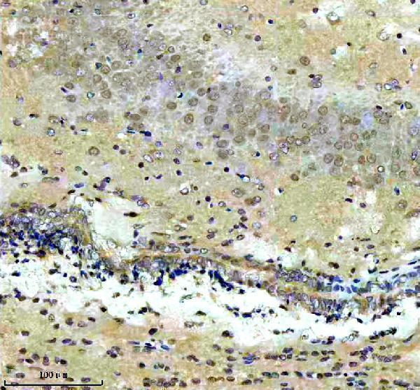
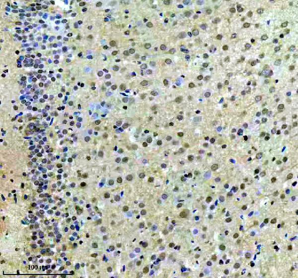
Real IHC data IHC analysis of CXXC5 using anti-CXXC5 antibody (A06865-1). CXXC5 was detected in a paraffin-embedded section of mouse brain tissue. Heat mediated antigen retrieval was performed in EDTA buffer (pH 8.0, epitope retrieval solution). The tissue section was blocked with 10% goat serum. The tissue section was then incubated with 2 μg/ml rabbit anti-CXXC5 Antibody (A06865-1) overnight at 4°C. Peroxidase Conjugated Goat Anti-rabbit IgG was used as secondary antibody and incubated for 30 minutes at 37°C. The tissue section was developed using HRP Conjugated Rabbit IgG Super Vision Assay Kit (Catalog # SV0002) with DAB as the chromogen.
Anti-CXXC5 Antibody ®
Cat # A06865-1
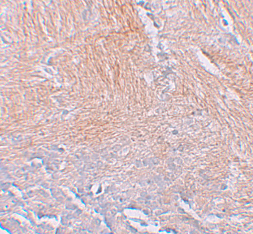
Real IHC data Immunohistochemistry of CXXC5 in human brain tissue with CXXC5 antibody at 2.5 μg/mL.
Anti-CXXC5 Antibody
Cat # A06865

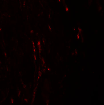
A06865-1 will render with IHC data from a paraffin-embedded mouse brain section (A06865-1 IHC caption). A06865 will render with IHC data from human brain tissue and also lists IF with a human brain image (A06865 IHC and IF captions; catalog applications).

Which to pick: For paraffin-section IHC, choose A06865-1 for its documented mouse brain workflow, or A06865 for its human brain IHC image; the fixative is unreported for both (respective IHC captions). For tissue IF, choose A06865; ICC requires separate validation because its IF image shows human brain tissue (A06865 IF caption). Both list human, mouse and rat reactivity for cross-species planning, but the pictured IHC evidence is mouse for A06865-1 and human for A06865; only A06865-1 is identified as polyclonal (catalog reactivity and dilution text; respective IHC captions).

Each figure is that product's own IHC / IF validation image from its datasheet.

References

  1. UniProt Consortium. UniProt entry Q7LFL8 (CXXC5_HUMAN, CXXC-type zinc finger protein 5).
  2. Human Protein Atlas. CXXC5 tissue IHC expression (reliability: Approved).
  3. Human Protein Atlas. CXXC5 subcellular location (ICC-IF): Mainly localized to the nucleoplasm. In addition localized to the cytosol..
  4. Human Protein Atlas. CXXC5 antibody validation summary (2 antibodies).
  5. Blockade of CXXC5-dishevelled interaction inhibits adipogenic differentiation, obesity, and insulin resistance in mice. Scientific reports 2022 — PMC9712602.
  6. Novel small molecules downregulate CDK1 expression and inhibit Wnt/β-catenin signaling in cutaneous squamous cell carcinoma by targeting its distinct tumor-specific cellular landscape. Experimental & molecular medicine 2025 — PMC12508097.
  7. The Dishevelled-binding protein CXXC5 negatively regulates cutaneous wound healing. The Journal of experimental medicine 2015 — PMC4493411.
  8. CXXC5 mediates growth plate senescence and is a target for enhancement of longitudinal bone growth. Life science alliance 2019 — PMC6458850.
  9. PubMed PMID:19557330 — UniProt-cited evidence.
  10. PubMed PMID:12761501 — UniProt-cited evidence.
  11. PubMed PMID:14702039 — UniProt-cited evidence.