This website uses cookies to ensure you get the best experience on our website.


- Table of Contents
Plan CYB5A chromogenic IHC around cytoplasmic staining most abundant in kidney and liver (HPA tissue IHC). Use the IHC-validated antibody at a starting dilution within 1:50–1:200, then optimize locally (datasheet).

Expected localisation, validated protocols, controls and antibodies — the at-a-glance facts below, then the full design guide.
| Expected localisation | Cytoplasmic tissue staining (HPA tissue IHC) | |
| Staining pattern | Cytoplasmic staining most abundant in kidney and liver (HPA tissue IHC) | |
| Antigen retrieval | Citrate pH 6.0 HIER, 95–98 °C, 20 min (rule: cytoplasmic / membrane antigen) | |
| Positive control | Lung+4 more · see all | |
| Negative control | Adipose tissue+4 more · see all |
| Fixation | Keep fixation consistent across sections. (standard IHC practice; not target-specific) | |
| Caveat | Antibody staining and RNA show medium consistency (HPA tissue IHC) | |
| Regulation | Kidney- and liver-enhanced RNA expression (HPA tissue RNA) | |
| Isoform / epitope | 3 isoforms; epitope coverage is unknown (UniProt) |
Compare the catalog antibody’s IHC-P protocol with three published CYB5A IHC workflows (PMC4448130; PMC8948269; PMC8488225).
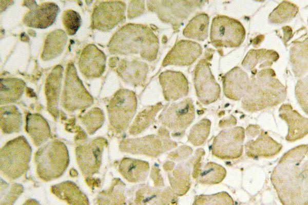
| Sample | Paraffin-embedded human heart tissue; fixative not specified (datasheet A04464-1) |
| Fixation | Image fixative and duration unreported (datasheet A04464-1); verify before use. |
| Sectioning | 4–5 µm sections on charged slides (standard) |
| Deparaffinisation | Xylene, graded ethanol series to water (standard) |
| Antigen retrieval | Heat-induced epitope retrieval in citrate buffer, pH 6.0, 20 min at 95–98 °C (standard rule: cytoplasmic / membrane antigen) |
| Peroxidase block | 3% H2O2, 10 min, room temperature (standard) |
| Blocking | 10% normal serum of the secondary host, 30 min, room temperature (standard) |
| Primary antibody | Rabbit anti-CYB5A, 1:50-1:200 (datasheet A04464-1) |
| Primary incubation | Overnight at 4 °C (standard) |
| Detection | HRP-polymer secondary, DAB chromogen 5–10 min (standard) |
| Counterstain | Hematoxylin, blue, dehydrate and mount (standard) |
| Expected result | CYB5A-positive staining in alveolar cells type II of lung (HPA tissue IHC: High). HPA tissue profile: Cytoplasmic expression most abundant in kidney and liver. No signal in the no-primary control. |
CYB5A is associated with the endoplasmic reticulum and microsome membranes and is also annotated in the cytoplasm; its transmembrane segment spans residues 109–131 (UniProt P00167 topology). Expect cytoplasmic IHC staining in selected cells, especially kidney and liver overall, with high staining reported in lung alveolar type II cells and testicular Leydig cells (HPA: tissue IHC). HPA rates the tissue profile Enhanced, with medium agreement between staining and RNA data (HPA: reliability).
| Cytoplasmic staining in alveolar type II or Leydig cells, with nuclei comparatively clear. | This fits the reported High staining in those cell populations (HPA: lung and testis IHC). Score the named cells separately from neighboring cells: staining intensity across an entire tissue section can hide whether the expected population carries the signal (standard IHC practice). |
| Cytoplasmic staining in kidney proximal tubule cell bodies, with a less intense result than the high-staining examples. | A Medium proximal tubule result matches the listed kidney observation (HPA: kidney IHC). The broader profile says expression is most abundant in kidney and liver; it does not assign every kidney cell or a specific liver cell an intensity (HPA: tissue IHC profile). |
| Predominantly nuclear staining, or a crisp cell-surface outline without convincing cytoplasmic signal. | Treat that compartment pattern as suspect: the reported tissue pattern is cytoplasmic (HPA: tissue IHC), while UniProt places CYB5A at endoplasmic reticulum and microsome membranes and in cytoplasm (UniProt P00167 subcellular location). Review controls and detection conditions before assigning biological meaning (standard IHC practice). |
| Strong staining in a cell population listed as not detected, such as appendix glandular cells. | This conflicts with that specific HPA observation (HPA: appendix glandular cells, Not detected). Consider cross-reactivity or endogenous detection activity and compare a known-positive section processed alongside it (standard IHC practice). HPA entries describe sampled cell populations; they are not a universal negative-cell atlas (HPA: tissue IHC). |
| Diffuse color across tissue and empty spaces, or no staining in a known-positive section. | Color outside cells is background rather than a localizable CYB5A pattern (standard IHC practice). A blank high-staining reference, such as alveolar type II or Leydig cells, makes a technical failure plausible; it cannot establish CYB5A absence in an experimental specimen (HPA: lung and testis IHC; standard IHC practice). |
| Membrane location and topology | CYB5A has one transmembrane segment at residues 109–131 and an annotated endoplasmic reticulum and microsome location (UniProt P00167 topology and subcellular location). Interpret chromogenic signal within the cytoplasmic compartment; these annotations alone do not establish an exact microscopic texture (HPA: cytoplasmic tissue IHC). |
| Reference tissue and cell selection | Lung alveolar type II and testicular Leydig cells are listed as High; kidney proximal tubule cell bodies are Medium (HPA: tissue IHC). Use the cell identity and its listed level together when judging a control; a medium result need not match a high result in intensity (standard IHC practice). |
| Strength of the tissue evidence | The tissue profile is Enhanced, with medium consistency between antibody staining and RNA expression (HPA: reliability). Three listed antibodies have Enhanced IHC status (HPA: antibody validation). These ratings support the reported pattern but do not guarantee that every specimen or assay condition reproduces it (standard IHC practice). |
| Context within a named cell type | Breast adipocytes are listed as Medium, whereas adipocytes in adipose tissue are listed as Not detected (HPA: breast and adipose tissue IHC). Preserve the tissue context when choosing controls or assessing an apparent exception; the shared cell label alone does not predict staining across sites (HPA: tissue IHC). |
| Isoforms and antigen retrieval | UniProt lists 3 isoforms, but the supplied record does not map antibody epitopes to them (UniProt P00167 isoforms). No target-specific retrieval or fixation effect is supplied (UniProt P00167; HPA: tissue IHC). Any retrieval comparison is assay optimization, not evidence that CYB5A requires a particular condition (standard IHC practice). |
| Situation | Likely cause | Next action |
|---|---|---|
| No signal in the experimental section or its known-positive control. | A blank positive reference suggests an assay or specimen-processing problem; it does not establish absent CYB5A (HPA: lung alveolar type II and testis Leydig cells, High; standard IHC practice). | Check section integrity, antibody and detection steps, then compare retrieval conditions using matched sections (standard IHC practice). Reassess the experimental section only after the reference cells show their expected cytoplasmic pattern (HPA: tissue IHC). |
| A reference stains, but the signal is mainly nuclear. | The compartment conflicts with HPA cytoplasmic tissue staining and UniProt membrane and cytoplasm annotations (HPA: tissue IHC; UniProt P00167 subcellular location). This raises an assay-specificity concern rather than proving nuclear CYB5A. | Compare the same cell population with another listed Enhanced IHC antibody if available, and check control and detection behavior (HPA: antibody validation; standard IHC practice). Record the discordant compartment instead of scoring it as expected staining. |
| Broad brown haze obscures cell boundaries. | Diffuse noncellular color prevents a confident compartment or cell-type call (standard IHC practice). It cannot be matched to the reported cytoplasmic pattern in named cells (HPA: tissue IHC). | Inspect a detection-only control and optimize blocking, washing, antibody concentration and detection development on matched sections (standard IHC practice). Re-score only when the expected cells can be distinguished from their surroundings. |
| An HPA-listed negative cell population stains strongly. | For example, staining in appendix glandular cells conflicts with their Not detected entry (HPA: appendix IHC). Cross-reactivity or endogenous detection activity is possible; a single discrepant section cannot distinguish them (standard IHC practice). | Compare a listed high-staining cell population in the same run and use a detection-only control to probe reagent-derived color (HPA: lung and testis IHC; standard IHC practice). Confirm the cell identity before calling the result discrepant. |
| The kidney section is weaker than a lung or testis reference. | That difference can fit the listed levels: kidney proximal tubule cell bodies are Medium, while alveolar type II and Leydig cells are High (HPA: tissue IHC). | Judge each reference against its own reported cell population and level, with matched staining conditions (HPA: tissue IHC; standard IHC practice). Investigate weak kidney staining only if its expected proximal tubule pattern is absent or unreadable. |
| What if an IF/ICC image looks vesicular or cytosolic? | HPA reports vesicles and cytosol as enhanced IF/ICC locations, while its tissue IHC description is cytoplasmic (HPA: subcellular ICC-IF; HPA: tissue IHC). The image types resolve and report location differently (standard microscopy practice). | Use the IF/ICC observation as a location cross-check, and judge the paraffin IHC section by cell identity and cytoplasmic staining (HPA: subcellular ICC-IF; HPA: tissue IHC). Consult the separate IF/ICC guide for that assay. |


Comprehensive Human Protein Atlas IHC scoring per tissue (reliability: Enhanced — Medium consistency between antibody staining and RNA expression data.). Rows are taken directly from the HPA tissue chart — click any row's HPA link to view the source.
| Tissue | Cell type | Level | Evidence | Source |
|---|---|---|---|---|
| Lung | Alveolar cells type II | High | Protein (IHC) | HPA → |
| Testis | Leydig cells | High | Protein (IHC) | HPA → |
| Adrenal gland | Glandular cells | Medium | Protein (IHC) | HPA → |
| Breast | Adipocytes | Medium | Protein (IHC) | HPA → |
| Bronchus | Respiratory epithelial cells | Medium | Protein (IHC) | HPA → |
| Tissue | Cell type | Level | Evidence | Source |
|---|---|---|---|---|
| Adipose tissue | Adipocytes | Not detected | Protein (IHC) | HPA → |
| Appendix | Glandular cells | Not detected | Protein (IHC) | HPA → |
| Bone marrow | Hematopoietic cells | Not detected | Protein (IHC) | HPA → |
| Caudate | Glial cells | Not detected | Protein (IHC) | HPA → |
| Cerebellum | Cells in granular layer | Not detected | Protein (IHC) | HPA → |
Troubleshoot CYB5A chromogenic IHC in paraffin sections using matched controls, compartment-aware scoring, and the antibody’s documented tissue evidence.
A04464-1 has an IHC image from paraffin-embedded human heart (IHC image caption) and is listed for IF and for human, mouse, and rat reactivity (catalog: applications and reactivity).
A04464-1 has an IHC image from paraffin-embedded human heart (A04464-1 IHC image caption). It is also listed for IF and human, mouse, and rat reactivity; no IF image is supplied (catalog: applications, reactivity, IF image alts).
Which to pick: For tissue IHC, choose A04464-1 for paraffin sections; its own image shows human heart, and the fixative is unreported (A04464-1 IHC image caption). For IF, A04464-1 lists a 1:50–1:200 dilution, but ICC is not listed and no IF image is supplied (catalog: applications, IF dilution, IF image alts). For cross-species work, A04464-1 is a rabbit antibody listed as reactive with human, mouse, and rat; the supplied IHC image documents human tissue only (catalog: host and reactivity; A04464-1 IHC image caption).