CYB5B / Cytochrome b5 type B · IHC design guide

Design Immunohistochemistry for CYB5B

Plan CYB5B paraffin IHC using adrenal gland glandular cells or testis Leydig cells as high-staining references (HPA tissue IHC). This guide covers staining interpretation and the catalog antibody’s 2–5 μg/ml IHC concentration (datasheet A09841-1).

Evidence assembled Oct 2026 · For research use; verify linked source records and product datasheet before use
Immunohistochemistry protocol sheet for CYB5B (IHC for CYB5B): expected localisation Cytoplasmic tissue staining; outer mitochondrial membrane expected (HPA tissue IHC; UniProt), antibody A09841-1, validated IHC image, and IHC protocol steps
Printable CYB5B IHC protocol sheet — expected localisation Cytoplasmic tissue staining; outer mitochondrial membrane expected (HPA tissue IHC; UniProt), antibody A09841-1, controls and protocol steps. Open the full CYB5B IHC guide →

CYB5B Immunohistochemistry Experimental Design Guide

Expected localisation, validated protocols, controls and antibodies — the at-a-glance facts below, then the full design guide.

Must know before staining
Expected localisation Cytoplasmic tissue staining; outer mitochondrial membrane expected (HPA tissue IHC; UniProt)
Staining pattern General cytoplasmic staining (HPA tissue IHC)
Antigen retrieval EDTA pH 8.0 HIER, heat-mediated (datasheet A09841-1)
Positive control ⓘ Adrenal gland+4 more · see all
Negative control ⓘ Ovary+2 more · see all
Important caveats
Reasons your staining may differ from the expected pattern.
Fixation Keep fixation consistent across sections (standard IHC practice; not target-specific)
Caveat RNA levels may not predict IHC intensity reliably (HPA tissue IHC)
Regulation Specific regulation unannotated (UniProt)
Isoform / epitope No annotated isoforms; residues 1–15 form a propeptide (UniProt)
Section 1

Recommended CYB5B IHC & IF Protocols

The catalog antibody’s IHC-P protocol uses EDTA retrieval (datasheet: pH 8.0); one published CYB5B IHC protocol uses citrate retrieval (PMC9100579).

Recommended immunohistochemistry (IHC-P) protocol parameters
SampleParaffin-embedded human gall bladder adenosquamous carcinoma tissue; fixative not specified (datasheet A09841-1)
FixationImage fixative and duration unreported (datasheet A09841-1); verify before use.
Sectioning4–5 µm sections on charged slides (standard)
DeparaffinisationXylene, graded ethanol series to water (standard)
Antigen retrievalHeat retrieval: EDTA pH 8.0 (datasheet A09841-1); 20 min, 95–100 °C (standard)
Peroxidase block3% H2O2, 10 min, room temperature (standard)
Blocking10% goat serum (datasheet A09841-1)
Primary antibodyRabbit anti-CYB5B, 2-5 μg/ml (datasheet A09841-1)
Primary incubationOvernight at 4 °C (datasheet A09841-1)
DetectionStreptavidin-biotin complex (SABC), DAB chromogen (datasheet A09841-1)
CounterstainHematoxylin, blue, dehydrate and mount (standard)
Expected resultCYB5B-positive staining in glandular cells of adrenal gland (HPA tissue IHC: High). HPA tissue profile: General cytoplasmic expression. No signal in the no-primary control.
💡Decision noteStart with heat-mediated EDTA retrieval at pH 8.0 (datasheet: A09841-1); try citrate at pH 6 when comparing with the published HNSCC protocol (PMC9100579).
Section 2

What Is the Expected CYB5B Staining Pattern?

CYB5B is an outer mitochondrial membrane protein with a transmembrane segment at residues 123–140 (UniProt O43169 topology). In paraffin section IHC, expect predominantly cytoplasmic staining in glandular cells of the adrenal gland, duodenum, parathyroid gland and small intestine, and in testicular Leydig cells and bladder urothelial cells (HPA: High in each). HPA calls its tissue IHC reliability Approved, with medium consistency between staining and RNA expression (HPA tissue IHC).

What am I looking at on my slide?
Cytoplasmic staining in expected cells, strongest in a known positive tissue.This fits HPA's general cytoplasmic IHC profile. Adrenal, duodenal, parathyroid and small intestinal glandular cells, Leydig cells and urothelial cells are reported High (HPA tissue IHC). A cytoplasmic chromogen signal cannot, by itself, resolve the outer mitochondrial membrane (UniProt O43169 localisation; general IHC practice).
Predominantly nuclear staining, or a sharply different compartment from the expected cytoplasmic pattern.Treat this as suspect localisation and review the control, antibody and detection conditions before assigning it to CYB5B (HPA: general cytoplasmic IHC profile; UniProt O43169: outer mitochondrial membrane). HPA's ICC-IF record reports other compartments, so the IHC and IF observations should be interpreted in their respective assay contexts (HPA subcellular).
Strong staining confined to a cell type reported as undetected.Investigate cross-reactivity or endogenous detection activity if ovary follicle cells, skeletal myocytes or smooth muscle cells stain strongly (HPA: Not detected in those cells; general IHC practice). Their HPA calls are reference observations, not absolute guarantees for every specimen or detection system (HPA tissue IHC).
Diffuse chromogen over tissue and cell-free areas, obscuring cell boundaries.This is more consistent with background than an interpretable CYB5B pattern; inspect the no-primary control and detection reagents (general IHC practice). The expected comparison is cell-associated cytoplasmic staining in the HPA positive populations, not uniform deposition across a section (HPA tissue IHC).
No convincing staining in a selected known-positive tissue.An absent signal in adrenal glandular cells or testicular Leydig cells conflicts with their reported High staining, but does not alone prove CYB5B absence (HPA tissue IHC). Check section quality, antibody compatibility and the IHC detection workflow against controls before interpreting the experimental specimen (general IHC practice).
💡Expected CYB5B appearanceA convincing positive is cell-associated cytoplasmic chromogen in HPA High populations, such as adrenal glandular cells or Leydig cells; dominant nuclear staining or diffuse, cell-independent color is suspect (HPA tissue IHC; UniProt O43169 localisation; general IHC practice).
How each factor affects the staining
Choice of tissue and cell populationUse a reported High population to judge whether the assay detects the expected pattern; follicle cells, skeletal myocytes and smooth muscle cells provide reported Not detected comparisons (HPA tissue IHC). Medium staining in endometrial and stomach glandular cells and Low staining in several other populations should not be scored against a High benchmark (HPA tissue IHC).
Antibody validation and strength of evidenceHPA lists antibody HPA007893 as IHC Approved, and rates tissue IHC reliability Approved with medium staining-to-RNA consistency (HPA tissue IHC; HPA antibodies). These labels support use of the reference pattern but do not establish perfect specificity in a new specimen (general IHC interpretation).
Membrane topology and epitope uncertaintyCYB5B has a transmembrane segment at residues 123–140 and a cytochrome b5 heme-binding domain at 24–100 (UniProt O43169). The supplied record does not map this antibody's epitope; do not infer its accessibility or choose a target-specific retrieval condition from topology alone (UniProt O43169 topology; supplied antibody record).
Endogenous chromogen activityFor peroxidase-based chromogenic IHC, endogenous activity can produce color unrelated to primary-antibody binding; assess it with an appropriate control and the detection system's recommended blocking step (general IHC practice). A widespread deposit must not be counted as CYB5B-positive cells merely because the tissue contains an HPA High population (HPA tissue IHC).
IF/ICC Q&A: should fluorescence be exclusively mitochondrial?No. HPA's approved ICC-IF localisation is mainly endoplasmic reticulum, with additional cytosol and centriolar satellites, while UniProt places CYB5B at the mitochondrial outer membrane (HPA subcellular; UniProt O43169). Record the marker and compartment used for comparison and acknowledge this source-level difference when interpreting IF/ICC.
Why is my staining missing, weak or wrong?
SituationLikely causeNext action
Known-positive glandular cells have no signal.The run may have inadequate primary-antibody or detection signal; absence alone cannot establish target loss (HPA: High in adrenal glandular cells; general IHC practice).Check a known-positive section alongside the sample, then review antibody dilution, retrieval and detection steps under the assay's validated IHC conditions (general IHC practice).
Most of the section has a uniform brown haze.Nonspecific reagent deposition or endogenous peroxidase activity can obscure cellular localisation (general IHC practice).Inspect a no-primary control, blocking and washes; score only distinct cell-associated signal after background is controlled (general IHC practice; HPA: general cytoplasmic profile).
Follicle cells or muscle cells stain strongly.Cross-reactivity or endogenous detection activity is plausible where HPA reports Not detected staining (HPA tissue IHC; general IHC practice).Compare the same run with a reported High tissue and a no-primary control; withhold a CYB5B call if the unexpected signal persists without support (HPA tissue IHC; general IHC practice).
Signal is mainly nuclear.The distribution conflicts with HPA's general cytoplasmic IHC profile and UniProt's membrane localisation (HPA tissue IHC; UniProt O43169).Recheck controls, slide identity and detection conditions before interpreting the nuclear color as CYB5B (general IHC practice).
A weak positive population is being scored as negative.Some populations are reported Medium or Low rather than High; a single intensity threshold can misclassify them (HPA tissue IHC).Compare like cell types and include a High reference population on the run; describe weak staining separately from an absence of detectable signal (HPA tissue IHC; general IHC practice).
IHC and IF suggest different compartments.The sources differ: HPA describes cytoplasmic tissue IHC and mainly ER localisation in ICC-IF, whereas UniProt assigns outer mitochondrial membrane localisation (HPA tissue IHC; HPA subcellular; UniProt O43169).Report each assay's observed pattern and its controls separately; avoid forcing one compartment assignment from chromogenic IHC alone (general IHC practice).

Sample controls for CYB5B IHC & IF

🧪Run adrenal gland first: glandular cells should stain (HPA: High in adrenal gland glandular cells). Use skeletal muscle myocytes as the negative tissue (HPA: Not detected in skeletal muscle myocytes); any independently verified CYB5B-negative cells on the adrenal slide should show only counterstain and background, without specific chromogen (standard IHC practice).
Positive control tissue: Adrenal gland (Glandular cells, HPA High)
Negative control tissue: Ovary (HPA Not detected)
ICC-IF cell lines (HPA subcellular resource): HPA ICC-IF images show CYB5B in A-431, U-251MG, U2OS, with annotated localisation: Endoplasmic reticulum (approved) (HPA subcellular).
Technical controls: Include a no-primary, secondary-only control; a rabbit IgG isotype control matched to the primary antibody; and a CYB5B-knockout biological negative (selected tissue-IHC caption: rabbit primary; standard IHC practice). Block endogenous peroxidase and biotin in adrenal sections when using the reported biotin–SABC/DAB detection (selected tissue-IHC caption: biotin–SABC/DAB; standard IHC practice).
⚠️Feasibility: No target-specific fixation window or fixation effect is reported, and the selected paraffin-section caption does not state a fixative (selected tissue-IHC caption: fixative unreported). The example uses heat retrieval in EDTA at pH 8.0, but whether retrieval is required is unreported (selected tissue-IHC caption: EDTA retrieval). No supplied comparison establishes that frozen sections or IF are easier; in adrenal sections, check endogenous pigment against the DAB signal using the no-primary control (standard IHC practice).

HPA tissue IHC evidence for CYB5B

Comprehensive Human Protein Atlas IHC scoring per tissue (reliability: Approved — Medium consistency between antibody staining and RNA expression data.). Rows are taken directly from the HPA tissue chart — click any row's HPA link to view the source.

Positive expression · recommended positive controls

TissueCell typeLevelEvidenceSource
Adrenal gland Glandular cells High Protein (IHC) HPA →
Duodenum Glandular cells High Protein (IHC) HPA →
Endometrium Glandular cells Medium Protein (IHC) HPA →
Parathyroid gland Glandular cells High Protein (IHC) HPA →
Small intestine Glandular cells High Protein (IHC) HPA →

Undetected expression · recommended negative controls

TissueCell typeLevelEvidenceSource
Ovary Follicle cells Not detected Protein (IHC) HPA →
Skeletal muscle Myocytes Not detected Protein (IHC) HPA →
Smooth muscle Smooth muscle cells Not detected Protein (IHC) HPA →
Section 3

Advanced CYB5B IHC Tips

Troubleshoot CYB5B staining in paraffin sections by checking retrieval, controls, compartmental pattern and cell-level scoring.

What retrieval should I try first when CYB5B staining is weak?
Start with heat-mediated retrieval in EDTA pH 8.0 for paraffin sections (datasheet A09841-1). The selected image used that retrieval before 2 μg/ml primary antibody overnight at 4°C, so keep those conditions together in the first comparison (caption A09841-1). If staining remains weak, compare retrieval time and heating intensity on adjacent sections while keeping detection and exposure to DAB consistent. Inspect tissue morphology and background alongside the expected cytoplasmic staining, since the tissue IHC profile describes general cytoplasmic expression (HPA tissue IHC). Include a known positive cell population from the HPA profile in each run (HPA tissue IHC).
How should I assess whether fixation is limiting CYB5B IHC?
The selected paraffin-section caption does not state a fixative, so target-specific fixation sensitivity is unknown (caption A09841-1). Do not infer fixation tolerance from CYB5B topology, post-translational modifications or tissue staining patterns (UniProt O43169; HPA tissue IHC). Compare sections with documented processing histories using the same EDTA pH 8.0 retrieval and primary incubation, then assess staining and morphology together (datasheet A09841-1). Record fixation type and duration for each specimen; uneven preservation can complicate comparisons within a staining run. If only poorly preserved areas are negative, repeat the assessment in intact tissue before assigning a biological absence.
Where should CYB5B staining appear, and what if it looks nuclear?
Score the selected chromogenic sections for cytoplasmic staining, the broad pattern reported in tissue IHC (HPA tissue IHC). CYB5B is annotated at the mitochondrial outer membrane, with a transmembrane segment at residues 123–140 (UniProt O43169 topology). Subcellular IF data instead place it mainly in the endoplasmic reticulum, with additional cytosol and centriolar-satellite localisation, so chromogenic cytoplasm alone cannot resolve the organelle (HPA subcellular). Predominantly nuclear DAB should prompt review of counterstain, background and the no-primary control before scoring. Compare the suspect field with an intact positive cell population on the same run (HPA tissue IHC).
Could epitope position change how I retrieve or interpret CYB5B?
CYB5B has no annotated isoforms, and its chain spans residues 16–150 after a reported 1–15 propeptide (UniProt O43169). Its heme-binding domain spans 24–100, while the membrane anchor spans 123–140 (UniProt O43169 domains and topology). The supplied antibody caption does not identify the immunogen, so the epitope’s position and fixation accessibility cannot be assigned (caption A09841-1). If a second antibody is available, compare staining only after checking its stated epitope and IHC validation. Keep retrieval at EDTA pH 8.0 for the catalog antibody’s initial comparison (datasheet A09841-1).
How can IF help check an ambiguous chromogenic CYB5B pattern?
Use IF on a separately validated specimen to compare CYB5B with an organelle marker, while keeping this page’s primary readout chromogenic IHC. Multiplex with a marker for the expected cell population, such as glandular cells in adrenal gland, and verify each channel with single-stain controls (HPA tissue IHC). Select fluorophores whose emission separates from the specimen’s autofluorescence, and inspect an unstained section before interpreting faint signal. Because CYB5B has a membrane anchor at residues 123–140, choose permeabilisation after establishing which side contains the antibody epitope; that side is unspecified here (UniProt O43169 topology; caption A09841-1). Compare compartment patterns cautiously: HPA IF reports mainly endoplasmic-reticulum localisation (HPA subcellular).
What should I check when DAB obscures CYB5B-positive cells?
Run a no-primary section to assess signal from the secondary, detection reagents and tissue itself. The selected workflow used a biotinylated goat anti-rabbit secondary, streptavidin-biotin complex and DAB, making reagent-associated background relevant to investigate (caption A09841-1). Apply an endogenous-peroxidase block as part of the chromogenic workflow, then compare with a detection control appropriate to the kit. The caption reports a 10% goat-serum block and 2 μg/ml primary antibody overnight at 4°C; check those starting conditions before changing concentration (caption A09841-1). Exclude diffuse edge staining and damaged areas when judging whether the reported cytoplasmic pattern remains visible (HPA tissue IHC).
How should I quantify CYB5B staining across paraffin sections? ⚠ ANSWER MARKED FOR VERIFICATION
Define the target cell population before scoring, since HPA reports different staining levels across cell types and tissues (HPA tissue IHC). For a chromogenic readout, record the percentage of positive target cells and staining intensity, then calculate an H-score using the same intensity thresholds across sections. Alternatively, report positive-cell density per mm² of viable tissue when cell counts and area are more reproducible. Normalise to the number of evaluable target cells or viable tissue area, and document how necrosis, edges and background were excluded. Include a reference section in each run to monitor changes in retrieval and DAB development.
How do I distinguish true CYB5B staining from artefact?
A credible result has cytoplasmic staining in intact cells, consistent with the tissue IHC profile (HPA tissue IHC). High staining in adrenal gland glandular cells or testis Leydig cells can serve as contextual positives, while follicle cells in ovary were reported as undetected (HPA tissue IHC). Predominantly nuclear signal, staining restricted to cut edges or necrosis, and colour in a no-primary control warrant investigation before calling cells positive. Check endogenous-peroxidase blocking and DAB development when pigment-like signal persists in the detection control. Treat an unexpected organelle assignment cautiously: UniProt annotates the mitochondrial outer membrane, whereas HPA IF reports mainly endoplasmic-reticulum localisation (UniProt O43169; HPA subcellular).
Boster reagents

Best CYB5B / Cytochrome b5 type B IHC Antibodies

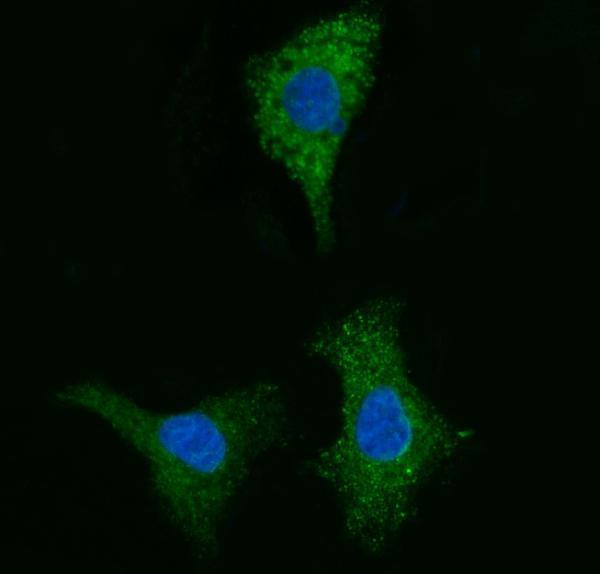
A09841-1 has IHC images from human paraffin sections and an IF image from A549 cells (catalog image captions); its listed reactivity covers human and mouse (catalog applications/reactivity).

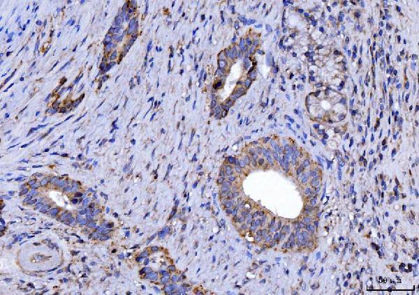
Real IHC data IHC analysis of CYB5B using anti-CYB5B antibody (A09841-1). CYB5B was detected in a paraffin-embedded section of human gall bladder adenosquamous carcinoma tissue. Heat mediated antigen retrieval was performed in EDTA buffer (pH 8.0, epitope retrieval solution). The tissue section was blocked with 10% goat serum. The tissue section was then incubated with 2 μg/ml rabbit anti-CYB5B Antibody (A09841-1) overnight at 4°C. Biotinylated goat anti-rabbit IgG was used as secondary antibody and incubated for 30 minutes at 37°C. The tissue section was developed using Strepavidin-Biotin-Complex (SABC) (Catalog # SA1022) with DAB as the chromogen.
Anti-CYB5B Antibody ®
Cat # A09841-1

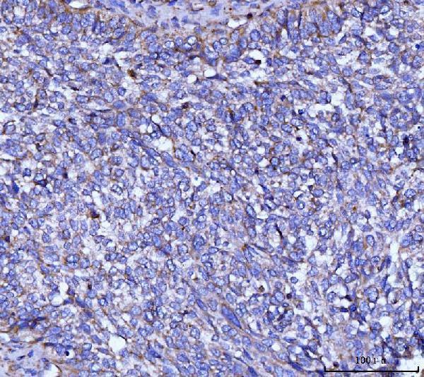
A09841-1 is listed for IHC and IF/ICC, with human and mouse reactivity (catalog applications/reactivity). Its images show IHC in human gall bladder adenosquamous carcinoma, lung cancer, placenta and breast cancer paraffin sections, and IF in A549 cells (catalog image captions).

Which to pick: Choose A09841-1 for paraffin-section IHC: its IHC captions document human paraffin sections stained at 2 μg/ml after EDTA pH 8.0 retrieval; the fixative is unreported (catalog IHC captions). For IF/ICC, the same SKU has an A549 cell image at 5 μg/ml (catalog IF caption). It also lists mouse reactivity, but the supplied IHC and IF images show human samples only; clonality is unreported (catalog applications/reactivity; catalog image captions; catalog clone field).

Each figure is that product's own IHC / IF validation image from its datasheet.

References

  1. UniProt Consortium. UniProt entry O43169 (CYB5B_HUMAN, Cytochrome b5 type B).
  2. Human Protein Atlas. CYB5B tissue IHC expression (reliability: Approved).
  3. Human Protein Atlas. CYB5B subcellular location (ICC-IF): Mainly localized to the endoplasmic reticulum. In addition localized to the cytosol and centriolar satellites..
  4. Human Protein Atlas. CYB5B antibody validation summary (1 antibodies).
  5. Transcriptomic and Proteomic Analysis of Clear Cell Foci (CCF) in the Human Non-Cirrhotic Liver Identifies Several Differentially Expressed Genes and Proteins with Functions in Cancer Cell Biology and Glycogen Metabolism. Molecules (Basel, Switzerland) 2020 — PMC7570661.
  6. Comprehensive Analysis of mTORC1 Signaling Pathway-Related Genes in the Prognosis of HNSCC and the Response to Chemotherapy and Immunotherapy. Frontiers in molecular biosciences 2022 — PMC9100579.
  7. PubMed PMID:14702039 — UniProt-cited evidence.
  8. PubMed PMID:15616553 — UniProt-cited evidence.
  9. PubMed PMID:15489334 — UniProt-cited evidence.