This website uses cookies to ensure you get the best experience on our website.


- Table of Contents
Plan CYP11A1 chromogenic IHC in paraffin sections using granular cytoplasmic staining and documented positive cell populations as reference points (HPA tissue IHC). The guide covers fixation, detection and interpretation for a protein located at the matrix side of the mitochondrial inner membrane (UniProt).

Expected localisation, validated protocols, controls and antibodies — the at-a-glance facts below, then the full design guide.
| Expected localisation | Granular cytoplasm (HPA tissue IHC); mitochondrial inner membrane, matrix side (UniProt). | |
| Staining pattern | Granular cytoplasm in zona fasciculata, cytotrophoblasts and Leydig cells (HPA tissue IHC). | |
| Antigen retrieval | Citrate pH 6.0 HIER, 95–98 °C, 20 min (rule: cytoplasmic / membrane antigen) | |
| Positive control | Adrenal gland+2 more · see all | |
| Negative control | Adipose tissue+4 more · see all |
| Fixation | Keep paraffin-section fixation consistent across samples. (standard IHC practice; not target-specific) | |
| Caveat | Expression varies by tissue and cell type (HPA tissue IHC). | |
| Regulation | High in adrenal gland, placenta and testis (HPA tissue IHC). | |
| Isoform / epitope | Two isoforms; whether PA1698 detects both is unknown (UniProt; catalog). |
The catalog antibody’s IHC-P protocol is followed by four published CYP11A1 staining protocols for adrenal or testis sections (PMC11586580; PMC12375238; PMC7541273; PMC8072358).
| Sample | Tissue sections; selected-image fixative not specified (standard IHC workflow) |
| Fixation | Image fixative and duration unreported (datasheet PA1698); verify before use. |
| Sectioning | 4–5 µm sections on charged slides (standard) |
| Deparaffinisation | Xylene, graded ethanol series to water (standard) |
| Antigen retrieval | Heat-induced epitope retrieval in citrate buffer, pH 6.0, 20 min at 95–98 °C (standard rule: cytoplasmic / membrane antigen) |
| Peroxidase block | 3% H2O2, 10 min, room temperature (standard) |
| Blocking | 10% normal serum of the secondary host, 30 min, room temperature (standard) |
| Primary antibody | Rabbit anti-CYP11A1, 0.5-1μg/ml (datasheet PA1698) |
| Primary incubation | Overnight at 4 °C (standard) |
| Detection | HRP-polymer secondary, DAB chromogen 5–10 min (standard) |
| Counterstain | Hematoxylin, blue, dehydrate and mount (standard) |
| Expected result | CYP11A1-positive staining in cells in zona fasciculata of adrenal gland (HPA tissue IHC: High). HPA tissue profile: Granular cytoplasmic expression, most abundant in adrenal gland, placenta and testis. No signal in the no-primary control. |
In paraffin-section IHC, expect CYP11A1 as granular cytoplasmic staining in adrenal zona fasciculata cells, placental cytotrophoblasts, and testicular Leydig cells (HPA: High in each). The pattern is consistent with localization on the matrix side of the mitochondrial inner membrane; individual membranes need not be resolved by light microscopy (UniProt P05108 topology). HPA rates the tissue staining profile Enhanced, citing consistency with RNA expression (HPA: Enhanced).
| Granular cytoplasmic staining in the expected cells, strongest in an appropriate positive tissue. | This fits the reported tissue pattern and mitochondrial localization (HPA: High in zona fasciculata cells, cytotrophoblasts, and Leydig cells; UniProt P05108 topology). Compare cells within the section: a correct result should preserve recognizable cell-specific staining rather than tint every cell equally (general IHC practice). |
| Predominantly nuclear staining, or a uniform stain unrelated to cytoplasmic granules. | This does not match the expected mitochondrial, granular cytoplasmic pattern (UniProt P05108 topology; HPA: granular cytoplasmic expression). Treat it as suspect and examine antibody specificity, counterstain, and detection controls before interpreting it as CYP11A1 (general IHC practice). |
| Strong staining in a cell type reported as undetected, such as adipocytes or appendix glandular cells. | HPA reports CYP11A1 as Not detected in those cells (HPA: adipocytes and appendix glandular cells). Check morphology and repeat with appropriate controls; cross-reactivity or endogenous chromogenic activity is possible, but an unexpected result alone does not identify its cause (general IHC practice). |
| Pale, diffuse color over cells and surrounding tissue, with little cellular distinction. | Diffuse color lacks the reported granular cytoplasmic distribution (HPA: granular cytoplasmic expression). Assess background with a no-primary control and review blocking, washing, and detection conditions before assigning a positive score (general IHC practice). |
| No staining in a section containing clearly identifiable adrenal zona fasciculata cells. | That conflicts with the reported High staining in these cells (HPA: adrenal zona fasciculata cells, High). Verify tissue identity and assay controls, then review the antibody’s validated IHC-P conditions; this record gives no target-specific fixation or retrieval sensitivity (general IHC practice; HPA: tissue IHC). |
| Cell type and tissue | Use adrenal zona fasciculata cells, placental cytotrophoblasts, or testicular Leydig cells as expected positives (HPA: High). Adipocytes and appendix glandular cells are reported Not detected, making them useful comparison cells, not absolute proof of specificity (HPA: Not detected; general IHC practice). |
| Subcellular resolution | CYP11A1 lies on the matrix side of the mitochondrial inner membrane, with no annotated transmembrane segment (UniProt P05108 topology). Interpret the HPA granular cytoplasmic pattern at light-microscope resolution; it does not establish that individual mitochondrial membranes can be seen (HPA: granular cytoplasmic expression; general IHC practice). |
| Antibody validation and protein forms | HPA lists antibody HPA016436 as IHC Enhanced (HPA: antibody validation). UniProt lists 2 isoforms and a mature chain spanning residues 40–521 (UniProt P05108 processing). Neither record specifies which form an IHC stain detects, so avoid isoform-specific calls. |
| IF/ICC Q&A: Is an IF localization pattern established here? | No HPA ICC-IF image set or main location is available (HPA: subcellular record). The mitochondrial location is a UniProt annotation (UniProt P05108 topology); this tissue IHC profile does not validate an IF/ICC pattern or provide an IF protocol (HPA: tissue IHC and subcellular records). |
| Situation | Likely cause | Next action |
|---|---|---|
| Known-positive adrenal cells show no chromogenic signal. | Possible assay failure, tissue-identification error, or staining conditions unsuitable for the antibody; HPA reports these cells as High (HPA: adrenal zona fasciculata cells). | Confirm morphology and a working detection control, then check the IHC-validated antibody’s instructions and run the intended positive tissue alongside the sample (general IHC practice). No target-specific fixation effect is established here. |
| Positive tissue shows color, but staining is mainly nuclear. | Nuclear predominance conflicts with mitochondrial localization and the reported granular cytoplasmic profile (UniProt P05108 topology; HPA: granular cytoplasmic expression). | Inspect counterstain and no-primary controls, then reassess antibody specificity and detection background before scoring the section (general IHC practice). |
| Nearly every cell and the extracellular space appear colored. | Diffuse deposition may reflect background or endogenous chromogenic activity; it lacks the reported cell-specific granular pattern (HPA: granular cytoplasmic expression; general IHC practice). | Use a no-primary control, inspect detection and blocking steps, and compare expected positive cells with nearby cells before interpreting signal (general IHC practice). |
| Reported negative cells stain strongly alongside expected positive cells. | Unexpected staining may arise from cross-reactivity or endogenous activity; HPA reports adipocytes and appendix glandular cells as Not detected (HPA: tissue IHC; general IHC practice). | Confirm cell identity, compare a no-primary control, and seek independent antibody or orthogonal evidence before calling the unexpected signal CYP11A1 (general IHC practice). |
| Adrenal, placenta, and testis sections give very different apparent intensity. | HPA reports High staining in specified cell populations, not an identical chromogen intensity across tissues or preparations (HPA: tissue IHC). | Score the identified positive cell population within each section and compare sections stained under matched assay conditions (general IHC practice). |
| An IF image appears to disagree with the IHC slide. | HPA supplies no ICC-IF images or main subcellular location for this target (HPA: subcellular record). | Interpret the IHC slide against HPA’s tissue pattern and UniProt’s mitochondrial annotation; establish any IF-specific conclusion with its own controls (HPA: tissue IHC; UniProt P05108 topology; general IF practice). |
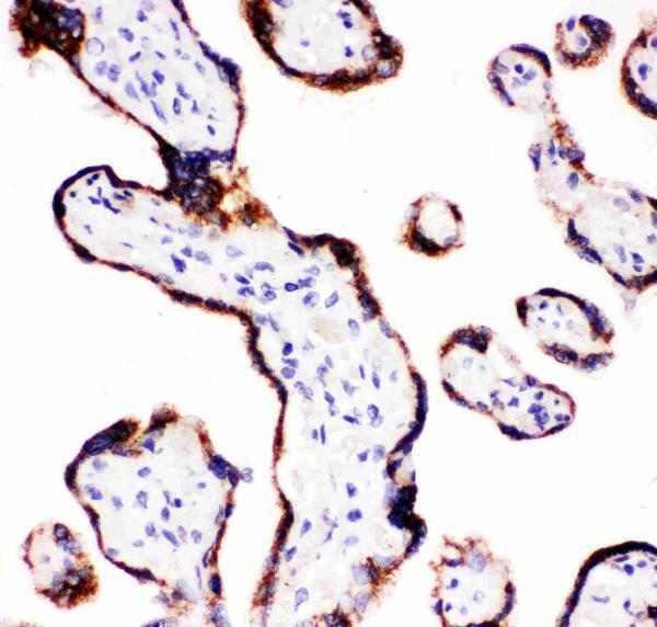
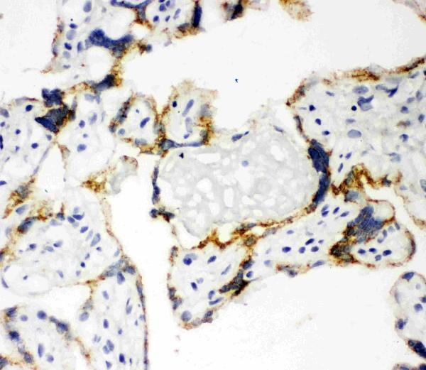


Comprehensive Human Protein Atlas IHC scoring per tissue (reliability: Enhanced — High consistency between antibody staining and RNA expression data.). Rows are taken directly from the HPA tissue chart — click any row's HPA link to view the source.
| Tissue | Cell type | Level | Evidence | Source |
|---|---|---|---|---|
| Adrenal gland | Cells in zona fasciculata | High | Protein (IHC) | HPA → |
| Placenta | Cytotrophoblasts | High | Protein (IHC) | HPA → |
| Testis | Leydig cells | High | Protein (IHC) | HPA → |
| Tissue | Cell type | Level | Evidence | Source |
|---|---|---|---|---|
| Adipose tissue | Adipocytes | Not detected | Protein (IHC) | HPA → |
| Appendix | Glandular cells | Not detected | Protein (IHC) | HPA → |
| Bone marrow | Hematopoietic cells | Not detected | Protein (IHC) | HPA → |
| Breast | Adipocytes | Not detected | Protein (IHC) | HPA → |
| Bronchus | Respiratory epithelial cells | Not detected | Protein (IHC) | HPA → |
Troubleshoot CYP11A1 staining in paraffin section IHC by checking retrieval, compartment, cell identity and controls before comparing signal intensity.
PA1698 has human placenta IHC images for paraffin and frozen sections (PA1698 image captions). The catalog provides no IF image (catalog: PA1698 IF image alts).
PA1698 is listed for human IHC and WB (catalog: PA1698 applications and reactivity). Its images show human placenta IHC in paraffin and frozen sections (PA1698 image captions).
Which to pick: For tissue IHC, choose PA1698 for human paraffin sections; its own placenta caption documents IHC(P), with a listed concentration of 0.5–1 μg/ml (PA1698 IHC(P) image caption; catalog: PA1698 IHC dilution). The paraffin caption does not report the fixative (PA1698 IHC(P) image caption). There is no catalog-supported IF/ICC or nonhuman choice here: PA1698 lists Human and IHC/WB, has no IF image or dilution, and reports no clone (catalog: PA1698 reactivity, applications, IF fields and clone).