CYP26B1 / Cytochrome P450 26B1 · IHC design guide

Design Immunohistochemistry for CYP26B1

Plan CYP26B1 paraffin IHC using the catalog antibody at 2–5 μg/ml (datasheet A02691-2). Compare staining with documented tissue cell patterns while accounting for uncertain staining reliability (HPA tissue IHC).

Evidence assembled Oct 2026 · For research use; verify linked source records and product datasheet before use
Immunohistochemistry protocol sheet for CYP26B1 (IHC for CYP26B1): expected localisation Cytoplasmic tissue staining (HPA tissue IHC); ER membrane localisation (UniProt), antibody A02691-2, validated IHC image, and IHC protocol steps
Printable CYP26B1 IHC protocol sheet — expected localisation Cytoplasmic tissue staining (HPA tissue IHC); ER membrane localisation (UniProt), antibody A02691-2, controls and protocol steps. Open the full CYP26B1 IHC guide →

CYP26B1 Immunohistochemistry Experimental Design Guide

Expected localisation, validated protocols, controls and antibodies — the at-a-glance facts below, then the full design guide.

Must know before staining
Expected localisation Cytoplasmic tissue staining (HPA tissue IHC); ER membrane localisation (UniProt)
Staining pattern Cytoplasmic staining in hepatocytes and Purkinje cells (HPA tissue IHC)
Antigen retrieval EDTA pH 8.0 HIER, heat-mediated (datasheet A02691-2)
Positive control ⓘ Adrenal gland+4 more · see all
Negative control ⓘ Adipose tissue+4 more · see all
Important caveats
Reasons your staining may differ from the expected pattern.
Fixation Keep fixation consistent across paraffin sections (standard IHC practice; not target-specific)
Caveat Staining has low concordance with RNA expression (HPA tissue IHC)
Regulation Expression regulation unreported (UniProt)
Isoform / epitope 3 isoforms; epitope effect unknown (UniProt)
Section 1

Recommended CYP26B1 IHC & IF Protocols

Compare the catalog antibody’s IHC-P protocol (datasheet A02691-2) with published protocols for liver, rat infarct, and oral mucosa sections (PMC11210240; PMC3595277; PMC13304650).

Recommended immunohistochemistry (IHC-P) protocol parameters
SampleParaffin-embedded human liver cancer tissue; fixative not specified (datasheet A02691-2)
FixationImage fixative and duration unreported (datasheet A02691-2); verify before use.
Sectioning4–5 µm sections on charged slides (standard)
DeparaffinisationXylene, graded ethanol series to water (standard)
Antigen retrievalHeat retrieval: EDTA pH 8.0 (datasheet A02691-2); 20 min, 95–100 °C (standard)
Peroxidase block3% H2O2, 10 min, room temperature (standard)
Blocking10% goat serum (datasheet A02691-2)
Primary antibodyRabbit anti-CYP26B1, 2-5 μg/ml (datasheet A02691-2)
Primary incubationOvernight at 4 °C (datasheet A02691-2)
DetectionHRP-conjugated secondary, DAB chromogen (datasheet A02691-2)
CounterstainHematoxylin, blue, dehydrate and mount (standard)
Expected resultCYP26B1-positive staining in glandular cells of adrenal gland (HPA tissue IHC: Medium). HPA tissue profile: Cytoplasmic expression in several tissues. No signal in the no-primary control.
💡Decision noteStart with EDTA pH 8.0 heat retrieval for the catalog antibody (datasheet A02691-2). Published liver and rat infarct protocols use citrate retrieval (PMC11210240; PMC3595277).
Section 2

What Is the Expected CYP26B1 Staining Pattern?

CYP26B1 is assigned to the endoplasmic reticulum and microsome membranes, with no annotated transmembrane segment (UniProt Q9NR63 topology). In paraffin IHC, expect cytoplasmic staining in selected cells, including cerebellar Purkinje cells, cortical neurons and hepatocytes (HPA: medium staining). Interpret the pattern cautiously: HPA rates the tissue IHC profile Uncertain because antibody staining and RNA expression show low consistency (HPA: tissue IHC reliability).

What am I looking at on my slide?
Cytoplasmic chromogen in Purkinje cells or cortical neurons, with identifiable cell boundaries and limited background (HPA: medium staining in these cells).This matches the reported tissue IHC distribution and the broad cytoplasmic appearance expected for an ER-associated enzyme (HPA: tissue IHC; UniProt Q9NR63 subcellular location). It supports a plausible positive result, but HPA's Uncertain tissue reliability means morphology alone does not establish antibody specificity (HPA: tissue IHC reliability).
Predominantly nuclear staining, with little cytoplasmic signal in the same cells.Nuclear staining does not match UniProt's ER and microsome membrane assignment or HPA's cytoplasmic tissue profile (UniProt Q9NR63 subcellular location; HPA: tissue IHC profile). Treat it as suspect and assess controls and detection background before scoring it as CYP26B1 (general IHC practice).
Strong chromogen in cells reported as undetected, such as adipocytes or cardiomyocytes (HPA: not detected in these cells).That distribution conflicts with the reported HPA tissue observations; cross-reactivity or endogenous detection activity are possibilities, not diagnoses (HPA: tissue IHC; general IHC practice). Compare the same run with reported positive cells and a no-primary control, while allowing for HPA's Uncertain reliability (HPA: tissue IHC reliability).
Uniform haze across several cell types, extracellular spaces or blank areas, obscuring cell boundaries.A broad background pattern cannot be assigned confidently to the reported cell-specific cytoplasmic staining (HPA: tissue IHC profile). Nonspecific binding, incomplete blocking or detection background can produce this appearance; inspect the no-primary control and slide morphology (general IHC practice).
No visible cytoplasmic signal in cerebellar Purkinje cells despite an interpretable section (HPA: medium staining in Purkinje cells).This misses a reported positive population, but one negative slide cannot establish absence of CYP26B1 because HPA rates tissue IHC reliability Uncertain (HPA: tissue IHC reliability). Check whether the detection control worked and review the catalog antibody's IHC-P conditions before interpreting the biological result (general IHC practice).
💡Expected CYP26B1 appearanceCall a result provisionally positive when medium cytoplasmic staining is cell-associated in reported populations such as Purkinje cells or hepatocytes (HPA: tissue IHC); predominantly nuclear staining or uniform haze is suspect (UniProt Q9NR63 subcellular location; general IHC practice).
How each factor affects the staining
Compartment assignmentUniProt assigns CYP26B1 to ER and microsome membranes and annotates no transmembrane segment (UniProt Q9NR63 topology). HPA describes cytoplasmic tissue IHC; routine chromogenic IHC may show cytoplasmic signal without resolving individual ER membranes (HPA: tissue IHC profile; general IHC practice).
Tissue and cell selectionHPA reports medium staining in cerebellar Purkinje cells, cortical neurons, hepatocytes and pancreatic exocrine glandular cells, while adipocytes and cardiomyocytes are not detected (HPA: tissue IHC). Score the named cell population rather than treating every cell in a tissue as equivalent (general IHC practice).
Evidence strengthHPA labels the tissue IHC profile Uncertain because antibody staining has low consistency with RNA expression and awaits external verification (HPA: tissue IHC reliability). The listed HPA012567 antibody is IHC Uncertain and ICC Approved; ICC approval does not upgrade tissue IHC reliability (HPA: antibody validation).
Isoforms and epitope coverageUniProt lists 3 isoforms but provides no epitope information in this record (UniProt Q9NR63 isoforms). Their recognition by a particular IHC antibody therefore cannot be inferred; do not explain a cell-level difference by isoform choice without separate evidence (UniProt Q9NR63 isoforms; general IHC interpretation).
Why is my staining missing, weak or wrong?
SituationLikely causeNext action
Reported positive cells are blank while the counterstain and section morphology are usable (HPA: tissue IHC positive cells).Possible weak primary or detection signal, or a run-level failure; the slide alone does not identify the cause (general IHC practice).Review the catalog antibody's IHC-P instructions and run controls; compare staining in a reported positive cell population on the same run (HPA: tissue IHC; general IHC practice). Avoid assigning target-specific fixation sensitivity because it is unreported in these sources.
Nuclear chromogen dominates the section.The compartment conflicts with the reported ER/microsome and cytoplasmic assignments; nonspecific staining is possible (UniProt Q9NR63 subcellular location; HPA: tissue IHC profile).Inspect no-primary and detection controls, then reassess whether any cell-associated cytoplasmic staining remains (general IHC practice). Do not score nuclear signal as CYP26B1 without independent support.
Chromogen appears in HPA-undetected adipocytes or cardiomyocytes (HPA: tissue IHC negative cells).The distribution may reflect nonspecific antibody binding or endogenous detection activity; HPA's Uncertain profile also limits the certainty of a negative comparator (HPA: tissue IHC reliability; general IHC practice).Compare reported positive and undetected cell populations and inspect a no-primary control. If using enzyme-based detection, check the relevant endogenous activity control (HPA: tissue IHC; general IHC practice).
Diffuse haze prevents cell-level scoring.Background from blocking, washing or detection can obscure the expected cytoplasmic profile (HPA: tissue IHC profile; general IHC practice).Check no-primary background, blocking and wash steps, and whether the chromogen development obscures cell boundaries; rescore only interpretable cells (general IHC practice).
One reported positive tissue stains, but another does not (HPA: tissue IHC positive cells).HPA reports staining by specific cell population, and its tissue IHC reliability is Uncertain; absence in one sample has several possible interpretations (HPA: tissue IHC reliability).Confirm that the named cell type is present and assess it separately from adjacent cells. Review run controls before calling a biological difference (HPA: tissue IHC; general IHC practice).
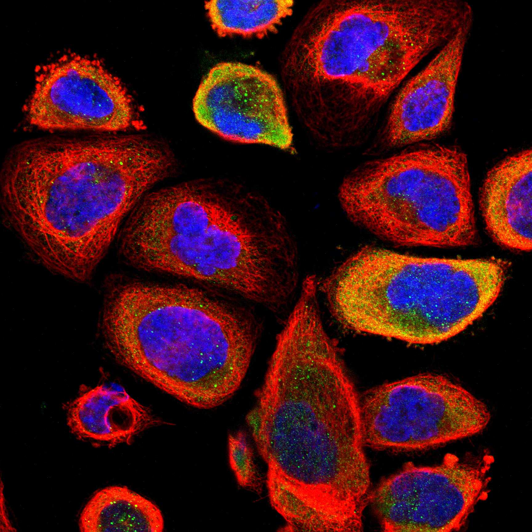
Can an ICC-IF cytosolic image establish the expected paraffin IHC pattern?HPA approves a cytosolic ICC-IF location, while UniProt assigns ER/microsome membranes and HPA rates tissue IHC Uncertain (HPA: subcellular ICC-IF and antibody validation; UniProt Q9NR63 subcellular location).Use the ICC-IF result as context only; interpret paraffin sections against the tissue IHC cell populations and IHC controls. Consult the separate IF/ICC guide for that application (HPA: tissue IHC; general IHC practice).

Sample controls for CYP26B1 IHC & IF

🧪Run cerebellum first: Purkinje cells should stain (HPA: Medium in cerebellar Purkinje cells). Use heart muscle cardiomyocytes as the negative tissue (HPA: Not detected in cardiomyocytes); neighboring non-Purkinje cells on the cerebellar slide should show little or no specific DAB signal as background comparators, without assuming they are CYP26B1-negative (standard IHC control practice).
Positive control tissue: Adrenal gland (Glandular cells, HPA Medium)
Negative control tissue: Adipose tissue (HPA Not detected)
ICC-IF cell lines (HPA subcellular resource): HPA ICC-IF images show CYP26B1 in A-431, U-251MG, U2OS, with annotated localisation: Cytosol (approved) (HPA subcellular).
Technical controls: Include a no-primary, secondary-only control, a rabbit IgG isotype control matched to the primary antibody’s clonality, and CYP26B1-knockout tissue if available (caption: rabbit primary and anti-rabbit secondary; standard IHC control practice). Quench endogenous peroxidase before HRP/DAB detection and assess background on the cerebellar control slide (caption: HRP/DAB detection; standard IHC practice).
⚠️Feasibility: A target-specific fixation window or fixation effect is unreported, and the selected A02691-2 paraffin-section caption does not state the fixative (selected SKU caption). That caption uses heat retrieval in EDTA at pH 8.0, but it does not establish whether retrieval is required for all specimens (selected SKU caption). The supplied evidence does not establish that frozen sections or IF are easier; for cerebellum, assess neuronal background against the no-primary slide before scoring (HPA: Medium in cerebellar Purkinje cells; standard IHC practice).

HPA tissue IHC evidence for CYP26B1

Comprehensive Human Protein Atlas IHC scoring per tissue (reliability: Uncertain — Low consistency between antibody staining and RNA expression data. Pending external verification.). Rows are taken directly from the HPA tissue chart — click any row's HPA link to view the source.

Positive expression · recommended positive controls

TissueCell typeLevelEvidenceSource
Adrenal gland Glandular cells Medium Protein (IHC) HPA →
Caudate Neuronal cells Medium Protein (IHC) HPA →
Cerebellum Purkinje cells Medium Protein (IHC) HPA →
Cerebral cortex Neuronal cells Medium Protein (IHC) HPA →
Cervix Glandular cells Medium Protein (IHC) HPA →

Undetected expression · recommended negative controls

TissueCell typeLevelEvidenceSource
Adipose tissue Adipocytes Not detected Protein (IHC) HPA →
Endometrium Cells in endometrial stroma Not detected Protein (IHC) HPA →
Esophagus Squamous epithelial cells Not detected Protein (IHC) HPA →
Fallopian tube Glandular cells Not detected Protein (IHC) HPA →
Heart muscle Cardiomyocytes Not detected Protein (IHC) HPA →
Section 3

Advanced CYP26B1 IHC Tips

Use matched controls and cell-level scoring to assess CYP26B1 staining in paraffin sections, especially where tissue staining and RNA expression disagree (HPA: reliability uncertain).

How should I optimize antigen retrieval when CYP26B1 staining is weak?
Start with heat-mediated antigen retrieval in EDTA at pH 8.0 for paraffin sections (datasheet A02691-2). The selected tissue-IHC image used this retrieval before overnight incubation at 4°C with 2 μg/ml antibody (A02691-2 IHC caption). If staining remains weak, compare a modestly longer heating period with the original condition on adjacent sections, keeping detection and imaging settings matched. Check tissue integrity and background after each change, since excessive heating can make a faint result harder to interpret. Run a no-primary control alongside the comparison so increased chromogen deposition is not mistaken for recovered CYP26B1 signal.
Could fixation explain inconsistent CYP26B1 staining across paraffin blocks?
Target-specific fixation sensitivity is unknown: the selected paraffin-section image does not state its fixative (A02691-2 IHC caption). Record each block’s fixative, fixation duration, and processing history before comparing CYP26B1 staining across cases. When material permits, stain sections from differently processed blocks together using the documented EDTA pH 8.0 retrieval and identical antibody incubation (datasheet A02691-2; A02691-2 IHC caption). Include a consistently staining reference section in each run, then compare its signal and background before attributing a case difference to biology. Do not infer a fixation effect from tissue staining patterns or protein topology alone.
Should CYP26B1 appear cytosolic or membrane-associated in chromogenic IHC?
Expect predominantly cytoplasmic staining at light-microscope resolution: tissue IHC describes cytoplasmic expression, while the protein is assigned to the endoplasmic reticulum and microsome membranes (HPA: tissue profile; UniProt Q9NR63: subcellular location). A separate cell-imaging annotation calls the cytosol its approved location, so compartment labels do not resolve the discrepancy by themselves (HPA: subcellular summary). Score granular or diffuse cytoplasmic signal separately from nuclear staining, and compare both with a no-primary control. Do not require a crisp membrane outline in paraffin sections to call a cell positive. Treat a predominantly nuclear pattern as a reason to recheck staining specificity and morphology.
Can this stain distinguish CYP26B1 isoforms or reveal epitope masking?
Do not assign isoform identity from this chromogenic stain alone: 3 CYP26B1 isoforms are listed, but the supplied product evidence does not map the antibody epitope (UniProt Q9NR63: isoforms; A02691-2 IHC caption). Compare the antibody immunogen or epitope documentation with the isoform sequences before claiming coverage of a specific variant. The record lists no transmembrane segment, glycosylation sites, or modified residues, which offers no direct test of epitope accessibility after processing (UniProt Q9NR63: topology and modifications). If a control stains inconsistently, revisit retrieval and tissue preservation first, using adjacent sections and identical detection conditions. Report the result as CYP26B1 immunoreactivity unless isoform specificity is independently established.
How can I use IF to check the CYP26B1 cell pattern seen by IHC?
Use IF on a separately optimized section to compare CYP26B1 with a marker for the cell population you intend to score, such as a neuronal marker when examining neuronal staining (HPA: medium staining in caudate and cerebral-cortex neuronal cells). Choose fluorophores after checking the tissue’s autofluorescence, and reserve a spectrally distinct channel for the cell marker. Because CYP26B1 is assigned to endoplasmic reticulum and microsome membranes, choose permeabilisation after establishing which side of the membrane contains the antibody epitope (UniProt Q9NR63: subcellular location; A02691-2 IHC caption: epitope not stated). Include single-stain and no-primary controls to assess bleed-through and background. Interpret IF alongside the chromogenic section’s cell morphology rather than assuming identical staining intensity.
What should I check when CYP26B1 DAB staining looks widespread?
First compare the stained section with a no-primary control and inspect whether color follows cells or pools at damaged edges. The selected paraffin-section image used 10% goat-serum blocking, a peroxidase-conjugated secondary antibody, and DAB development (A02691-2 IHC caption). For routine chromogenic IHC, verify the peroxidase block, wash thoroughly, and shorten chromogen development if background obscures cell boundaries. Check a secondary-only control when nonspecific secondary binding is suspected, keeping exposure to the detection reagents matched. Reassess the 2 μg/ml primary condition only after separating antibody-associated signal from endogenous enzyme activity and detection-system background (A02691-2 IHC caption).
How should I quantify CYP26B1 across heterogeneous tissue sections? ⚠ ANSWER MARKED FOR VERIFICATION
Define the scored cell population and viable tissue area before examining CYP26B1 intensity, then apply those boundaries consistently across sections. For chromogenic IHC, record the percentage of positive cells and staining intensity; an H-score combines percentages at intensity grades 0–3 into a 0–300 score (standard IHC scoring practice). Alternatively, report positive-cell density per mm² of viable tissue when cellularity varies substantially. Normalize cell counts to the number of eligible cells, or density to measured viable area, and exclude folds, edges, and necrotic regions. Keep thresholds and reference sections fixed because tissue-IHC agreement with RNA is reported as uncertain (HPA: reliability uncertain).
How do I distinguish genuine CYP26B1 staining from artefact?
A credible result should localize to identifiable cytoplasm in intact cells and persist across comparable sections, consistent with the reported tissue-IHC pattern (HPA: cytoplasmic expression). Check whether staining instead concentrates at section edges, necrotic areas, or sites of endogenous enzyme activity; those patterns warrant control-based review before scoring. Compare the cell population with the tissue context: medium staining is reported in cerebellar Purkinje cells and liver hepatocytes, while several other cell populations are reported as not detected (HPA: tissue IHC). Treat predominantly nuclear staining as discordant with the endoplasmic reticulum and microsome assignment (UniProt Q9NR63: subcellular location). Because HPA rates tissue-IHC reliability uncertain, require morphology and controls before making a biological claim (HPA: reliability uncertain).
Boster reagents

Best CYP26B1 / Cytochrome P450 26B1 IHC Antibodies

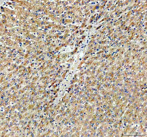
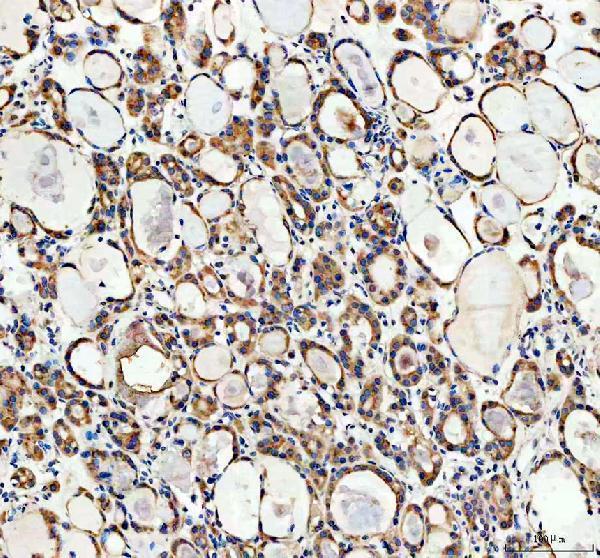
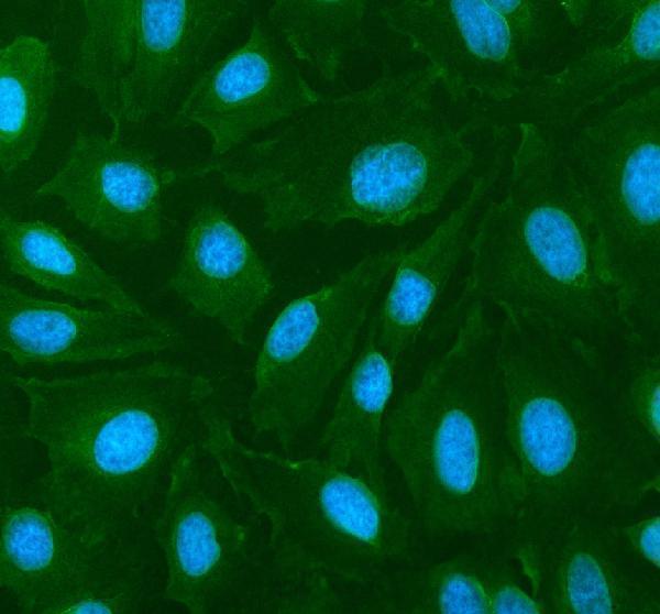
A02691-2 has IHC images from paraffin sections of human liver and thyroid cancer tissue and an IF/ICC image from U2OS cells (catalog image captions). Its listed reactivity covers human, mouse and rat (catalog).

Real IHC data IHC analysis of CYP26B1 using anti-CYP26B1 antibody (A02691-2). CYP26B1 was detected in a paraffin-embedded section of human liver cancer tissue. Heat mediated antigen retrieval was performed in EDTA buffer (pH 8.0, epitope retrieval solution). The tissue section was blocked with 10% goat serum. The tissue section was then incubated with 2 μg/ml rabbit anti-CYP26B1 Antibody (A02691-2) overnight at 4°C. Peroxidase Conjugated Goat Anti-rabbit IgG was used as secondary antibody and incubated for 30 minutes at 37°C. The tissue section was developed using HRP Conjugated Rabbit IgG Super Vision Assay Kit (Catalog # SV0002) with DAB as the chromogen.
Anti-CYP26B1 Antibody ®
Cat # A02691-2

A02691-2 shows IHC staining in paraffin sections of human liver and thyroid cancer tissue (IHC image captions). A02691-2 also shows IF/ICC staining in U2OS cells (IF image caption).

Which to pick: Choose A02691-2 for tissue IHC: its paraffin-section captions document EDTA retrieval at pH 8.0 and 2 μg/ml primary antibody; the fixative is unreported (IHC image captions). For IF/ICC, A02691-2 has a U2OS image using 5 μg/ml primary antibody (IF image caption). A02691-2 lists mouse and rat reactivity, although the supplied IHC and IF images show human samples; clonality is unreported (catalog reactivity; image captions; catalog clone field).

Each figure is that product's own IHC / IF validation image from its datasheet.

References

  1. UniProt Consortium. UniProt entry Q9NR63 (CP26B_HUMAN, Cytochrome P450 26B1).
  2. Human Protein Atlas. CYP26B1 tissue IHC expression (reliability: Uncertain).
  3. Human Protein Atlas. CYP26B1 subcellular location (ICC-IF): Localized to the cytosol..
  4. Human Protein Atlas. CYP26B1 antibody validation summary (1 antibodies).
  5. Prognostic implication and immunotherapy response prediction of a novel ubiquitination-related gene signature in liver cancer. Aging 2024 — PMC11210240.
  6. Retinoic acid metabolism related gene CYP26B1 promotes tumor stemness and tumor microenvironment remodeling in bladder cancer. Journal of Cancer 2025 — PMC12170498.
  7. 5-Methoxyleoligin, a lignan from Edelweiss, stimulates CYP26B1-dependent angiogenesis in vitro and induces arteriogenesis in infarcted rat hearts in vivo. PloS one 2013 — PMC3595277.
  8. Connective tissue profiling in keratinized and non-keratinized oral mucosa reveals distinct extracellular and intracellular features. Frontiers in cell and developmental biology 2026 — PMC13304650.
  9. PubMed PMID:10823918 — UniProt-cited evidence.
  10. PubMed PMID:14702039 — UniProt-cited evidence.
  11. PubMed PMID:15815621 — UniProt-cited evidence.