This website uses cookies to ensure you get the best experience on our website.


- Table of Contents
Plan CYP7B1 chromogenic IHC on paraffin sections using the catalog antibody’s 2–5 μg/ml range (datasheet A03545-3). Assess cytoplasmic and nuclear staining with caution because the tissue IHC evidence flags possible off-target binding (HPA tissue IHC).

Expected localisation, validated protocols, controls and antibodies — the at-a-glance facts below, then the full design guide.
| Expected localisation | Cytoplasmic/nuclear staining (HPA tissue IHC); ER membrane (UniProt) | |
| Staining pattern | Glandular, neuronal and hematopoietic cells: cytoplasmic/nuclear (HPA tissue IHC) | |
| Antigen retrieval | EDTA pH 8.0 HIER, heat-mediated (datasheet A03545-3) | |
| Positive control | Adrenal gland+4 more · see all | |
| Negative control | None in HPA (detected in all 45 tissues); use no-primary + isotype controls |
| Fixation | Keep fixation consistent across paraffin sections (standard IHC practice; not target-specific) | |
| Caveat | Possible off-target binding; staining reliability is uncertain (HPA tissue IHC) | |
| Regulation | No expression regulator specified (UniProt) | |
| Isoform / epitope | No annotated isoforms or processing; epitope side unspecified (UniProt) |
The catalog antibody’s IHC-P protocol (datasheet A03545-3) is accompanied by three published CYP7B1 staining protocols (PMC6993528; PMC9200159; PMC10494299).
| Sample | Paraffin-embedded human breast cancer tissue; fixative not specified (datasheet A03545-3) |
| Fixation | Image fixative and duration unreported (datasheet A03545-3); verify before use. |
| Sectioning | 4–5 µm sections on charged slides (standard) |
| Deparaffinisation | Xylene, graded ethanol series to water (standard) |
| Antigen retrieval | Heat retrieval: EDTA pH 8.0 (datasheet A03545-3); 20 min, 95–100 °C (standard) |
| Peroxidase block | 3% H2O2, 10 min, room temperature (standard) |
| Blocking | 10% goat serum (datasheet A03545-3) |
| Primary antibody | Rabbit anti-CYP7B1, 2-5 μg/ml (datasheet A03545-3) |
| Primary incubation | Overnight at 4 °C (datasheet A03545-3) |
| Detection | HRP-conjugated secondary, DAB chromogen (datasheet A03545-3) |
| Counterstain | Hematoxylin, blue, dehydrate and mount (standard) |
| Expected result | CYP7B1-positive staining in glandular cells of adrenal gland (HPA tissue IHC: High). HPA tissue profile: Cytoplasmic and nuclear expression in most tissues. No signal in the no-primary control. |
CYP7B1 is an endoplasmic reticulum and microsome membrane protein with transmembrane segments at residues 17–37 and 289–309 (UniProt O75881 topology). In paraffin-section IHC, assess cytoplasmic staining in the cell populations HPA reports as positive, including adrenal glandular cells, bone marrow hematopoietic cells and cerebral cortex neurons (HPA tissue IHC). HPA also reports nuclear staining in many tissues, but rates its tissue IHC evidence Uncertain because presumed off-target binding was observed (HPA tissue IHC).
| Cytoplasmic staining appears in adrenal glandular cells, bone marrow hematopoietic cells or cerebral cortex neurons. | These are reported high-staining cell populations (HPA: High in each). A cell-associated cytoplasmic pattern is compatible with CYP7B1 at the endoplasmic reticulum and microsome membranes (UniProt O75881 subcellular location). HPA rates the tissue IHC evidence Uncertain, so a matching pattern supports interpretation but does not establish antibody specificity (HPA tissue IHC). |
| Signal is predominantly nuclear, with little or no cytoplasmic staining. | Interpret cautiously: UniProt places CYP7B1 at endoplasmic reticulum and microsome membranes (UniProt O75881 subcellular location), while HPA describes both cytoplasmic and nuclear staining in most tissues (HPA tissue IHC). Because HPA disregarded presumed off-target binding and assigns Uncertain reliability, nuclear staining alone is insufficient to identify specific CYP7B1 signal (HPA tissue IHC). |
| A different cell population stains more strongly than the reported positive cells. | Compare cell identities before assigning the signal to CYP7B1: HPA reports high staining in defined glandular, hematopoietic and neuronal populations, among others (HPA tissue IHC). Unexpected staining may reflect cross-reactivity or endogenous detection activity (general IHC practice). The HPA list is illustrative, so staining outside those examples is not, by itself, proof of an artefact (HPA tissue IHC). |
| Color is widespread across the section without clear cell boundaries or a compartment pattern. | Diffuse deposition is difficult to reconcile with the endoplasmic reticulum and microsome membrane location (UniProt O75881 subcellular location). Nonspecific antibody binding or detection background can produce diffuse chromogenic signal (general IHC practice). Judge the pattern against a matched negative detection control and the reported positive cells before scoring it as CYP7B1 (general IHC practice; HPA tissue IHC). |
| No signal appears in an expected high-staining population, such as adrenal glandular cells. | HPA reports High staining in adrenal glandular cells (HPA tissue IHC), so an absent signal warrants a check of section quality, retrieval, antibody incubation and chromogenic detection (general IHC practice). It does not alone establish that the tissue lacks CYP7B1; HPA's tissue IHC reliability is Uncertain (HPA tissue IHC). |
| Membrane location and epitope access | CYP7B1 has two annotated transmembrane segments and localizes to endoplasmic reticulum and microsome membranes (UniProt O75881 topology and subcellular location). Epitope accessibility depends on the antibody's epitope and the staining conditions (general IHC practice); neither an epitope position nor a CYP7B1-specific antigen retrieval requirement is supplied here. |
| Strength of tissue-pattern evidence | HPA reports high staining across several cell populations but rates its tissue IHC evidence Uncertain after disregarding presumed off-target binding (HPA tissue IHC). Its listed antibody, HPA017761, has IHC status Uncertain (HPA antibodies). Use the cell pattern as a comparison, with controls, rather than as independent proof of specificity (general IHC practice). |
| IF/ICC Q&A | Q: Can the IHC pattern be carried over to IF/ICC? A: UniProt supports a membrane-associated expectation (UniProt O75881 subcellular location), and HPA's subcellular summary says “Membrane” (HPA subcellular). HPA supplies no ICC-IF image cell lines, and its listed antibody has no ICC status (HPA subcellular; HPA antibodies), so an IF/ICC staining pattern cannot be validated from these records. |
| Situation | Likely cause | Next action |
|---|---|---|
| Adrenal glandular cells show no chromogenic signal. | The section or staining workflow may have failed (general IHC practice); HPA reports these cells as High staining (HPA tissue IHC). | Confirm tissue identity and section integrity, then check retrieval, antibody incubation and detection against appropriate controls (general IHC practice). Treat a persistently negative result cautiously because HPA tissue IHC reliability is Uncertain (HPA tissue IHC). |
| Nuclei stain strongly while cytoplasm is faint. | HPA reports nuclear as well as cytoplasmic staining, but also flags presumed off-target binding (HPA tissue IHC); UniProt places CYP7B1 at intracellular membranes (UniProt O75881 subcellular location). | Record the nuclear and cytoplasmic signals separately. Compare with a negative detection control and a reported positive cell population before assigning nuclear staining to CYP7B1 (general IHC practice; HPA tissue IHC). |
| Color appears in unexpected cells or across the whole section. | Cross-reactivity, nonspecific binding or endogenous detection activity can create misleading signal (general IHC practice). HPA reports specific high-staining cell populations but assigns Uncertain reliability (HPA tissue IHC). | Check cell identity and review matched negative controls; if using enzyme-based chromogenic detection, assess endogenous activity with an appropriate control (general IHC practice). Reassess any signal that does not track recognizable cells (general IHC practice). |
| Background obscures the difference between cells and surrounding tissue. | Nonspecific binding, excessive antibody signal or detection background can reduce contrast (general IHC practice); the supplied records do not identify a CYP7B1-specific cause. | Review blocking, washes, antibody concentration and detection exposure using the established IHC workflow and negative controls (general IHC practice). Score only signal that remains clearly cell-associated and interpretable (general IHC practice). |
| Bone marrow or cerebral cortex stains, but the expected cells are unclear. | HPA attributes High staining to bone marrow hematopoietic cells and cerebral cortex neuronal cells (HPA tissue IHC); mixed cell populations can complicate assignment on a section (general IHC practice). | Identify the staining cells from section morphology and counterstain before comparing intensity with HPA's cell-specific observations (general IHC practice; HPA tissue IHC). Avoid scoring the entire tissue as uniformly positive when the relevant cells cannot be resolved (general IHC practice). |
| An IF/ICC image seems to show a different compartment. | HPA gives a “Membrane” subcellular summary but provides no ICC-IF image cell lines or ICC status for its listed antibody (HPA subcellular; HPA antibodies). | Keep the IF/ICC observation separate from this paraffin IHC assessment. Compare it with UniProt's endoplasmic reticulum and microsome membrane location, and require assay-specific controls before assigning the fluorescent signal to CYP7B1 (UniProt O75881 subcellular location; general IF practice). |


Comprehensive Human Protein Atlas IHC scoring per tissue (reliability: Uncertain — Presumed off target binding observed and disregarded. Pending external verification.). Rows are taken directly from the HPA tissue chart — click any row's HPA link to view the source.
| Tissue | Cell type | Level | Evidence | Source |
|---|---|---|---|---|
| Adrenal gland | Glandular cells | High | Protein (IHC) | HPA → |
| Appendix | Glandular cells | High | Protein (IHC) | HPA → |
| Bone marrow | Hematopoietic cells | High | Protein (IHC) | HPA → |
| Breast | Glandular cells | High | Protein (IHC) | HPA → |
| Bronchus | Respiratory epithelial cells | High | Protein (IHC) | HPA → |
| Tissue | Cell type | Level | Evidence | Source |
|---|---|---|---|---|
| None in HPA: CYP7B1 is detected in all 45 scored tissues. Use a no-primary (secondary-only) and an isotype control instead. | ||||
Troubleshoot CYP7B1 staining in paraffin sections with attention to retrieval, membrane localisation, controls, and the limits of available tissue evidence.
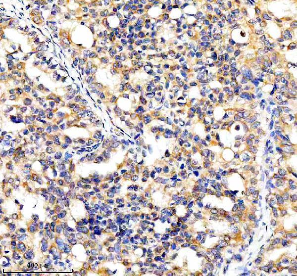
A03545-3 has real IHC data from a human breast cancer paraffin section (image caption). Human, mouse and rat reactivity is listed (catalog); IF/ICC validation is unreported (catalog).
A03545-3 was demonstrated by IHC on a paraffin-embedded human breast cancer section (image caption). Its listed applications include IHC, and its listed reactivity includes human, mouse and rat (catalog).
Which to pick: Choose A03545-3 for paraffin-section IHC: its own figure shows EDTA retrieval, 2 μg/ml primary antibody and DAB detection; the fixative is unreported (image caption). IF/ICC validation is unreported, so there is no validated IF/ICC pick here (catalog). For mouse or rat studies, A03545-3 lists reactivity with both species, but its pictured IHC example is human; it is rabbit-hosted, and clonality is unreported (catalog; image caption).