This website uses cookies to ensure you get the best experience on our website.
- Table of Contents
Facts about C-C chemokine receptor type 1.
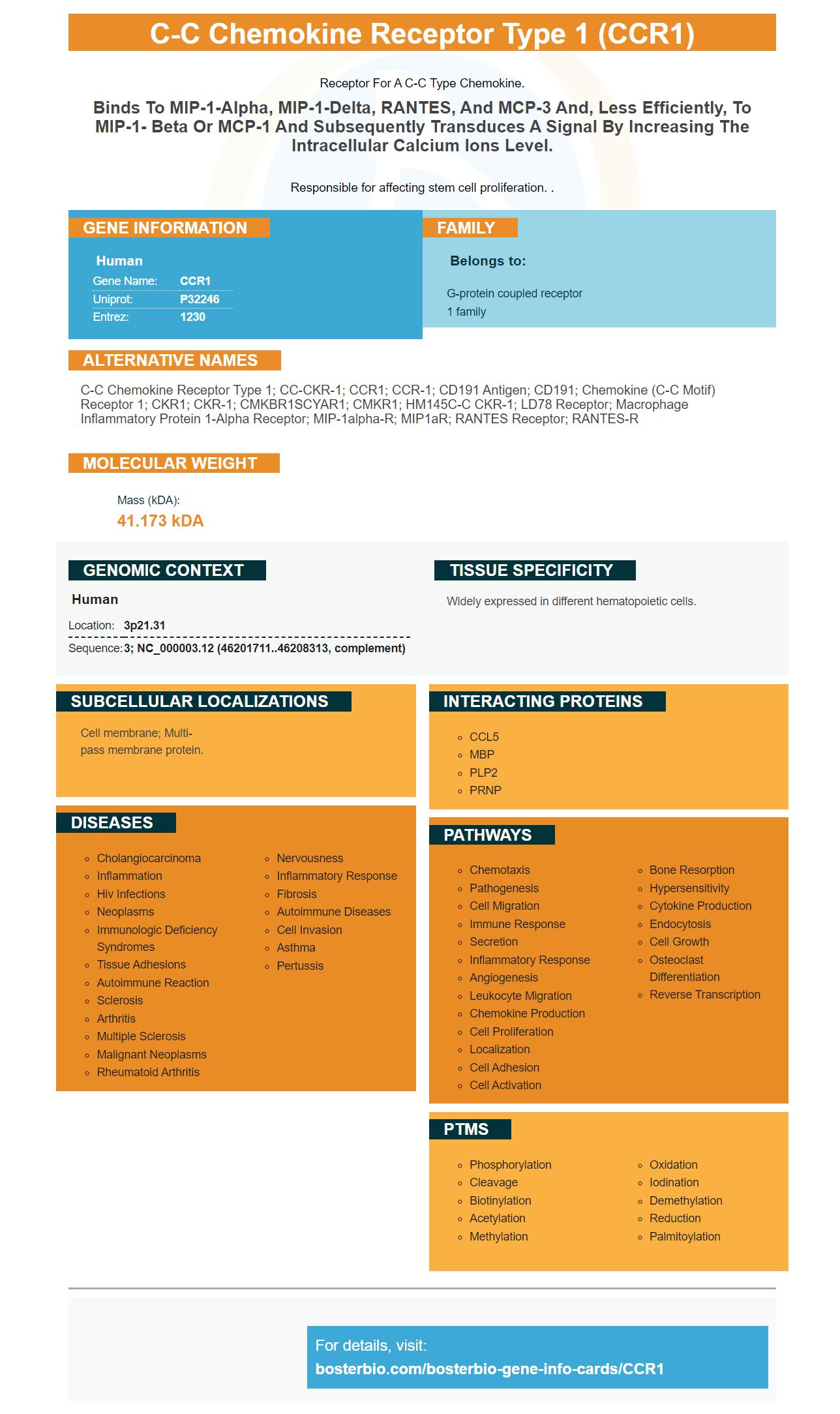
Receptor for a C-C type chemokine.
Binds to MIP-1-alpha, MIP-1-delta, RANTES, and MCP-3 and, less efficiently, to MIP-1- beta or MCP-1 and subsequently transduces a signal by increasing the intracellular calcium ions level.Responsible for affecting stem cell proliferation. .
Human | |
---|---|
Gene Name: | CCR1 |
Uniprot: | P32246 |
Entrez: | 1230 |
Belongs to: |
---|
G-protein coupled receptor 1 family |
C-C chemokine receptor type 1; CC-CKR-1; CCR1; CCR-1; CD191 antigen; CD191; chemokine (C-C motif) receptor 1; CKR1; CKR-1; CMKBR1SCYAR1; CMKR1; HM145C-C CKR-1; LD78 receptor; Macrophage inflammatory protein 1-alpha receptor; MIP-1alpha-R; MIP1aR; RANTES receptor; RANTES-R
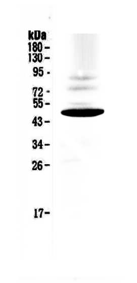
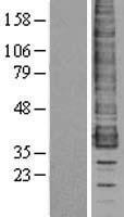
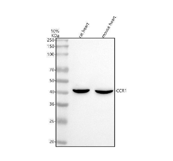
Mass (kDA):
41.173 kDA
Human | |
---|---|
Location: | 3p21.31 |
Sequence: | 3; NC_000003.12 (46201711..46208313, complement) |
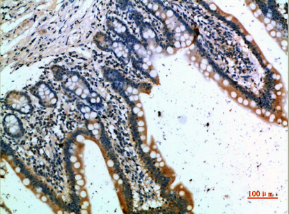
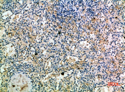
Widely expressed in different hematopoietic cells.
Cell membrane; Multi-pass membrane protein.
There are many benefits of using the CCR1 marker in biological research. These include enhanced DGCR8 modification, improved ERK phosphorylation, and activation of spinal cord microglia. Additionally, CCR1 increased the sensitivity of SNL rats to pain. Thus, CCR1 could be a promising therapeutic target in neuropathic pain. In addition, CCR1 promotes the activation of DGCR8 and the inflammatory response of spinal cord microglia.
The CCR1 marker is an antibody to the cell surface receptor on the bovine chorioallantoic membrane (CXCL12). This specific antibody recognizes a molecule known as TARC, which is a functional ligand for CCR1. In this study, we used polyclonal antibodies to CCR1 and CCR4 produced by Santa Cruz Biotechnology and antiphosphotyrosine antibodies from Upstate Biochemicals.
CCR4 is expressed on T cells and peripheral blood leukocytes. It is expressed at low levels in B cells, monocytes, and spleen cells. Although CCR4 is expressed on T cells, other leukocytes may also express it at very low levels, which are not detectable by reverse transcriptase PCR. Mutant mice with defective expression of CCR4 will greatly facilitate the elucidation of their physiological functions.
TARC and CCR4 are two of the markers expressed in the thymus. They play an important role in thymocyte education and trafficking. They are both found on a high percentage of T cells and low concentrations on Th2 cells. This means that CCR3 is more sensitive than CCR4 in the differentiation of T cells. As such, it can be used to distinguish between two types of T cells, eotaxin and TARC.
Chemokines can be classified into two categories: b and c. The b-chemokines contain the CC motif. Examples of b-chemokines are monocyte chemotactic protein 1 (MCP-1), macrophage inflammatory peptide 1 (MIP-1a), and TARC. They are all characterized by their ligands, but the CCR1 marker in Boster bio is unique, as it recognizes two C chemokine receptors and a large number of CC chemokines.
In addition to cCR1 and cCR2, the CCR4 marker is expressed on memory T cells in peripheral blood. These cells express CCR4 at a high level, according to unpublished data from F. Sallusto and others. CCR4 is expressed on the hCR6-CXCR1 gene. When CCR1 is expressed in T cells, it is thought that Th2 T cells express it selectively.
The human gene for the CCR1 marker is expressed in many different types of tissues. Among these are bone marrow-derived mast cells, osteoclasts, and IL-13+ T cells. It has been found to bind to multiple chemokines, including CCL15, CCL9, and CCL23, and is reported to be membrane-subcellularly localized. It may also be referred to as RANTES-R or the LD78 receptor.
The CCR1 marker is expressed by osteoblasts, and in the presence of wild-type or CCR1-/ osteoblasts, the precursors did not differentiate into differentiation-resistant osteoclasts. The CCR1 chemokine receptor is likely involved in osteoblast differentiation, function, and cellular interactions. Because CCR1 expression is present in many cell types, the ELISA assay may provide useful information about the underlying mechanisms that regulate bone formation.
The CCR1 gene is expressed on some human colon cancer cells. In addition, some tumor cells express the CCR1 ligand. These cells may represent potential therapeutic targets for neuropathic pain. These mice also express CCL15, which may be the result of tumor cells. For this reason, CCR1-expressing mice are a great alternative for humans with colon cancer. And they're compatible with each other, indicating that cancer cells that express CCR1 are in a way that promotes cancer growth.
The ELISA for the CCR1 marker can be used to monitor RANK expression in osteoblastic cells. The mice expressing the RANK marker have a greater risk of developing osteoclasts than mice lacking the gene. The ELISA also provides information about the cytotoxic activity of RANK on osteoclast cells. The CCR1 marker is a good marker for osteoclast differentiation in mice, and further study will help determine whether the CCR1 axis plays a role in this process.
Although the CCR1 gene has been implicated in chronic renal failure, the expression pattern in mice lacking it has been determined. This study provides a model for further interventional studies. In addition, studies have shown that CCR1-/ mice do not display a reduced response to sheep IgG. Therefore, the gene may play an autocrine loop in the control of osteoclast function. The CCR1 gene is a potential candidate for drug development in the treatment of rheumatoid arthritis.
A variety of scientific applications are possible with the use of a CCR1 marker. These markers are used to identify the presence of specific proteins in a variety of samples. These markers can be used for a variety of applications and special samples. Boster scientists can submit their results for species-based analysis or application-based testing, and receive product credits for their work. They can also use this marker for screening drug candidates for their effectiveness in killing CSCs.
For scientists, the CCR1 marker can help them find and understand the functions of specific molecules and pathways in the body. By identifying these molecules, scientists can target them and find new treatments for diseases. In addition, the CCR1 marker can identify human fibroblasts and improve the clinical outcomes of patients with EBV-infected cancer. A self-powered smart patch that serves as a reservoir of chemokine/neural directed exosomes is also designed to stimulate tissue regeneration and neuro-engineering.
Researchers have identified CCR1 as a promising therapeutic target for neuropathic pain. The CCR1 gene is located on cells of the peripheral nervous system and affects neurotransmission. The CCR1 gene encodes the chemokine receptor type CCR1. In this case, the gene is a receptor for chemokines that inhibit the activity of neurons. CCR1 is expressed in the peripheral nervous system in most animals, including humans.
PMID: 7679328 by Neote K., et al. Molecular cloning, functional expression, and signaling characteristics of a C-C chemokine receptor.
PMID: 7683036 by Gao J.-L., et al. Structure and functional expression of the human macrophage inflammatory protein 1 alpha/RANTES receptor.
*More publications can be found for each product on its corresponding product page