This website uses cookies to ensure you get the best experience on our website.


- Table of Contents
Plan DAB1 IHC in paraffin sections using small intestine as a strong-staining reference (HPA tissue IHC). The guide covers expected staining, fixation consistency, antibody dilution, and interpretation; the catalog antibody lists an IHC dilution of 1:50–1:200 (datasheet).

Expected localisation, validated protocols, controls and antibodies — the at-a-glance facts below, then the full design guide.
| Expected localisation | Cytoplasmic (UniProt); intestinal membranous (HPA tissue IHC) | |
| Staining pattern | Intestinal microvilli stain strongly; cytoplasmic and membranous pattern (HPA tissue IHC) | |
| Antigen retrieval | Citrate pH 6.0 HIER, 95–98 °C, 20 min (rule: cytoplasmic / membrane antigen) | |
| Positive control | Duodenum+4 more · see all | |
| Negative control | Adipose tissue+4 more · see all |
| Fixation | Keep fixation consistent across sections. (standard IHC practice; not target-specific) | |
| Caveat | CNS staining is low despite brain-enhanced RNA (HPA tissue IHC) | |
| Regulation | Tissue-dependent expression (HPA tissue IHC) | |
| Isoform / epitope | Six splice isoforms; check epitope coverage (UniProt) |
The catalog antibody’s IHC-P protocol is followed by four article-derived DAB1 IHC protocols, including one for phospho-DAB1 Tyr232 (PMC5881691 methods).
| Sample | Paraffin-embedded human small intestine tissue; fixative not specified (datasheet A03459) |
| Fixation | Image fixative and duration unreported (datasheet A03459); verify before use. |
| Sectioning | 4–5 µm sections on charged slides (standard) |
| Deparaffinisation | Xylene, graded ethanol series to water (standard) |
| Antigen retrieval | Heat-induced epitope retrieval in citrate buffer, pH 6.0, 20 min at 95–98 °C (standard rule: cytoplasmic / membrane antigen) |
| Peroxidase block | 3% H2O2, 10 min, room temperature (standard) |
| Blocking | 10% normal serum of the secondary host, 30 min, room temperature (standard) |
| Primary antibody | Rabbit anti-DAB1, 1:50-1:200 (datasheet A03459) |
| Primary incubation | Overnight at 4 °C (standard) |
| Detection | HRP-polymer secondary, DAB chromogen 5–10 min (standard) |
| Counterstain | Hematoxylin, blue, dehydrate and mount (standard) |
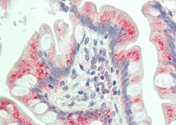
| Expected result | DAB1-positive staining in enterocytes - Microvilli of duodenum (HPA tissue IHC: High). HPA tissue profile: Cytoplasmic and membranous expression in small intestines. Low expression in CNS and retina. No signal in the no-primary control. |
DAB1 is a cytoplasmic protein with no transmembrane segment (UniProt O75553 topology). In paraffin-section IHC, expect cytoplasmic and membranous staining in small intestine, especially duodenal enterocyte microvilli and intestinal endocrine cells; selected neuronal populations also stain (HPA: tissue IHC). HPA rates the tissue pattern Enhanced, citing high consistency between antibody staining and RNA expression (HPA: reliability).
| Strong staining along duodenal enterocyte microvilli, with staining of small-intestinal endocrine cells. | This matches HPA's high staining in both cell populations and its cytoplasmic and membranous small-intestine profile (HPA: tissue IHC). Assess the cellular pattern alongside the section's morphology; intensity alone does not establish specificity (general IHC practice). |
| Staining in cerebellar molecular-layer cells, cortical neurons or retinal ganglion cells is weaker than in the intestinal positive control. | These populations are reported at medium staining, while HPA describes low expression across CNS and retina overall (HPA: tissue IHC). Compare the same named cell populations, rather than treating an entire brain or retinal section as uniformly positive (HPA: tissue IHC). |
| A strong, predominantly nuclear signal replaces the expected cytoplasmic pattern. | UniProt places DAB1 in the cytoplasm, and HPA describes intestinal staining as cytoplasmic and membranous (UniProt O75553: subcellular location; HPA: tissue IHC). Treat isolated nuclear staining as suspect; inspect the counterstain and detection controls before scoring it as DAB1 (general IHC practice). |
| Strong staining appears in a cell population HPA lists as undetected, such as adipocytes in adipose tissue. | HPA reports no detectable staining in those adipocytes (HPA: tissue IHC). Check tissue identity and cell morphology, then investigate antibody cross-reactivity or endogenous detection activity with appropriate controls (general IHC practice). An unexpected result warrants validation rather than automatic rejection. |
| No signal appears in duodenal enterocyte microvilli or small-intestinal endocrine cells. | Both are high-staining populations in HPA's tissue record (HPA: tissue IHC). Verify that the expected cells are present, then check primary-antibody and detection controls and the retrieval conditions used for the IHC assay (general IHC practice). A blank section alone cannot establish absent DAB1. |
| Cellular location and topology | DAB1 is cytoplasmic and has no transmembrane segment (UniProt O75553: subcellular location and topology). HPA nevertheless reports membranous intestinal staining, including high signal at enterocyte microvilli (HPA: tissue IHC). Interpret that observed pattern in its cell context; it does not imply that DAB1 spans a membrane. |
| Choice of reference tissue and cells | Duodenal enterocyte microvilli and small-intestinal endocrine cells are high-staining references; cerebellar molecular-layer cells, cortical neurons and retinal ganglion cells are medium (HPA: tissue IHC). HPA lists several undetected cell populations, including adipocytes in adipose tissue and endocrine cells in colon (HPA: tissue IHC). Match the named cells when comparing sections. |
| Evidence tied to the antibody | HPA marks IHC staining Enhanced for HPA052033 and CAB032329; its Enhanced tissue profile reflects consistency with RNA expression (HPA: antibodies; HPA: reliability). HPA067495 has Enhanced ICC validation but no IHC status in the supplied antibody record (HPA: antibodies). Do not transfer an ICC validation label to paraffin-section IHC. |
| Isoforms and antibody epitope | UniProt lists six DAB1 isoforms and a PID domain at residues 36–189 (UniProt O75553: isoforms and domains). The supplied record gives no epitope for the catalog antibody, so these facts cannot predict which isoforms it detects or whether retrieval exposes its epitope. Review antibody-specific IHC documentation when interpreting an unexpected pattern (general IHC practice). |
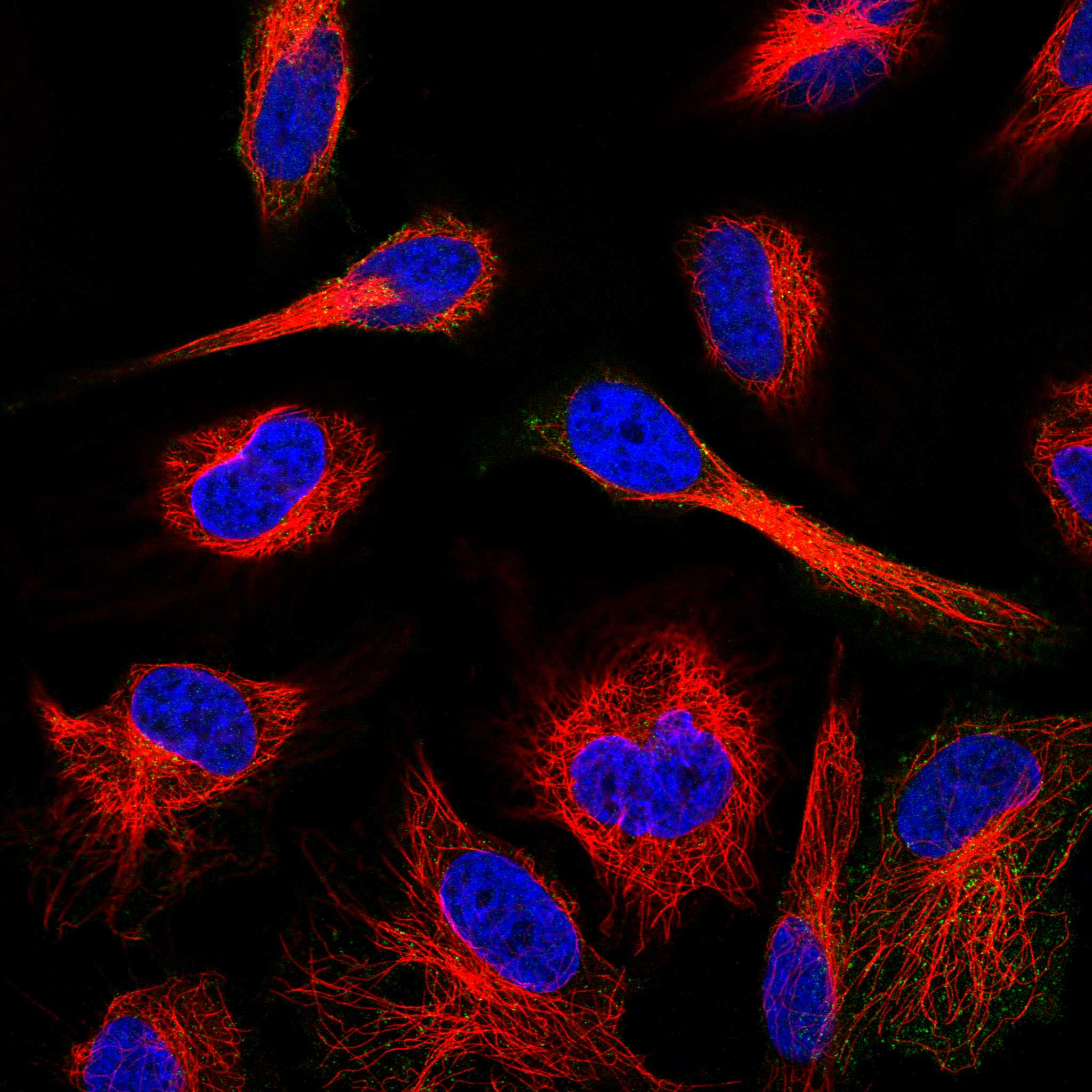
| IF/ICC interpretation | What should IF/ICC show? HPA reports an enhanced vesicular location and images from HeLa and RT-4 cells (HPA: subcellular ICC-IF). That observation can guide interpretation of an IF image, but it does not replace the tissue-specific IHC pattern or establish an IF protocol (HPA: tissue IHC; HPA: subcellular ICC-IF). |
| Situation | Likely cause | Next action |
|---|---|---|
| The intestinal positive control is blank. | The slide may lack the named positive cells, or an IHC reagent or retrieval step may have failed (general IHC practice). HPA expects high staining in duodenal enterocyte microvilli and small-intestinal endocrine cells (HPA: tissue IHC). | Confirm cell identity and section integrity, then check the assay's primary-antibody, retrieval and detection controls (general IHC practice). No DAB1-specific fixation sensitivity is reported in the supplied sources. |
| The slide shows strong nuclear staining. | The compartment conflicts with UniProt's cytoplasmic location and HPA's intestinal cytoplasmic and membranous profile (UniProt O75553: subcellular location; HPA: tissue IHC). Counterstain overlap or nonspecific detection may confuse interpretation (general IHC practice). | Inspect the counterstain separately, compare a known positive section and check a primary-antibody omission control (general IHC practice). Score DAB1 only where the cell and compartment pattern is supported. |
| An HPA-undetected cell population stains strongly. | Cross-reactivity or endogenous detection activity is possible (general IHC practice); HPA lists, for example, adipose-tissue adipocytes as undetected (HPA: tissue IHC). | Verify the cell type, examine a primary-antibody omission control and check the detection-system controls (general IHC practice). Record any reproducible departure from HPA's pattern rather than assigning it to DAB1 solely by color. |
| Diffuse brown haze obscures cell boundaries. | Nonspecific background or endogenous chromogenic detection activity can reduce contrast in IHC (general IHC practice). HPA's named-cell staining pattern cannot be assessed reliably through haze (HPA: tissue IHC). | Review blocking, wash and detection controls and adjust assay conditions according to the antibody's IHC instructions (general IHC practice). Reassess microvilli and endocrine cells only after cellular boundaries are interpretable (HPA: tissue IHC). |
| Neural staining seems faint beside a strong intestinal control. | HPA rates the specified cerebellar, cortical and retinal cells medium, while the two intestinal reference populations are high (HPA: tissue IHC). HPA also summarizes overall CNS and retinal expression as low (HPA: tissue IHC). | Compare the named neural cells with local background and their morphology; avoid requiring intestinal-level intensity in every neural cell (HPA: tissue IHC). Use the high-staining intestinal cells to check that the assay worked. |
| An antibody with ICC evidence gives an unexpected IHC pattern. | ICC and IHC validation are application specific (general IHC practice). HPA067495 has Enhanced ICC status but no supplied IHC status, whereas HPA052033 and CAB032329 have Enhanced IHC status (HPA: antibodies). | Check the antibody identifier and its IHC validation before interpreting the paraffin section (HPA: antibodies). Compare the result with HPA's tissue and cell pattern, using appropriate IHC controls (HPA: tissue IHC; general IHC practice). |


Comprehensive Human Protein Atlas IHC scoring per tissue (reliability: Enhanced — High consistency between antibody staining and RNA expression data.). Rows are taken directly from the HPA tissue chart — click any row's HPA link to view the source.
| Tissue | Cell type | Level | Evidence | Source |
|---|---|---|---|---|
| Duodenum | Enterocytes - Microvilli | High | Protein (IHC) | HPA → |
| Small intestine | Endocrine cells | High | Protein (IHC) | HPA → |
| Cerebellum | Cells in molecular layer | Medium | Protein (IHC) | HPA → |
| Cerebral cortex | Neuronal cells | Medium | Protein (IHC) | HPA → |
| Retina | Ganglion cells | Medium | Protein (IHC) | HPA → |
| Tissue | Cell type | Level | Evidence | Source |
|---|---|---|---|---|
| Adipose tissue | Adipocytes | Not detected | Protein (IHC) | HPA → |
| Adrenal gland | Glandular cells | Not detected | Protein (IHC) | HPA → |
| Appendix | Endocrine cells | Not detected | Protein (IHC) | HPA → |
| Bone marrow | Hematopoietic cells | Not detected | Protein (IHC) | HPA → |
| Breast | Adipocytes | Not detected | Protein (IHC) | HPA → |
Troubleshoot DAB1 staining in paraffin sections using the documented tissue pattern, the selected antibody image, and standard chromogenic IHC controls.
A03459 has a human paraffin-section IHC figure (catalog image caption) and listed IF use; its reported reactivity covers human, mouse, and rat (catalog: applications and reactivity).
Only A03459 renders an IHC card: its figure shows paraffin-embedded human small intestine at 1:100 (catalog image caption). IF is listed for A03459, but no IF figure is supplied (catalog: applications and image alts).
Which to pick: Choose A03459 for tissue IHC: its own figure supports paraffin-embedded human small intestine, with the fixative unreported (catalog image caption). For IF, A03459 and A03459Y232 both list human, mouse, and rat reactivity; A03459Y232 targets phospho-DAB1 Y232, while neither has an IF figure or an explicit ICC listing (catalog: titles, applications, reactivity, and image alts).