DARS2 / Aspartate--tRNA ligase, mitochondrial · IHC design guide

Design Immunohistochemistry for DARS2

Plan DARS2 IHC in paraffin sections around granular cytoplasmic staining reported across most tissues (HPA tissue IHC). This guide covers tissue controls, fixation, detection and scoring for the IHC-validated antibody.

Evidence assembled Oct 2026 · For research use; verify linked source records and product datasheet before use
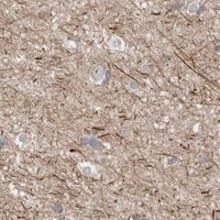
Immunohistochemistry protocol sheet for DARS2 (IHC for DARS2): expected localisation Granular cytoplasm (HPA tissue IHC); mitochondrial matrix and membrane (UniProt), antibody A06034-1, validated IHC image, and IHC protocol steps
Printable DARS2 IHC protocol sheet — expected localisation Granular cytoplasm (HPA tissue IHC); mitochondrial matrix and membrane (UniProt), antibody A06034-1, controls and protocol steps. Open the full DARS2 IHC guide →

DARS2 Immunohistochemistry Experimental Design Guide

Expected localisation, validated protocols, controls and antibodies — the at-a-glance facts below, then the full design guide.

Must know before staining
Expected localisation Granular cytoplasm (HPA tissue IHC); mitochondrial matrix and membrane (UniProt)
Staining pattern Granular cytoplasm in most tissues, including glandular cells (HPA tissue IHC)
Antigen retrieval Citrate pH 6 HIER, heat-mediated (datasheet A06034-1)
Positive control ⓘ Appendix+4 more · see all
Negative control ⓘ Heart muscle+4 more · see all
Important caveats
Reasons your staining may differ from the expected pattern.
Fixation Keep tissue fixation consistent between samples. (standard IHC practice; not target-specific)
Caveat Lung macrophages stain strongly and may dominate scoring (HPA tissue IHC)
Regulation Expression regulation not annotated (UniProt)
Isoform / epitope No isoforms annotated; mature chain spans residues 48–645 (UniProt)
Section 1

Recommended DARS2 IHC & IF Protocols

Compare the catalog antibody’s IHC-P protocol (datasheet: A06034-1) with published DARS2 IHC methods for lung adenocarcinoma tissue and nude mouse tumors (PMC10733159; PMC11535832).

Recommended immunohistochemistry (IHC-P) protocol parameters
SampleParaffin-embedded human intestinal cancer tissue; fixative not specified (datasheet A06034-1)
FixationImage fixative and duration unreported (datasheet A06034-1); verify before use.
Sectioning4–5 µm sections on charged slides (standard)
DeparaffinisationXylene, graded ethanol series to water (standard)
Antigen retrievalHeat retrieval: Citrate pH 6, 20 min (datasheet A06034-1)
Peroxidase block3% H2O2, 10 min, room temperature (standard)
Blocking10% goat serum (datasheet A06034-1)
Primary antibodyRabbit anti-DARS2, 0.5-1μg/ml (datasheet A06034-1)
Primary incubationOvernight at 4 °C (datasheet A06034-1)
DetectionStreptavidin-biotin complex (SABC), DAB chromogen (datasheet A06034-1)
CounterstainHematoxylin, blue, dehydrate and mount (standard)
Expected resultDARS2-positive staining in glandular cells of appendix (HPA tissue IHC: High). HPA tissue profile: Granular cytoplasmic expression in most tissues. No signal in the no-primary control.
💡Decision noteStart with heat-mediated citrate retrieval at pH 6 (datasheet: citrate pH 6 HIER); PMC10733159 reports 97°C for 20 min (PMC10733159).
Section 2

What Is the Expected DARS2 Staining Pattern?

DARS2 is assigned to the mitochondrial matrix and membrane, with no transmembrane segment (UniProt Q6PI48). In paraffin sections, expect granular cytoplasmic staining in many tissues, including glandular cells, kidney tubular cells and lung macrophages (HPA tissue IHC). HPA rates its tissue staining reliability Enhanced, while reporting medium consistency between antibody staining and RNA expression (HPA tissue IHC). Interpret intensity by cell type rather than expecting uniform staining across a section.

What am I looking at on my slide?
Granular cytoplasmic staining is prominent in colon glandular cells or kidney tubular cells (HPA tissue IHC).This matches the reported DARS2 tissue pattern and High staining in those cell types (HPA tissue IHC). Mitochondrial localisation provides a compatible compartment assignment, though chromogenic granules alone do not prove mitochondrial identity (UniProt Q6PI48; HPA subcellular ICC-IF).
The dominant signal is sharply nuclear or confined to the plasma membrane, without granular cytoplasm.That pattern conflicts with the main mitochondrial localisation and tissue IHC profile (UniProt Q6PI48; HPA tissue IHC). Check for artefact before scoring it as DARS2; HPA also reports additional nucleoplasmic localisation by ICC-IF, so nuclear signal alone is not conclusive (HPA subcellular ICC-IF).
Strong staining appears in cardiomyocytes or skeletal myocytes while an expected positive cell type is weak.HPA reports DARS2 as Not detected in those muscle cell types (HPA tissue IHC). Consider cross-reactivity or endogenous detection activity, then compare the same run with a negative control and an HPA High cell type (general IHC practice; HPA tissue IHC).
Diffuse colour covers multiple compartments and obscures cell boundaries across the section.This does not resemble HPA’s granular cytoplasmic profile (HPA tissue IHC). Assess background with a negative control and review blocking, washes and chromogen development before interpreting cell-specific staining (general IHC practice).
No staining is visible in colon glandular cells or kidney tubular cells.These are HPA High cell types, so a blank result warrants a technical check (HPA tissue IHC). Verify tissue morphology and the detection controls, then review the IHC-validated antibody’s specified dilution and retrieval conditions (general IHC practice).
💡Expected DARS2 appearanceCall a result positive when granular cytoplasmic staining is evident in an HPA High cell type such as colon glandular cells or kidney tubular cells; widespread diffuse colour without that cell-specific pattern is suspect (HPA tissue IHC; general IHC practice).
How each factor affects the staining
Compartment and topology (UniProt Q6PI48; HPA subcellular ICC-IF)DARS2 is assigned to the mitochondrial matrix and membrane and has no transmembrane segment (UniProt Q6PI48). Use HPA’s granular cytoplasmic tissue pattern as the chromogenic readout; morphology alone cannot identify individual mitochondria (HPA tissue IHC; general IHC practice).
Cell-type reference range (HPA tissue IHC)HPA reports High staining in several glandular cell populations, kidney tubular cells and lung macrophages, but Not detected in cardiomyocytes, skeletal myocytes and several other listed cell types (HPA tissue IHC). Score the identified cell type, not the organ as a whole.
Antibody validation and evidence limits (HPA tissue IHC; HPA antibodies)Two listed antibodies have Enhanced IHC validation, and the tissue profile has medium staining-to-RNA consistency (HPA antibodies; HPA tissue IHC). These support a reference pattern, not a guarantee that every section or antibody will reproduce each intensity (general IHC practice).
IF/ICC localisation? (HPA subcellular ICC-IF)HPA reports mainly mitochondrial signal, with additional approved nucleoplasmic localisation, in ICC-IF (HPA subcellular ICC-IF). Use that observation to qualify a nuclear finding; this IHC section does not establish an IF/ICC protocol.
Why is my staining missing, weak or wrong?
SituationLikely causeNext action
A known HPA High cell type has no visible signal.The result conflicts with the reported cell-type pattern; the failed step is not identified by that observation alone (HPA tissue IHC; general IHC practice).Confirm the cell type on the counterstain, inspect the run controls, and review the antibody’s IHC-P retrieval, dilution and detection instructions (general IHC practice).
Staining appears in nearly every cell, including HPA Not detected reference cell types.The distribution is broader than the reported tissue profile; nonspecific binding or endogenous chromogenic activity is possible (HPA tissue IHC; general IHC practice).Compare negative controls, check blocking and wash steps, and verify the detection system before assigning DARS2 positivity (general IHC practice).
The section shows flat, diffuse cytoplasmic colour rather than granules.The appearance differs from HPA’s granular cytoplasmic tissue staining (HPA tissue IHC). Excess background or overdevelopment can make the pattern hard to judge (general IHC practice).Review negative controls and chromogen development, then rescore only cells whose cytoplasmic pattern remains distinguishable (general IHC practice).
The dominant signal appears nuclear in a paraffin section.A nuclear-dominant pattern differs from the main mitochondrial localisation, although ICC-IF reports additional nucleoplasmic DARS2 (HPA subcellular ICC-IF; UniProt Q6PI48).Compare an HPA High tissue cell type and negative controls in the same run; record nuclear staining separately from granular cytoplasmic staining (HPA tissue IHC; general IHC practice).
Only a small population stains in a mixed tissue section.Cell types can differ within an organ: HPA reports High lung macrophage staining and Not detected liver cholangiocyte staining (HPA tissue IHC).Identify the stained cells using morphology and counterstain, then compare their compartment and intensity with the relevant HPA cell-type entry (HPA tissue IHC; general IHC practice).
Two IHC-validated antibodies give different intensity or distribution.HPA lists Enhanced IHC validation for two antibodies but reports medium consistency between tissue staining and RNA expression (HPA antibodies; HPA tissue IHC).Compare matched cell types and controls, document each antibody’s pattern, and avoid resolving the discrepancy by intensity alone (general IHC practice).

Sample controls for DARS2 IHC & IF

🧪Run appendix first: glandular cells should stain strongly (HPA: High in appendix glandular cells). Use heart muscle cardiomyocytes as the negative tissue (HPA: Not detected in cardiomyocytes); on the appendix slide, assess adjacent nonglandular cells for background staining, but do not assume they are DARS2-negative without validation.
Positive control tissue: Appendix (Glandular cells, HPA High)
Negative control tissue: Heart muscle (HPA Not detected)
ICC-IF cell lines (HPA subcellular resource): HPA ICC-IF images show DARS2 in A-431, U-251MG, U2OS, with annotated localisation: Mitochondria (supported) (HPA subcellular).
Technical controls: Include no-primary (secondary-only), host-matched rabbit IgG isotype, and DARS2-knockout material where available as a biological negative (caption: rabbit primary antibody; standard IHC controls). Block endogenous peroxidase and check for endogenous biotin before biotin-based DAB detection in the positive tissue (caption: biotinylated secondary, SABC and DAB; standard IHC practice).
⚠️Feasibility: A target-specific fixation window or fixation effect is unreported, and the selected A06034-1 tissue-IHC caption does not state the fixative (caption: fixative unreported). The demonstrated paraffin-section workflow used citrate retrieval at pH 6 for 20 minutes; retrieval dependence beyond that example is unreported (caption: citrate retrieval). The supplied evidence does not establish whether frozen sections or IF are easier; when scoring glandular cells, exclude luminal DAB deposits from cellular staining (standard IHC practice).

HPA tissue IHC evidence for DARS2

Comprehensive Human Protein Atlas IHC scoring per tissue (reliability: Enhanced — Medium consistency between antibody staining and RNA expression data.). Rows are taken directly from the HPA tissue chart — click any row's HPA link to view the source.

Positive expression · recommended positive controls

TissueCell typeLevelEvidenceSource
Appendix Glandular cells High Protein (IHC) HPA →
Colon Glandular cells High Protein (IHC) HPA →
Duodenum Glandular cells High Protein (IHC) HPA →
Epididymis Glandular cells High Protein (IHC) HPA →
Fallopian tube Glandular cells High Protein (IHC) HPA →

Undetected expression · recommended negative controls

TissueCell typeLevelEvidenceSource
Heart muscle Cardiomyocytes Not detected Protein (IHC) HPA →
Liver Cholangiocytes Not detected Protein (IHC) HPA →
Skeletal muscle Myocytes Not detected Protein (IHC) HPA →
Smooth muscle Smooth muscle cells Not detected Protein (IHC) HPA →
Soft tissue Fibroblasts Not detected Protein (IHC) HPA →
Section 3

Advanced DARS2 IHC Tips

Use the reported paraffin-section conditions as a starting point, then assess DARS2 staining against its expected cellular pattern and controls.

How should I adjust retrieval if DARS2 staining is weak in paraffin sections?
Start with heat-mediated citrate retrieval at pH 6 for 20 minutes on paraffin sections (datasheet A06034-1). Keep section thickness, heating, cooling, and DAB development consistent while assessing retrieval, since the reported example also used 1 μg/mL primary antibody overnight at 4°C (datasheet A06034-1). If staining remains weak, compare longer citrate treatment or another retrieval buffer on serial sections, checking morphology and a no-primary control (standard IHC practice). Look for clearer granular cytoplasmic staining in intact cells rather than an overall increase in diffuse DAB (HPA tissue IHC).
Can I infer which fixation conditions preserve DARS2 staining best?
Target-specific fixation sensitivity is unknown because the selected paraffin-section caption does not report a fixative (datasheet A06034-1). For a new fixation condition, compare matched sections with documented fixation intervals while holding citrate retrieval at pH 6 for 20 minutes and primary antibody at 1 μg/mL (datasheet A06034-1; standard IHC practice). Assess cellular preservation, granular cytoplasmic signal, and no-primary background before changing detection chemistry (HPA tissue IHC; standard IHC practice). Mitochondrial localisation and the annotated phosphorylation and acetylation sites do not establish a fixation effect; determine any sensitivity empirically (UniProt Q6PI48).
What DARS2 staining pattern should I expect in chromogenic IHC?
Expect a granular cytoplasmic pattern in many tissues, consistent with DARS2 being mainly mitochondrial (HPA tissue IHC; HPA subcellular). UniProt places DARS2 in the mitochondrial matrix and membrane but annotates no transmembrane segment, so a crisp cell-surface rim would warrant investigation (UniProt Q6PI48). HPA also reports an additional nucleoplasmic location from subcellular imaging; interpret nuclear DAB cautiously and compare it with the dominant cytoplasmic pattern (HPA subcellular; standard IHC practice). On serial sections, check that granules sit within intact cells and persist under matched retrieval and exposure conditions, rather than following damaged edges or precipitate (standard IHC practice).
How do DARS2 processing and modifications affect epitope troubleshooting?
The annotated DARS2 chain spans residues 48–645, and UniProt lists 0 isoforms and no transmembrane segment (UniProt Q6PI48). Because the antibody epitope is not specified in the supplied caption, do not assume that it recognizes the removed N-terminal region or a particular modification state (datasheet A06034-1; UniProt Q6PI48). Annotated sites include phosphothreonine 219, phosphoserine 242, and acetyllysines 368 and 382, but their effects on this antibody are untested (UniProt Q6PI48). If retrieval changes staining, compare serial sections and controls before attributing the difference to processing or modification (standard IHC practice).
How can I check the IHC pattern with multiplex IF?
Use IF as a separate validation experiment, pairing DARS2 with a mitochondrial marker and, where useful, a marker identifying the cells under study (HPA subcellular; standard IF practice). HPA reports high IHC staining in kidney tubular cells and lung macrophages, which can guide cell-type comparisons without establishing an IF protocol (HPA tissue IHC). Choose fluorophores in channels with low measured tissue autofluorescence, and include single-label and no-primary controls to assess bleed-through and background (standard IF practice). Because DARS2 is reported in the mitochondrial matrix and membrane without a transmembrane segment, optimise permeabilisation for access to an intracellular epitope whose exact position is unknown (UniProt Q6PI48; standard IF practice).
How can I distinguish DARS2 signal from chromogenic background?
The reported paraffin-section example used 10% goat serum, a biotinylated secondary antibody, streptavidin-biotin detection, and DAB (datasheet A06034-1). Include no-primary and secondary-only controls, apply a peroxidase block, and compare matched DAB development times to identify detection background (standard IHC practice). If background persists, check whether it follows tissue edges, folds, damaged areas, or endogenous biotin-associated staining rather than intact cellular granules (standard IHC practice). Genuine candidate staining should resemble the granular cytoplasmic pattern reported across most tissues; diffuse brown haze alone does not support DARS2 localisation (HPA tissue IHC; standard IHC practice).
What should I measure when comparing DARS2 IHC across sections? ⚠ ANSWER MARKED FOR VERIFICATION
Define the cell population and tissue region before scoring, then measure the percentage of positive cells and staining intensity or calculate an H-score from 0–300 (standard IHC practice). Score granular cytoplasmic signal within intact cells, reflecting the reported IHC pattern, and record nuclear staining separately if present (HPA tissue IHC; standard IHC practice). Normalise positive counts to the number of eligible cells or to sampled tissue area in mm², using the same rule across sections (standard IHC practice). Keep retrieval, primary concentration, DAB development, imaging, and threshold settings consistent so processing differences do not drive the comparison (standard IHC practice).
When should apparent DARS2 positivity be treated as suspect?
Treat granular cytoplasmic staining in intact cells as the expected IHC pattern, consistent with DARS2's mainly mitochondrial location (HPA tissue IHC; HPA subcellular). Check unexpected strong staining in cardiomyocytes or cholangiocytes carefully, since HPA reports DARS2 as not detected in those cell populations (HPA tissue IHC). A cell-surface rim, staining concentrated at section edges or necrotic areas, or signal reproduced in a no-primary control calls for further investigation (UniProt Q6PI48 topology; standard IHC practice). Use a peroxidase-block control and matched DAB development to assess endogenous enzyme signal before assigning unexpected brown deposits to DARS2 (standard IHC practice).
Boster reagents

Best DARS2 / Aspartate--tRNA ligase, mitochondrial IHC Antibodies

A06034-1 has IHC images from human paraffin-embedded cancer sections and IF images from U20S and A431 cells (catalog image captions); listed reactivity is human (catalog: reactivity).

Real IHC data IHC analysis of DARS2 using anti-DARS2 antibody (A06034-1). DARS2 was detected in paraffin-embedded section of human intestinal cancer tissue. Heat mediated antigen retrieval was performed in citrate buffer (pH6, epitope retrieval solution) for 20 mins. The tissue section was blocked with 10% goat serum. The tissue section was then incubated with 1μg/ml rabbit anti-DARS2 Antibody (A06034-1) overnight at 4°C. Biotinylated goat anti-rabbit IgG was used as secondary antibody and incubated for 30 minutes at 37°C. The tissue section was developed using Strepavidin-Biotin-Complex (SABC)(Catalog # SA1022) with DAB as the chromogen.
Anti-DARS2 Antibody ®
Cat # A06034-1

A06034-1 has IHC data from paraffin-embedded human intestinal, mammary and lung cancer sections (catalog: IHC image captions). The same SKU has IF data from U20S and A431 cells (catalog: IF image captions).

Which to pick: Choose A06034-1 for tissue IHC on paraffin sections; its IHC captions document that preparation, but do not report the fixative (catalog: IHC image captions). Choose the same SKU for IF/ICC, which are listed applications with cell-image data (catalog: applications and IF image captions). Its listed reactivity is human, so the payload provides no cross-species validation (catalog: reactivity).

Each figure is that product's own IHC / IF validation image from its datasheet.

References

  1. UniProt Consortium. UniProt entry Q6PI48 (SYDM_HUMAN, Aspartate--tRNA ligase, mitochondrial).
  2. Human Protein Atlas. DARS2 tissue IHC expression (reliability: Enhanced).
  3. Human Protein Atlas. DARS2 subcellular location (ICC-IF): Mainly localized to the mitochondria. In addition localized to the nucleoplasm..
  4. Human Protein Atlas. DARS2 antibody validation summary (2 antibodies).
  5. Prognostic biomarker DARS2 correlated with immune infiltrates in bladder tumor. Frontiers in immunology 2023 — PMC10827901.
  6. DARS2 promotes the occurrence of lung adenocarcinoma via the ERK/c-Myc signaling pathway. Thoracic cancer 2023 — PMC10733159.
  7. Upregulation of DARS2 by HBV promotes hepatocarcinogenesis through the miR-30e-5p/MAPK/NFAT5 pathway. Journal of experimental & clinical cancer research : CR 2017 — PMC5649064.
  8. Role of CENPL, DARS2, and PAICS in determining the prognosis of patients with lung adenocarcinoma. Translational lung cancer research 2024 — PMC11535832.
  9. PubMed PMID:16710414 — UniProt-cited evidence.
  10. PubMed PMID:15489334 — UniProt-cited evidence.
  11. PubMed PMID:15779907 — UniProt-cited evidence.