This website uses cookies to ensure you get the best experience on our website.


- Table of Contents
Plan chromogenic DAZL IHC-P using testis pachytene spermatocytes as a positive control (HPA tissue IHC). This guide covers expected localisation (UniProt) and the IHC-validated antibody’s 0.5–1 μg/ml working range (datasheet A02069-2).

Expected localisation, validated protocols, controls and antibodies — the at-a-glance facts below, then the full design guide.
| Expected localisation | Expected predominantly cytoplasmic; can be nuclear in spermatogonia (UniProt) | |
| Staining pattern | High in pachytene spermatocytes; compartment unreported (HPA tissue IHC) | |
| Antigen retrieval | EDTA pH 8.0 HIER, heat-mediated (datasheet A02069-2) | |
| Positive control | Testis | |
| Negative control | Adipose tissue+4 more · see all |
| Fixation | Keep fixation consistent across sections. (standard IHC practice; not target-specific) | |
| Caveat | Localisation changes with meiotic stage (UniProt) | |
| Regulation | Testis specific expression (UniProt) | |
| Isoform / epitope | 2 isoforms; epitope differences unreported (UniProt) |
The catalog antibody protocol uses EDTA retrieval (datasheet: A02069-2). The published chromogenic IHC option stains human glioblastoma tissue arrays (PMC7368788).
| Sample | Paraffin-embedded human testies cancer tissue; fixative not specified (datasheet A02069-2) |
| Fixation | Image fixative and duration unreported (datasheet A02069-2); verify before use. |
| Sectioning | 4–5 µm sections on charged slides (standard) |
| Deparaffinisation | Xylene, graded ethanol series to water (standard) |
| Antigen retrieval | Heat retrieval: EDTA pH 8.0 (datasheet A02069-2); 20 min, 95–100 °C (standard) |
| Peroxidase block | 3% H2O2, 10 min, room temperature (standard) |
| Blocking | 10% goat serum (datasheet A02069-2) |
| Primary antibody | Rabbit anti-DAZL, 0.5-1μg/ml (datasheet A02069-2) |
| Primary incubation | Overnight at 4 °C (datasheet A02069-2) |
| Detection | Streptavidin-biotin complex (SABC), DAB chromogen (datasheet A02069-2) |
| Counterstain | Hematoxylin, blue, dehydrate and mount (standard) |
| Expected result | DAZL-positive staining in pachytene spermatocytes of testis (HPA tissue IHC: High). HPA tissue profile: High expression in testis. No signal in the no-primary control. |
DAZL staining is expected in testicular germ cells, with high IHC signal reported in pachytene spermatocytes (HPA: testis IHC, Enhanced reliability). DAZL is predominantly cytoplasmic, while nuclear localisation can occur earlier in germ cell development (UniProt Q92904: subcellular location). It has no transmembrane segment, so a membrane outline is not the expected pattern (UniProt Q92904: topology).
| Strong cytoplasmic staining in pachytene spermatocytes, with little staining elsewhere in the section. | This matches the reported high testis signal (HPA: pachytene spermatocytes). Predominantly cytoplasmic localisation is consistent with UniProt Q92904. Assess the labelled cells within the seminiferous tubules; a dark field alone does not establish cell specific staining (general IHC practice). |
| Nuclear staining, or a sharp outline around cells, dominates the positive region. | Nuclear DAZL can occur in earlier germ cells, so interpret it in relation to cell stage (UniProt Q92904: subcellular location). A dominant membrane outline has no support from DAZL topology (UniProt Q92904: no transmembrane segment). Review morphology and detection controls before calling either pattern specific (general IHC practice). |
| Strong staining appears in adipocytes or other cells listed as negative. | HPA reports DAZL as not detected in adipocytes from adipose tissue and breast, and in several other sampled cell types (HPA: tissue IHC). Check for cross reactivity or endogenous detection activity; the HPA result applies to the listed cells and samples, not every cell in those organs (general IHC interpretation). |
| Diffuse colour covers tissue, stroma or blank regions without a clear germ cell pattern. | This does not reproduce the cell restricted testis pattern (HPA: testis IHC). Possible general IHC causes include nonspecific binding, incomplete blocking, residual endogenous enzyme activity or excessive development. Compare a no primary control and review wash and detection steps before assigning the colour to DAZL (general IHC practice). |
| Pachytene spermatocytes lack visible signal in a known positive testis section. | A negative result conflicts with the high signal reported for these cells (HPA: testis IHC). First confirm that the expected cells are present and that the control run worked. Then review antibody performance, retrieval and detection conditions as general IHC variables; no DAZL specific fixation sensitivity is established here. |
| Germ cell stage and compartment | DAZL is predominantly cytoplasmic; UniProt also describes nuclear localisation in spermatogonia until near the end of meiotic prophase, followed by cytoplasmic localisation (UniProt Q92904). Interpret nuclei alongside cell stage. |
| Choice of tissue control | Testis contains high staining pachytene spermatocytes and is the strongest supplied positive reference (HPA: testis IHC). HPA lists selected cells in other tissues as not detected; use their stated cell types when assessing negative staining. |
| Antibody validation | The tissue profile has Enhanced reliability, reflecting consistency between staining and RNA data (HPA: tissue IHC). HPA019777 has Enhanced IHC validation and HPA047113 has Supported IHC validation (HPA: antibodies); these labels do not validate every staining pattern. |
| Topology and interpretation | DAZL has no transmembrane segment and no signal peptide (UniProt Q92904: topology and processing). A membrane rim should prompt review of staining specificity; these annotations alone do not predict retrieval needs or fixation sensitivity. |
| IF/ICC: is a cellular pattern established? | HPA gives no main subcellular location or ICC-IF image set for DAZL (HPA: subcellular). UniProt describes stage dependent nuclear and cytoplasmic localisation, but this is not an HPA ICC-IF validation or an IF protocol (UniProt Q92904). |
| Situation | Likely cause | Next action |
|---|---|---|
| No staining in pachytene spermatocytes of a testis section. | The expected cell population may be absent from the examined area, or the IHC run may have failed (HPA: high in pachytene spermatocytes; general IHC practice). | Verify morphology and the positive control, then review the antibody, retrieval and detection workflow (general IHC practice). Do not infer a DAZL specific fixation effect from this result. |
| Signal appears mainly as a membrane outline. | A membrane dominant pattern conflicts with the absence of a DAZL transmembrane segment (UniProt Q92904: topology); nonspecific staining is possible (general IHC practice). | Compare the no primary control, inspect whether the outline tracks tissue edges, and reassess antibody specificity (general IHC practice). |
| Nuclear signal is treated as an automatic failure. | UniProt describes nuclear DAZL in spermatogonia and a later shift toward cytoplasmic localisation (UniProt Q92904: subcellular location). | Identify the labelled germ cell stage and compare the surrounding cytoplasmic pattern before rejecting the section (UniProt Q92904; general IHC interpretation). |
| Adipocytes or another HPA negative cell type stain strongly. | The signal conflicts with a cell specific HPA not detected observation; cross reactivity or endogenous detection activity is possible (HPA: tissue IHC; general IHC practice). | Confirm cell identity, compare a no primary control, and review blocking and detection chemistry (general IHC practice). |
| Diffuse chromogen obscures germ cell boundaries. | Background may reflect nonspecific antibody binding, residual endogenous enzyme activity or excessive chromogen development (general IHC practice). | Use the no primary control to locate background, then review blocking, washes, antibody concentration and development time (general IHC practice). |
| An IF/ICC result is used to justify the IHC compartment call. | HPA supplies no DAZL ICC-IF images or main subcellular location (HPA: subcellular); its reported testis pattern comes from tissue IHC (HPA: testis IHC). | Evaluate the paraffin section against the HPA testis IHC pattern and the stage dependent UniProt localisation; assess IF/ICC evidence separately (HPA: tissue IHC; UniProt Q92904). |
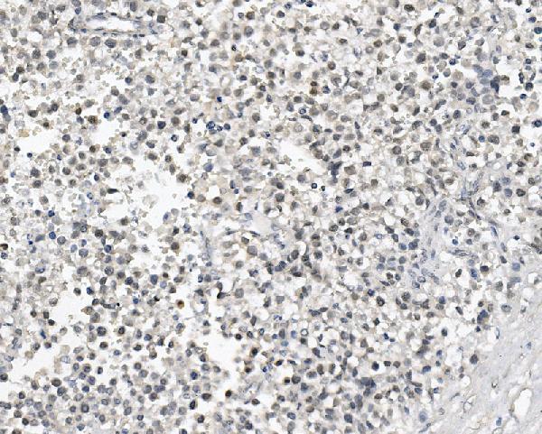
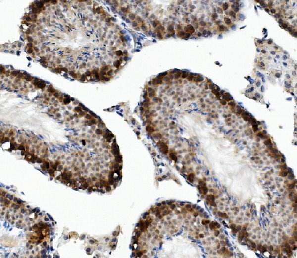


Comprehensive Human Protein Atlas IHC scoring per tissue (reliability: Enhanced — High consistency between antibody staining and RNA expression data.). Rows are taken directly from the HPA tissue chart — click any row's HPA link to view the source.
| Tissue | Cell type | Level | Evidence | Source |
|---|---|---|---|---|
| Testis | Pachytene spermatocytes | High | Protein (IHC) | HPA → |
| Tissue | Cell type | Level | Evidence | Source |
|---|---|---|---|---|
| Adipose tissue | Adipocytes | Not detected | Protein (IHC) | HPA → |
| Adrenal gland | Glandular cells | Not detected | Protein (IHC) | HPA → |
| Appendix | Glandular cells | Not detected | Protein (IHC) | HPA → |
| Bone marrow | Hematopoietic cells | Not detected | Protein (IHC) | HPA → |
| Breast | Adipocytes | Not detected | Protein (IHC) | HPA → |
Use the catalog antibody’s paraffin section evidence and cell resolved testis controls to troubleshoot DAZL staining (datasheet A02069-2; HPA tissue IHC).
A02069-2 has real IHC data from paraffin sections of human, mouse and rat testis cancer tissue (catalog image captions); IF/ICC data are not provided (catalog payload).
A02069-2 is listed for IHC in human, mouse and rat samples (catalog applications and reactivity). Its image captions show staining of paraffin sections of human, mouse and rat testis cancer tissue; no IF figure is listed (catalog image captions; catalog IF images).
Which to pick: For tissue IHC across human, mouse and rat samples, choose A02069-2: it is IHC listed, rabbit hosted and shown on paraffin sections from all three species (catalog applications, host and image captions). Its captions do not report the fixative (catalog image captions). No IF/ICC choice is supported by the supplied application list or figures, and the antibody’s clonality is unreported (catalog applications, IF images and clone field).