This website uses cookies to ensure you get the best experience on our website.


- Table of Contents
Plan DCK chromogenic IHC in paraffin sections with the IHC-validated antibody at 2–5 μg/ml (datasheet A01655-1). This guide covers nuclear and cytoplasmic staining in immune-cell subsets and high staining in tonsil germinal center cells (HPA tissue IHC).

Expected localisation, validated protocols, controls and antibodies — the at-a-glance facts below, then the full design guide.
| Expected localisation | Nuclear and cytoplasmic in immune-cell subsets (HPA tissue IHC) | |
| Staining pattern | Some immune cells show nuclear and cytoplasmic staining (HPA tissue IHC) | |
| Antigen retrieval | EDTA pH 8.0 HIER, heat-mediated (datasheet A01655-1) | |
| Positive control | Appendix+4 more · see all | |
| Negative control | Adipose tissue+4 more · see all |
| Fixation | Keep fixation consistent across paraffin sections. (standard IHC practice; not target-specific) | |
| Caveat | Presumed off-target staining was observed and disregarded (HPA tissue IHC) | |
| Regulation | RNA expression is enhanced in lymphoid tissue (HPA tissue RNA) | |
| Isoform / epitope | No annotated isoforms; one 1–260 chain (UniProt) |
Compare the catalog antibody’s IHC-P protocol with four published DCK IHC protocols in paraffin sections (datasheet A01655-1; PMC4998873; PMC7271117; PMC4819809; PMC3669607).
| Sample | Paraffin-embedded human breast cancer tissue; fixative not specified (datasheet A01655-1) |
| Fixation | Image fixative and duration unreported (datasheet A01655-1); verify before use. |
| Sectioning | 4–5 µm sections on charged slides (standard) |
| Deparaffinisation | Xylene, graded ethanol series to water (standard) |
| Antigen retrieval | Heat retrieval: EDTA pH 8.0 (datasheet A01655-1); 20 min, 95–100 °C (standard) |
| Peroxidase block | 3% H2O2, 10 min, room temperature (standard) |
| Blocking | 10% goat serum (datasheet A01655-1) |
| Primary antibody | Rabbit anti-DCK, 2-5μg/ml (datasheet A01655-1) |
| Primary incubation | Overnight at 4 °C (datasheet A01655-1) |
| Detection | HRP-conjugated secondary, DAB chromogen (datasheet A01655-1) |
| Counterstain | Hematoxylin, blue, dehydrate and mount (standard) |
| Expected result | DCK-positive staining in lymphoid tissue of appendix (HPA tissue IHC: High). HPA tissue profile: Nuclear and cytoplasmic expression in a subset of immune cells. No signal in the no-primary control. |
DCK should stain the nucleus and cytoplasm of a subset of immune cells in tissue sections (HPA tissue IHC); its annotated location is nuclear, with no transmembrane segment (UniProt P27707). HPA rates the tissue staining profile Enhanced, while noting medium agreement with RNA expression and presumed off-target staining that was disregarded (HPA tissue IHC).
| Distinct nuclear staining, sometimes accompanied by cytoplasmic staining, in selected immune cells; germinal center cells stain strongly. | This fits the reported subset-specific tissue pattern (HPA tissue IHC). Germinal center cells in lymph node and tonsil are rated High; lymphoid tissue in appendix is also High (HPA tissue IHC). Assess the named cell population rather than treating every cell in a positive tissue as positive. |
| Predominantly membrane-rim, extracellular, or vessel-lumen color, without a convincing immune-cell nuclear pattern. | Treat this as a questionable compartment pattern: DCK is annotated in the nucleus and has no transmembrane segment (UniProt P27707), while tissue IHC reports nuclear and cytoplasmic staining (HPA tissue IHC). Recheck morphology and detection controls before assigning the color to DCK (standard IHC practice). |
| Strong color in cells outside the reported immune-cell subset, especially where a listed cell population is undetected. | Consider off-target antibody binding or endogenous chromogenic detection activity (standard IHC practice). HPA explicitly notes presumed off-target staining in its tissue assessment (HPA tissue IHC). For example, adipocytes in adipose tissue and respiratory epithelial cells in bronchus are Not detected; those labels do not classify every cell in either tissue (HPA tissue IHC). |
| Diffuse color across the section, including spaces between cells, with poorly defined nuclei. | A field-wide haze is difficult to score as the reported cell-specific pattern (HPA tissue IHC). In chromogenic IHC, incomplete blocking, insufficient washing, or detection-reagent activity can produce background (standard IHC practice). Compare a matched negative control and examine whether the color follows recognizable cells. |
| No convincing signal in germinal center cells of lymph node or tonsil, while the control section also appears blank. | These cells are reported High and make useful positive reference populations (HPA tissue IHC). A blank reference suggests checking the staining run before calling the test tissue negative (standard IHC practice). Bone marrow hematopoietic cells and spleen white-pulp cells are only Medium references (HPA tissue IHC). |
| Which tissue and cell population is scored? | Expected intensity varies by annotated population: appendix lymphoid tissue and lymph node or tonsil germinal center cells are High, whereas bone marrow hematopoietic cells and spleen white-pulp cells are Medium (HPA tissue IHC). Compare like cell populations when judging a weak result. |
| What does antibody validation support? | The tissue profile is rated Enhanced, but HPA reports medium agreement with RNA and disregarded presumed off-target staining (HPA tissue IHC). Antibody HPA062773 has Enhanced IHC validation; HPA059609 has Supported ICC validation and no IHC status listed (HPA antibodies). Validation does not make unexpected staining automatically specific. |
| Does DCK processing suggest a second compartment? | The annotated protein is one 1–260 chain, with no signal peptide, propeptide, or transmembrane segment (UniProt P27707). These annotations give no basis for expecting a secreted or membrane-bound DCK staining pattern; assess such color against cellular morphology and controls. |
| Can retrieval conditions explain an unexpectedly weak section? | Antigen retrieval is a routine variable in paraffin-section IHC (standard IHC practice). No target-specific fixation or retrieval sensitivity is supplied for DCK, so adjust a retrieval condition only against a positive reference and controls; do not infer a DCK-specific masking effect from the HPA tissue intensities. |
| IF/ICC: where should DCK appear? | HPA reports supported nucleoplasmic localization in ICC-IF (HPA subcellular). That observation helps interpret fluorescent localization, while the tissue IHC profile permits nuclear and cytoplasmic staining in an immune-cell subset (HPA tissue IHC). IF/ICC experimental conditions belong in its separate guide. |
| Situation | Likely cause | Next action |
|---|---|---|
| The positive reference is blank or much weaker than expected. | The run may have a primary-antibody, retrieval, or chromogenic detection problem (standard IHC practice); germinal center cells are reported High references (HPA tissue IHC). | Confirm that the reference contains germinal centers, then check antibody application, retrieval settings, detection reagents, and counterstain visibility against run controls (standard IHC practice). Do not interpret a blank test section until the reference works. |
| A negative control develops color similar to the test section. | Nonspecific secondary-reagent binding or endogenous detection activity can mimic antibody signal in chromogenic IHC (standard IHC practice). | Inspect the omission or matched negative control, review blocking and detection steps, and reassess only the cell-associated pattern that remains distinguishable from control color (standard IHC practice). HPA's off-target caveat also warrants caution (HPA tissue IHC). |
| Most cells stain uniformly, including cells outside the reported immune-cell subset. | Excess background or cross-reactivity is plausible when staining loses the reported cell selectivity (standard IHC practice; HPA tissue IHC). | Compare cell morphology and a negative control; review antibody concentration and washes as general IHC variables (standard IHC practice). Use HPA's named cell populations as references, and avoid calling an entire tissue positive or negative from one cell type (HPA tissue IHC). |
| Color is concentrated on cell borders, extracellular material, or luminal surfaces. | That distribution conflicts with the nuclear annotation and lack of a transmembrane segment (UniProt P27707), and with the reported nuclear and cytoplasmic immune-cell pattern (HPA tissue IHC). | Check the counterstain, tissue boundaries, and negative control for deposits or detection background; score DCK only where a convincing cellular pattern is present (standard IHC practice). |
| Bone marrow or spleen looks weaker than lymph node or tonsil. | This difference can match HPA's ratings: marrow hematopoietic cells and spleen white-pulp cells are Medium; lymph node and tonsil germinal center cells are High (HPA tissue IHC). | Score the specified cell populations and compare sections within each tissue context (HPA tissue IHC). Investigate a technical failure if an appropriate high-staining reference also loses its expected signal (standard IHC practice). |
| Fluorescent ICC shows little nucleoplasmic signal despite apparent tissue IHC positivity. | HPA's supported ICC-IF localization is nucleoplasmic, whereas its tissue IHC summary includes nuclear and cytoplasmic staining in selected immune cells (HPA subcellular; HPA tissue IHC). The assays and sampled cells may differ. | Check the ICC-IF positive reference and imaging controls using the separate IF/ICC guide (standard IF practice). Do not use an IHC tissue rating alone to predict fluorescent intensity in an unspecified cell population (HPA tissue IHC). |
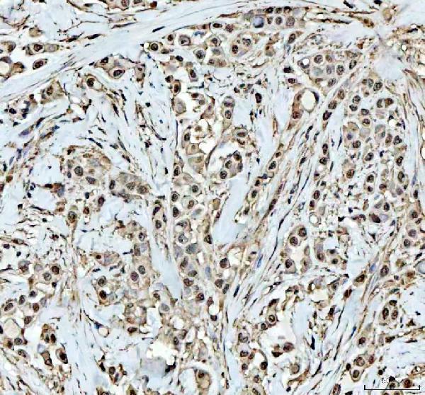
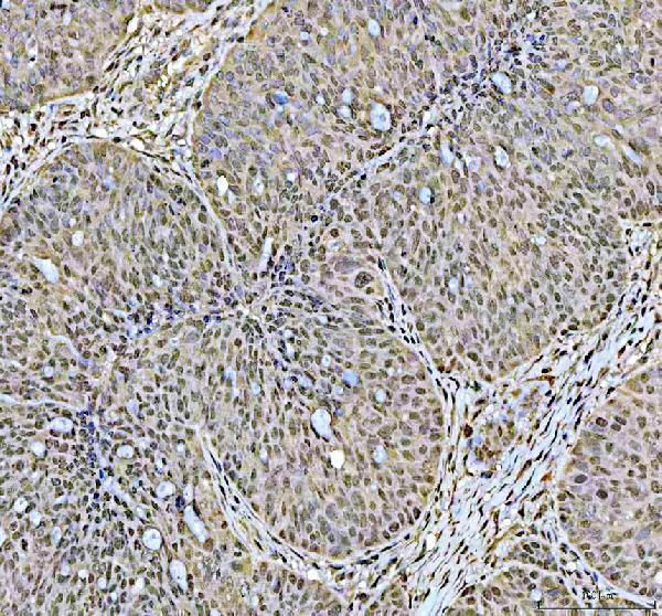
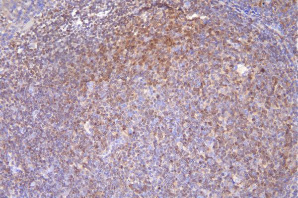
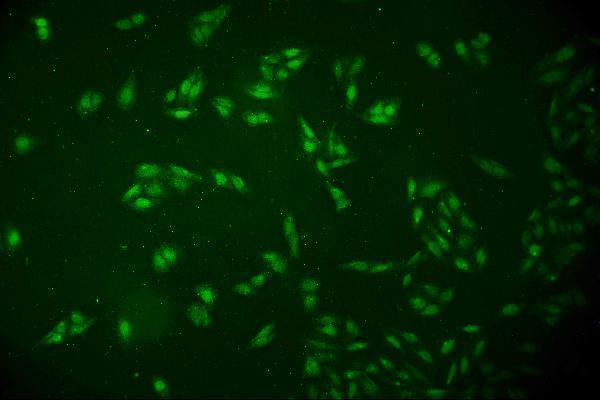


Comprehensive Human Protein Atlas IHC scoring per tissue (reliability: Enhanced — Medium consistency between antibody staining and RNA expression data. Presumed off target binding observed and disregarded. Antibody staining in cells/structures not annotated, view images.). Rows are taken directly from the HPA tissue chart — click any row's HPA link to view the source.
| Tissue | Cell type | Level | Evidence | Source |
|---|---|---|---|---|
| Appendix | Lymphoid tissue | High | Protein (IHC) | HPA → |
| Lymph node | Germinal center cells | High | Protein (IHC) | HPA → |
| Tonsil | Germinal center cells | High | Protein (IHC) | HPA → |
| Bone marrow | Hematopoietic cells | Medium | Protein (IHC) | HPA → |
| Spleen | Cells in white pulp | Medium | Protein (IHC) | HPA → |
| Tissue | Cell type | Level | Evidence | Source |
|---|---|---|---|---|
| Adipose tissue | Adipocytes | Not detected | Protein (IHC) | HPA → |
| Adrenal gland | Glandular cells | Not detected | Protein (IHC) | HPA → |
| Breast | Adipocytes | Not detected | Protein (IHC) | HPA → |
| Bronchus | Respiratory epithelial cells | Not detected | Protein (IHC) | HPA → |
| Caudate | Glial cells | Not detected | Protein (IHC) | HPA → |
Troubleshoot DCK staining in paraffin sections by checking retrieval, cell identity, compartment, controls, and scoring before interpreting chromogenic signal.
Two anti-DCK antibodies have IHC images from human paraffin sections; one also has a mouse liver IHC image and a human U2OS IF/ICC image (catalog image captions).
A01655-1 has IHC images from human breast cancer, ovary cancer and tonsil paraffin sections, plus mouse liver paraffin sections (A01655-1 IHC captions). M01655 has an IHC image from a human tonsil cancer paraffin section and an IF/ICC image from U2OS cells (M01655 IHC and IF captions).
Which to pick: For human tissue IHC, both A01655-1 and M01655 have paraffin-section DAB images using EDTA pH 8 retrieval, with primary antibody concentrations of 2 μg/ml and 1 μg/ml, respectively (A01655-1 IHC caption; M01655 IHC caption). Choose M01655 for human IF/ICC: it is monoclonal clone 3G10, lists IF and ICC applications, and has a U2OS IF image at 2 μg/ml (M01655 catalog; M01655 IF caption). Choose A01655-1 for cross-species IHC because it lists human, mouse and rat reactivity, although its IHC images show human and mouse samples only; the fixative is unreported in both antibodies’ paraffin-section captions (A01655-1 catalog and IHC captions; M01655 IHC caption).