This website uses cookies to ensure you get the best experience on our website.


- Table of Contents
Plan chromogenic DCTN1 IHC on paraffin sections with the catalog antibody at 2–5 μg/mL (datasheet: A02175-2). Compare staining with HPA’s high staining in fallopian tube glandular cells and undetected staining in lymph node germinal center cells (HPA tissue IHC).

Expected localisation, validated protocols, controls and antibodies — the at-a-glance facts below, then the full design guide.
| Expected localisation | Cytoplasmic and nuclear staining in most tissues (HPA tissue IHC) | |
| Staining pattern | Cells in most tissues show cytoplasmic and nuclear staining (HPA tissue IHC) | |
| Antigen retrieval | EDTA pH 8.0 HIER, heat-mediated (datasheet A02175-2) | |
| Positive control | Fallopian tube+4 more · see all | |
| Negative control | Lymph node+1 more · see all |
| Fixation | Keep fixation consistent across sections. (standard IHC practice; not target-specific) | |
| Caveat | Germinal center cells may lack detectable staining (HPA tissue IHC) | |
| Regulation | No expression regulator specified (UniProt) | |
| Isoform / epitope | 6 isoforms; antibody epitope coverage is unspecified (UniProt) |
The catalog antibody has an IHC-P protocol (datasheet). Published examples include direct DCTN1 staining in human paraffin sections (PMC3898179) and FLAG-based detection of human DCTN1 in mouse free-floating sections (PMC12730482).
| Sample | Paraffin-embedded human ovarian adenocarcinoma tissue; fixative not specified (datasheet A02175-2) |
| Fixation | Image fixative and duration unreported (datasheet A02175-2); verify before use. |
| Sectioning | 4–5 µm sections on charged slides (standard) |
| Deparaffinisation | Xylene, graded ethanol series to water (standard) |
| Antigen retrieval | Heat retrieval: EDTA pH 8.0 (datasheet A02175-2); 20 min, 95–100 °C (standard) |
| Peroxidase block | 3% H2O2, 10 min, room temperature (standard) |
| Blocking | 10% goat serum (datasheet A02175-2) |
| Primary antibody | Rabbit anti-DCTN1, 2-5μg/ml (datasheet A02175-2) |
| Primary incubation | Overnight at 4 °C (datasheet A02175-2) |
| Detection | Streptavidin-biotin complex (SABC), DAB chromogen (datasheet A02175-2) |
| Counterstain | Hematoxylin, blue, dehydrate and mount (standard) |
| Expected result | DCTN1-positive staining in glandular cells of fallopian tube (HPA tissue IHC: High). HPA tissue profile: Cytoplasmic and nuclear expression in most tissues. No signal in the no-primary control. |
DCTN1 is a non-transmembrane protein associated with the cytoplasm, cytoskeleton, centrosome, spindle, nuclear envelope and cell cortex (UniProt Q14203 topology and subcellular location). In tissue IHC, expect cytoplasmic and sometimes nuclear staining across many cell types, including glandular and neuronal cells (HPA: tissue IHC profile). HPA rates its tissue staining “Approved,” with medium consistency against RNA data and external verification pending (HPA: tissue IHC reliability).
| Cytoplasmic staining in fallopian tube or gallbladder glandular cells, with possible nuclear staining. | This fits the reported tissue pattern: both glandular populations stain at a high level, and the overall profile includes cytoplasmic and nuclear expression (HPA: tissue IHC). Compare cells within the section when judging distribution (general IHC practice). |
| Staining is confined to cell surfaces or extracellular spaces, with no convincing intracellular signal. | Treat this as a compartment mismatch and investigate artefact: DCTN1 has no transmembrane segment, while its reported locations are intracellular (UniProt Q14203 topology and subcellular location). Nuclear staining alone is not a mismatch because HPA reports nuclear expression (HPA: tissue IHC profile). |
| A strong reaction dominates a cell population reported as undetected, while expected positive cells are unstained. | Consider cross-reactivity or endogenous detection activity and check the assay controls (general IHC practice). HPA reports no detection in lymph-node germinal-center cells and spleen red-pulp cells, but these observations are assay-specific, not universal negative controls (HPA: tissue IHC). |
| Diffuse color covers cells and surrounding tissue without clear cellular boundaries. | This is difficult to score as DCTN1 because the reported pattern is cellular (HPA: tissue IHC profile). Review background in a control lacking primary antibody, blocking, washes and chromogen development; these are general IHC checks, not documented DCTN1-specific effects (general IHC practice). |
| No signal appears in fallopian tube or gallbladder glandular cells. | First check section quality, controls, retrieval and detection before calling DCTN1 absent (general IHC practice). HPA reports high staining in these cells, but its tissue evidence has medium consistency with RNA data and awaits external verification (HPA: tissue IHC and reliability). |
| Tissue and cell selection | HPA reports high staining in fallopian-tube and gallbladder glandular cells; medium staining in several other listed populations, including caudate neurons; and low staining in hippocampal neurons (HPA: tissue IHC). Select comparison cells by their reported level rather than treating all tissues as equally bright (general IHC practice). |
| Compartment used for scoring | HPA summarizes tissue staining as cytoplasmic and nuclear in most tissues (HPA: tissue IHC profile). UniProt also lists cytoskeletal, centrosomal, spindle, nuclear-envelope and cortical locations (UniProt Q14203 subcellular location). A fine structure seen in cells need not resolve as a distinct feature in chromogenic tissue IHC (general IHC practice). |
| Antibody evidence | Four listed antibodies have an “Approved” IHC status; the tissue profile has medium consistency with RNA expression and awaits external verification (HPA: antibodies and tissue IHC reliability). An Approved label supports use as a starting point but does not establish that every positive deposit is specific (general IHC practice). |
| Isoforms and epitope | UniProt lists six DCTN1 isoforms and a CAP-Gly domain at residues 48–90 (UniProt Q14203 isoforms and domains). The supplied evidence does not locate any catalog-antibody epitope, so isoform coverage and any retrieval dependence cannot be inferred from it (UniProt Q14203; HPA: supplied antibody records). |
| Processing and fixation evidence | UniProt lists one chain spanning residues 1–1278, with no signal peptide or propeptide and no reported glycosylation sites (UniProt Q14203 processing and glycosylation). Neither source supplied here reports a DCTN1-specific fixation effect; determine retrieval conditions with assay controls (UniProt Q14203; HPA: supplied records; general IHC practice). |
| Situation | Likely cause | Next action |
|---|---|---|
| Expected high-staining glandular cells are blank. | Section or detection failure is possible (general IHC practice); high staining is reported for these cells, though HPA verification remains pending (HPA: tissue IHC and reliability). | Check a positive section and detection controls, then review retrieval, antibody dilution and incubation against the chosen antibody's validated IHC conditions (general IHC practice). |
| Only a surface rim or extracellular material stains. | That distribution conflicts with the intracellular locations and absence of a transmembrane segment (UniProt Q14203 topology and subcellular location). | Inspect a control lacking primary antibody and reassess chromogen deposits and section morphology before scoring the rim as positive (general IHC practice). |
| Lymph-node germinal-center or spleen red-pulp cells stain strongly. | HPA reports these populations as not detected; cross-reactivity or endogenous detection activity is possible (HPA: tissue IHC; general IHC practice). | Compare a control lacking primary antibody and check endogenous-activity blocking; interpret the HPA negative observations within their assay limits (general IHC practice; HPA: tissue IHC). |
| Color is widespread, hazy or similar in cells and stroma. | High background can obscure a cellular pattern (general IHC practice); HPA reports cytoplasmic and nuclear tissue staining (HPA: tissue IHC profile). | Review blocking, washes, antibody concentration and development time alongside a control lacking primary antibody (general IHC practice). |
| Nuclear staining appears alongside cytoplasmic staining. | Nuclear expression is included in the HPA tissue profile, and UniProt lists the nuclear envelope as a location (HPA: tissue IHC; UniProt Q14203 subcellular location). | Score nuclear and cytoplasmic compartments separately; judge each against tissue morphology and controls (general IHC practice). |
| IF/ICC question: what pattern should be visible? | HPA supports microtubules as the main location and the cytokinetic bridge as an additional location; it marks spindle, primary-cilium and basal-body locations uncertain (HPA: subcellular ICC-IF). | Use the supported microtubule pattern as the primary localization check and interpret uncertain locations cautiously (HPA: subcellular ICC-IF). |
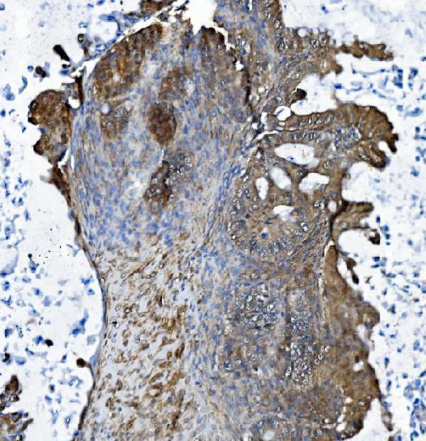
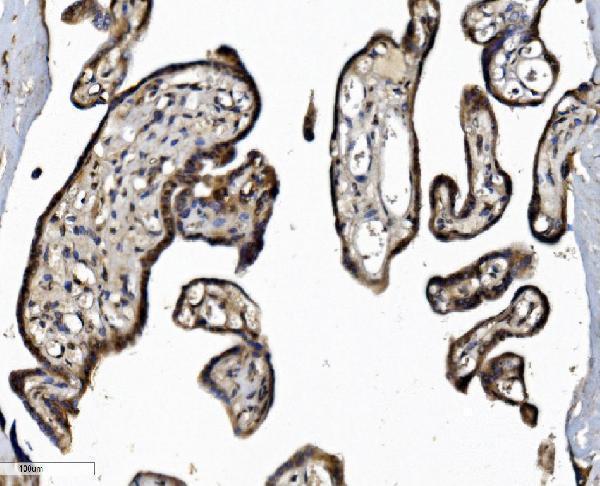
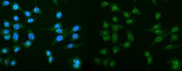


Comprehensive Human Protein Atlas IHC scoring per tissue (reliability: Approved — Medium consistency between antibody staining and RNA expression data. Pending external verification.). Rows are taken directly from the HPA tissue chart — click any row's HPA link to view the source.
| Tissue | Cell type | Level | Evidence | Source |
|---|---|---|---|---|
| Fallopian tube | Glandular cells | High | Protein (IHC) | HPA → |
| Gallbladder | Glandular cells | High | Protein (IHC) | HPA → |
| Adipose tissue | Adipocytes | Medium | Protein (IHC) | HPA → |
| Adrenal gland | Glandular cells | Medium | Protein (IHC) | HPA → |
| Appendix | Glandular cells | Medium | Protein (IHC) | HPA → |
Use the IHC-validated antibody’s paraffin-section example as a starting point, then judge staining against DCTN1’s documented distribution and appropriate controls.
A02175-2 has human paraffin-section IHC images (IHC captions) and a CACO-2 cell IF image (IF caption); catalog reactivity covers human, mouse and rat (catalog: reactivity).
A02175-2 has IHC images from human paraffin-embedded ovarian adenocarcinoma, placenta and breast cancer sections (IHC captions). The same SKU has an IF image from CACO-2 cells (IF caption) and lists IHC, ICC and IF applications (catalog: applications).
Which to pick: For paraffin-section IHC, choose A02175-2: its human tissue captions use EDTA retrieval at pH 8.0 and 2 μg/ml primary antibody (IHC captions); the fixative is unreported (IHC captions). For IF/ICC, A02175-2 has a CACO-2 cell IF image at 5 μg/ml (IF caption). For mouse or rat samples, A02175-2 lists those species as reactive (catalog: reactivity), while the supplied IHC and IF images show human samples (IHC and IF captions).