This website uses cookies to ensure you get the best experience on our website.
- Table of Contents
1 Citations 15 Q&As


1 Citations 5 Q&As
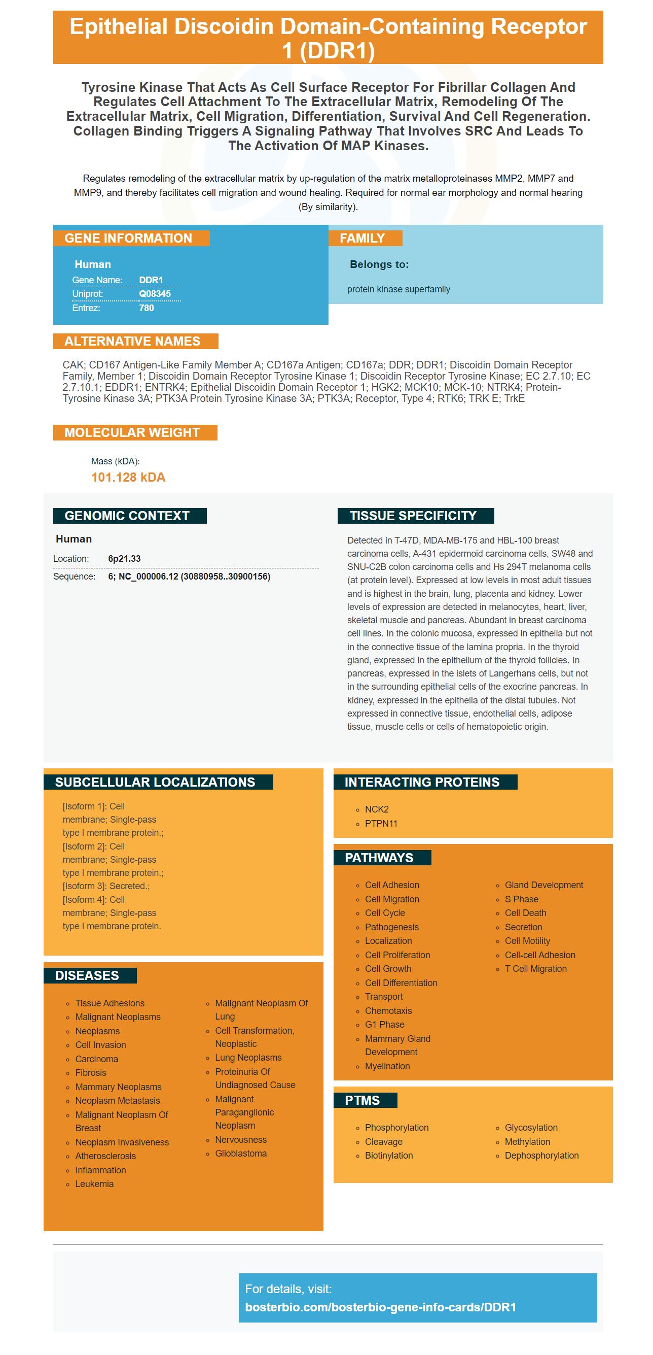
Facts about Epithelial discoidin domain-containing receptor 1.

Regulates remodeling of the extracellular matrix by up-regulation of the matrix metalloproteinases MMP2, MMP7 and MMP9, and thereby facilitates cell migration and wound healing. Required for normal ear morphology and normal hearing (By similarity).
| Human | |
|---|---|
| Gene Name: | DDR1 |
| Uniprot: | Q08345 |
| Entrez: | 780 |

| Belongs to: |
|---|
| protein kinase superfamily |

CAK; CD167 antigen-like family member A; CD167a antigen; CD167a; DDR; DDR1; discoidin domain receptor family, member 1; discoidin domain receptor tyrosine kinase 1; Discoidin receptor tyrosine kinase; EC 2.7.10; EC 2.7.10.1; EDDR1; ENTRK4; Epithelial discoidin domain receptor 1; HGK2; MCK10; MCK-10; NTRK4; Protein-tyrosine kinase 3A; PTK3A protein tyrosine kinase 3A; PTK3A; receptor, type 4; RTK6; TRK E; TrkE
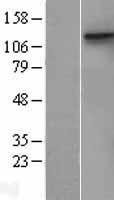
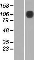
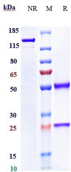
Mass (kDA):
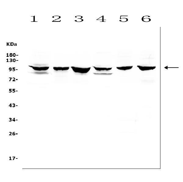
101.128 kDA

| Human | |
|---|---|
| Location: | 6p21.33 |
| Sequence: | 6; NC_000006.12 (30880958..30900156) |
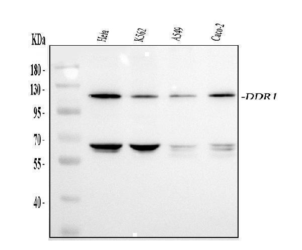
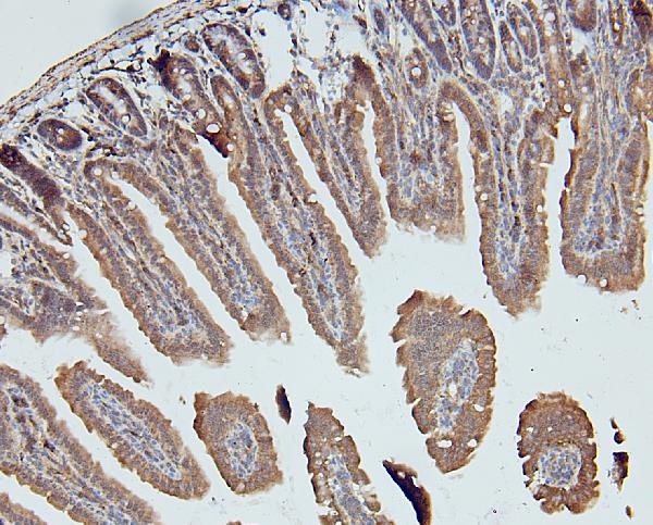
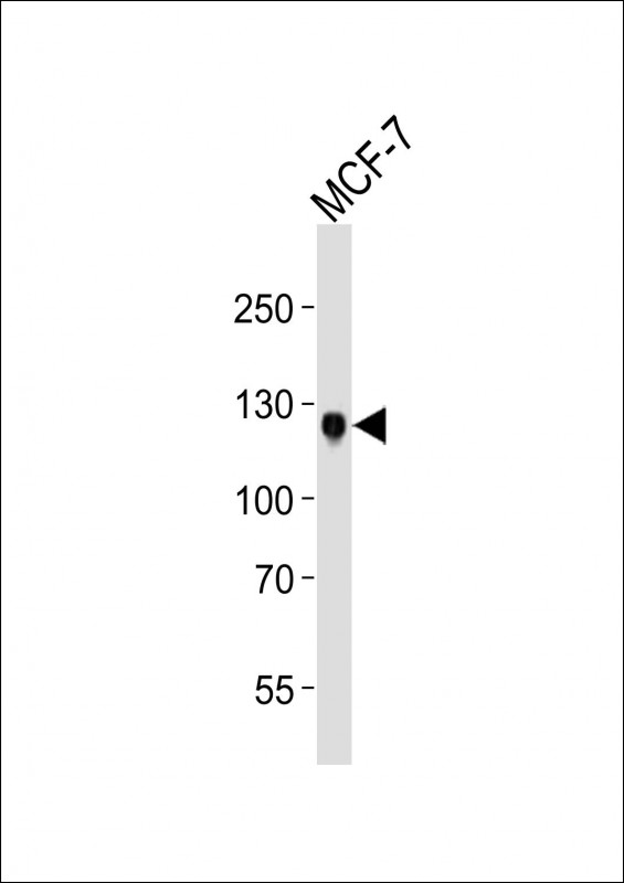
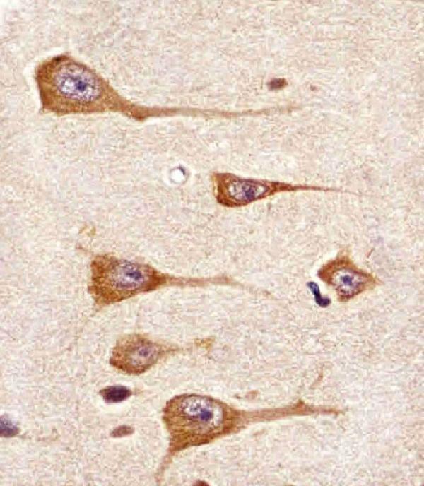
Detected in T-47D, MDA-MB-175 and HBL-100 breast carcinoma cells, A-431 epidermoid carcinoma cells, SW48 and SNU-C2B colon carcinoma cells and Hs 294T melanoma cells (at protein level). Expressed at low levels in most adult tissues and is highest in the brain, lung, placenta and kidney. Lower levels of expression are detected in melanocytes, heart, liver, skeletal muscle and pancreas. Abundant in breast carcinoma cell lines. In the colonic mucosa, expressed in epithelia but not in the connective tissue of the lamina propria. In the thyroid gland, expressed in the epithelium of the thyroid follicles. In pancreas, expressed in the islets of Langerhans cells, but not in the surrounding epithelial cells of the exocrine pancreas. In kidney, expressed in the epithelia of the distal tubules. Not expressed in connective tissue, endothelial cells, adipose tissue, muscle cells or cells of hematopoietic origin.
[Isoform 1]: Cell membrane; Single-pass type I membrane protein.; [Isoform 2]: Cell membrane; Single-pass type I membrane protein.; [Isoform 3]: Secreted.; [Isoform 4]: Cell membrane; Single-pass type I membrane protein.


PMID: 8226977 by di Marco E., et al. Molecular cloning of trkE, a novel trk-related putative tyrosine kinase receptor isolated from normal human keratinocytes and widely expressed by normal human tissues.
PMID: 8390675 by Johnson J.D., et al. A receptor tyrosine kinase found in breast carcinoma cells has an extracellular discoidin I-like domain.
*More publications can be found for each product on its corresponding product page