This website uses cookies to ensure you get the best experience on our website.


- Table of Contents
Plan DDX1 paraffin-section IHC around widespread nuclear staining (HPA tissue IHC). This guide covers fixation consistency, primary antibody titration at 0.5–1 µg/mL (datasheet A03727-1), DAB detection (datasheet A03727-1) and nuclear scoring.

Expected localisation, validated protocols, controls and antibodies — the at-a-glance facts below, then the full design guide.
| Expected localisation | Nuclear staining (HPA tissue IHC); cytosolic localization possible (UniProt) | |
| Staining pattern | Widespread nuclear staining across tissue cell types (HPA tissue IHC) | |
| Antigen retrieval | EDTA pH 8.0 HIER, heat-mediated (datasheet A03727-1) | |
| Positive control | Appendix+4 more · see all | |
| Negative control | None in HPA (detected in all 44 tissues); use no-primary + isotype controls |
| Fixation | Keep fixation consistent across sections (standard IHC practice; not target-specific); Matched tissue-IHC evidence does not establish the fixation claim. Validate the specimen-specific method before use. (selected-SKU IHC image A03727-1) | |
| Caveat | Active transcription can shift DDX1 into nuclei (UniProt) | |
| Regulation | Higher transcription in neuroectodermal tissues (UniProt) | |
| Isoform / epitope | 3 isoforms; antibody epitope coverage is unspecified (UniProt) |
The catalog antibody protocol uses EDTA pH 8.0 heat retrieval (datasheet: A03727-1); the published IHC protocols below cover hepatocellular carcinoma and colorectal cancer specimens (PMC10921000; PMC6113447).
| Sample | Paraffin-embedded rat testis tissue; fixative not specified (datasheet A03727-1) |
| Fixation | Image fixative and duration unreported (datasheet A03727-1); verify before use. |
| Sectioning | 4–5 µm sections on charged slides (standard) |
| Deparaffinisation | Xylene, graded ethanol series to water (standard) |
| Antigen retrieval | Heat retrieval: EDTA pH 8.0 (datasheet A03727-1); 20 min, 95–100 °C (standard) |
| Peroxidase block | 3% H2O2, 10 min, room temperature (standard) |
| Blocking | 10% goat serum (datasheet A03727-1) |
| Primary antibody | Rabbit anti-DDX1, 0.5-1μg/ml (datasheet A03727-1) |
| Primary incubation | Overnight at 4 °C (datasheet A03727-1) |
| Detection | Streptavidin-biotin complex (SABC), DAB chromogen (datasheet A03727-1) |
| Counterstain | Hematoxylin, blue, dehydrate and mount (standard) |
| Expected result | DDX1-positive staining in glandular cells of appendix (HPA tissue IHC: High). HPA tissue profile: Ubiquitous nuclear expression. No signal in the no-primary control. |
In paraffin-section IHC, expect DDX1 staining mainly in nuclei across many cell types, including appendix glandular cells and bone-marrow hematopoietic cells (HPA: ubiquitous nuclear expression; High in both cell types). HPA rates the tissue pattern Enhanced, with medium consistency between staining and RNA data (HPA: tissue IHC). DDX1 has no transmembrane segment, so a membrane pattern is unexpected (UniProt Q92499 topology).
| Clear nuclear staining in appendix glandular cells or bone-marrow hematopoietic cells. | This fits the expected compartment and two reported High-staining cell groups (HPA: ubiquitous nuclear expression; High in appendix glandular and bone-marrow hematopoietic cells). Compare staining within the identified cells; a dark section alone cannot establish the cellular pattern (general IHC practice). |
| Predominantly membranous staining, or cytoplasmic staining with little nuclear signal. | Reassess the result: HPA reports ubiquitous nuclear tissue staining, while UniProt reports no transmembrane segment (HPA: tissue IHC; UniProt Q92499 topology). Cytosolic DDX1 is also documented, so cytoplasmic signal alone is not proof of an artefact (HPA: ICC-IF supported cytosol). |
| Strong signal in an unexpected cell population, with weak signal in the identified HPA High cells. | Check cell identification, antibody specificity and chromogenic background before assigning the signal to DDX1 (general IHC practice). HPA reports High staining in specified cell groups but calls the overall nuclear pattern ubiquitous; its examples do not establish negative cell types (HPA: tissue IHC). |
| Diffuse brown haze across cells, extracellular spaces or the whole section. | A haze that obscures nuclear boundaries cannot be scored as the reported nuclear pattern (HPA: ubiquitous nuclear expression; general IHC practice). Check the no-primary control, blocking, washes and detection background; endogenous chromogenic activity is a possible technical cause (general IHC practice). |
| No nuclear signal in a section containing a reported High-staining cell group. | Treat this as an unresolved assay result before calling DDX1 absent (HPA: High in the listed cell groups; general IHC practice). Confirm that the relevant cells are present, then review the antibody's IHC validation, retrieval and detection controls (general IHC practice). |
| Cell population and tissue context | HPA reports ubiquitous nuclear expression and High staining in several identified populations, including caudate neuronal cells and cerebral-cortex glial cells (HPA: tissue IHC). These examples guide cell-level review; they do not define a negative tissue (HPA: no negative tissue listed). |
| Antibody validation | HPA034502 and HPA034503 have Enhanced IHC status; CAB012280 has Supported IHC status (HPA: antibody validation). These ratings describe those antibodies, so do not transfer them to an unidentified catalog antibody (HPA: antibody validation; general IHC practice). |
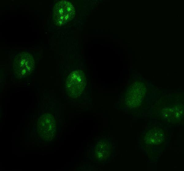
| IF/ICC question: can cytosolic signal be expected? | Yes. HPA reports mainly nucleoplasmic localization with additional supported cytosolic localization in ICC-IF (HPA: subcellular ICC-IF). That observation helps interpret IF/ICC images; it does not establish a separate paraffin-IHC staining threshold (HPA: tissue IHC; general IHC practice). |
| Isoform recognition and induced foci | UniProt lists 3 DDX1 isoforms and reports relocalization to foci after irradiation and to stress granules under stress (UniProt Q92499). Without an antibody epitope or treatment history, neither isoform coverage nor a punctate routine-IHC pattern can be predicted (general IHC practice). |
| Situation | Likely cause | Next action |
|---|---|---|
| Reported High-staining cells show no nuclear signal. | The assay may have failed, or the relevant cells may be absent from the examined area (general IHC practice). | Locate the identified cell group first; then check an IHC-validated antibody, the documented retrieval conditions and detection controls (HPA: High in listed cell groups; general IHC practice). |
| Most visible signal outlines cell membranes. | That distribution conflicts with the reported nuclear tissue pattern and lacks support from DDX1 topology (HPA: tissue IHC; UniProt Q92499 topology). | Review morphology and a no-primary control, then reassess antibody-specific signal and detection background (general IHC practice). |
| Unexpected cells stain strongly while HPA-listed cells stain weakly. | Cell misidentification, cross-reactivity or detection background may explain the discrepancy (general IHC practice). HPA lists no negative tissues (HPA: tissue IHC). | Identify cell types on the counterstained section and compare an appropriate antibody or control before interpreting the difference (general IHC practice). |
| Brown staining is widespread and nuclear edges are hard to distinguish. | Nonspecific reagent signal or endogenous chromogenic activity may obscure the pattern (general IHC practice). | Inspect a no-primary control; review blocking, washes and endogenous-activity controls appropriate to the detection system (general IHC practice). |
| Cytoplasmic signal accompanies clear nuclear staining. | DDX1 can also occur in cytosol, although HPA describes tissue IHC as ubiquitously nuclear (HPA: supported cytosol in ICC-IF; HPA: tissue IHC). | Score the nuclear component separately and check whether cytoplasmic staining is cell-associated and above control background (general IHC practice). |
| Punctate staining appears after a documented stress or irradiation treatment. | UniProt reports stress granules and irradiation-induced DDX1 foci (UniProt Q92499); the treatment alone does not identify every punctum. | Record the treatment and compartment, compare matched controls, and require corroboration before assigning puncta to DDX1 structures (general IHC practice). |
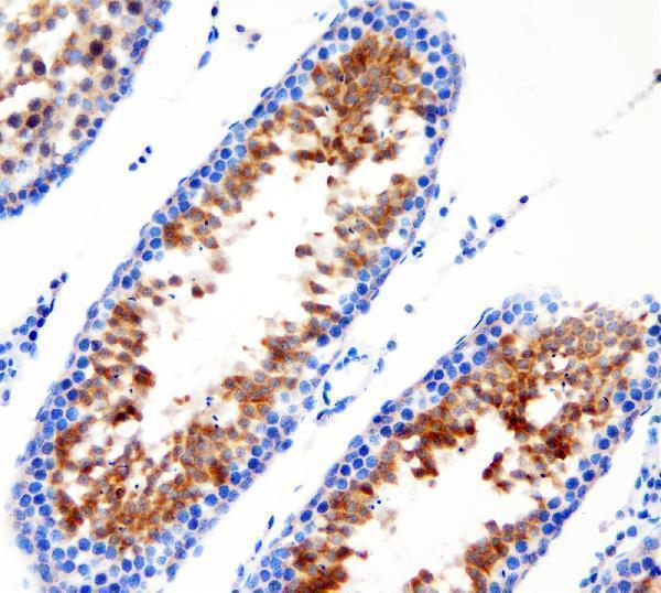
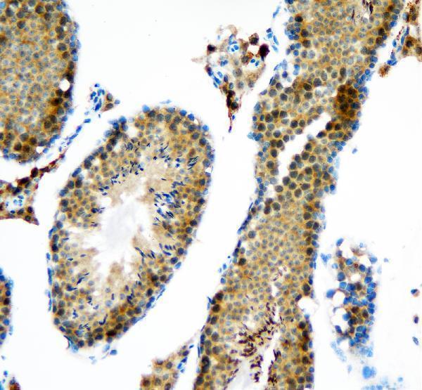
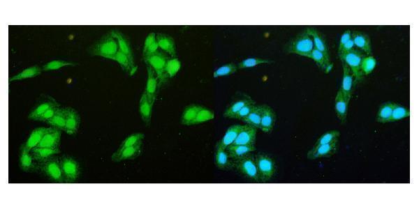
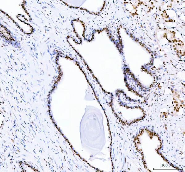
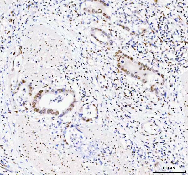
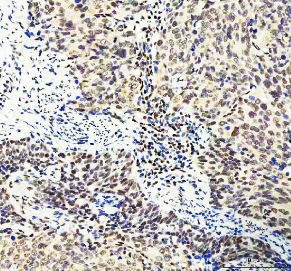
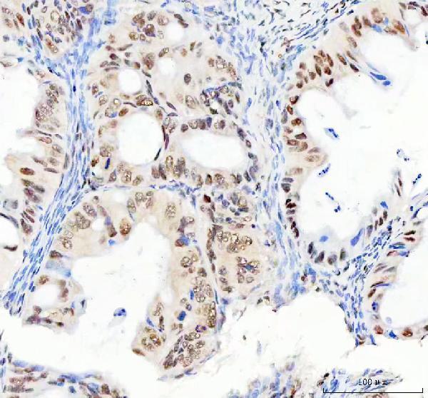


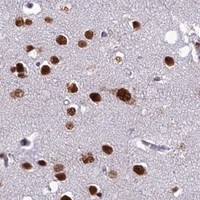
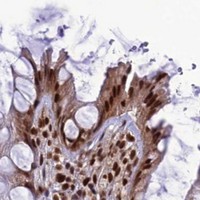


Comprehensive Human Protein Atlas IHC scoring per tissue (reliability: Enhanced — Medium consistency between antibody staining and RNA expression data.). Rows are taken directly from the HPA tissue chart — click any row's HPA link to view the source.
| Tissue | Cell type | Level | Evidence | Source |
|---|---|---|---|---|
| Appendix | Glandular cells | High | Protein (IHC) | HPA → |
| Bone marrow | Hematopoietic cells | High | Protein (IHC) | HPA → |
| Breast | Adipocytes | High | Protein (IHC) | HPA → |
| Caudate | Neuronal cells | High | Protein (IHC) | HPA → |
| Cerebellum | Cells in granular layer | High | Protein (IHC) | HPA → |
| Tissue | Cell type | Level | Evidence | Source |
|---|---|---|---|---|
| None in HPA: DDX1 is detected in all 44 scored tissues. Use a no-primary (secondary-only) and an isotype control instead. | ||||
Troubleshoot DDX1 staining in paraffin sections by checking retrieval, compartment, controls, and scoring before interpreting biological differences.
Catalog IHC images show DDX1 in rat testis and human cancer paraffin sections; IF images show human cells, human cancer sections and mouse brain (catalog image captions).
A03727-1 has rat testis IHC and human cell IF images (A03727-1 image captions); M03727 has human prostatic adenocarcinoma IHC and human cell IF images (M03727 image captions). M03727-2 has human cervical squamous carcinoma IHC images (M03727-2 image captions); M03727-1 has human gastric cancer IHC images (M03727-1 image captions).
Which to pick: For human tissue IHC, choose M03727: its own caption shows staining in a paraffin section, and it is monoclonal clone 3I10 (M03727 IHC caption; catalog: clone 3I10). For IF/ICC, choose A03727-1, which lists both applications and has IF images of human cells and paraffin sections; for cross-species IHC, its own IHC captions show mouse and rat paraffin sections, while human reactivity is listed separately (A03727-1 applications; IF and IHC captions; catalog reactivity). The fixative is unreported in these paraffin-section captions (catalog IHC captions).