DDX17 / Probable ATP-dependent RNA helicase DDX17 · IHC design guide

Design Immunohistochemistry for DDX17

Plan DDX17 IHC in paraffin sections with nuclear staining expected in most tissues (HPA tissue IHC). Lung alveolar type I cells show high staining and can serve as a positive reference (HPA tissue IHC); score signal by cell type.

Evidence assembled Oct 2026 · For research use; verify linked source records and product datasheet before use
Immunohistochemistry protocol sheet for DDX17 (IHC for DDX17): expected localisation Nuclear in most tissues (HPA tissue IHC), antibody A01656, validated IHC image, and IHC protocol steps
Printable DDX17 IHC protocol sheet — expected localisation Nuclear in most tissues (HPA tissue IHC), antibody A01656, controls and protocol steps. Open the full DDX17 IHC guide →

DDX17 Immunohistochemistry Experimental Design Guide

Expected localisation, validated protocols, controls and antibodies — the at-a-glance facts below, then the full design guide.

Must know before staining
Expected localisation Nuclear in most tissues (HPA tissue IHC)
Staining pattern Nuclear staining in cells of most tissues (HPA tissue IHC)
Antigen retrieval EDTA pH 8.0 HIER, heat-mediated (datasheet A01656)
Positive control ⓘ Lung+4 more · see all
Negative control ⓘ Adipose tissue+4 more · see all
Important caveats
Reasons your staining may differ from the expected pattern.
Fixation Keep fixation conditions consistent across paraffin sections. (standard IHC practice; not target-specific)
Caveat Staining intensity varies by cell type (HPA tissue IHC)
Regulation May rise with colon cancer progression (UniProt)
Isoform / epitope Four isoforms; epitope coverage is unreported (UniProt)
Section 1

Recommended DDX17 IHC & IF Protocols

The catalog antibody uses EDTA pH 8.0 heat retrieval (datasheet A01656). The published protocols below cover mouse heart sections (PMC13449089) and nasopharyngeal carcinoma tissue (PMC11736280).

Recommended immunohistochemistry (IHC-P) protocol parameters
SampleParaffin-embedded human liver cancer tissue; fixative not specified (datasheet A01656)
FixationImage fixative and duration unreported (datasheet A01656); verify before use.
Sectioning4–5 µm sections on charged slides (standard)
DeparaffinisationXylene, graded ethanol series to water (standard)
Antigen retrievalHeat retrieval: EDTA pH 8.0 (datasheet A01656); 20 min, 95–100 °C (standard)
Peroxidase block3% H2O2, 10 min, room temperature (standard)
Blocking10% goat serum (datasheet A01656)
Primary antibodyRabbit anti-DDX17, 1:50 recommended; image 2 μg/ml (datasheet A01656)
Primary incubationOvernight at 4 °C (datasheet A01656)
DetectionHRP-conjugated secondary, DAB chromogen (datasheet A01656)
CounterstainHematoxylin, blue, dehydrate and mount (standard)
Expected resultDDX17-positive staining in alveolar cells type I of lung (HPA tissue IHC: High). HPA tissue profile: Nuclear expression in most tissues. No signal in the no-primary control.
💡Decision noteStart with EDTA pH 8.0 heat retrieval for the catalog antibody (datasheet A01656); consider protease K retrieval for mouse heart sections (PMC13449089).
Section 2

What Is the Expected DDX17 Staining Pattern?

DDX17 should appear predominantly nuclear in paraffin sections, with staining assessed in the specific cell populations shown by HPA rather than across an entire tissue (HPA: nuclear expression in most tissues; IHC reliability Supported, with medium RNA–staining consistency). UniProt also places DDX17 in the nucleolus and cytosol and reports no transmembrane segment (UniProt Q92841: subcellular location and topology).

What am I looking at on my slide?
Clear nuclear staining in rectal or stomach glandular cells, or in lung alveolar type I cells.This matches cell populations scored High by tissue IHC (HPA: rectum, stomach and lung). Assess nuclear signal within the named cells; a positive tissue designation does not mean every cell on the section must stain equally (HPA: cell-level tissue observations).
Cytoplasmic staining dominates while nuclei are faint or unstained in an otherwise positive tissue.Recheck the staining pattern before calling this a routine positive: HPA reports predominantly nuclear tissue expression (HPA: tissue IHC profile). Cytosolic localization is listed by UniProt, including relocalization during bunyavirus infection, but that context does not establish a cytoplasmic pattern for an ordinary section (UniProt Q92841: subcellular location).
Strong staining appears in adipocytes, cholangiocytes or soft-tissue fibroblasts.These named populations were Not detected in the supplied HPA tissue observations (HPA: adipose tissue, liver and soft tissue). Consider nonspecific antibody binding or endogenous chromogenic activity before assigning the signal to DDX17 (general IHC practice); the HPA result does not prove those cells can never express it.
Colour spreads across nuclei, cytoplasm and cell-free areas without a clear cellular boundary.A diffuse deposit is difficult to score as DDX17 because the reference tissue pattern is nuclear (HPA: tissue IHC profile). Background from detection chemistry, blocking or washing is a plausible technical cause (general IHC practice); compare the deposit with a control lacking primary antibody.
No convincing nuclear signal appears in a section containing HPA high-staining cells.First confirm that the named cells are present and intact; examples include decidual cells and urothelial cells (HPA: placenta and urinary bladder, High). An absent signal may reflect the staining run or the sampled material (general IHC practice). HPA's Supported rating is not a guarantee for every specimen or workflow (HPA: IHC reliability Supported).
💡Expected DDX17 appearanceCall a section positive when the relevant cells show distinct, predominantly nuclear DDX17 staining—potentially High in HPA-listed rectal glandular or urothelial cells—while diffuse colour or dominant staining in HPA-listed undetected cells prompts a false-positive check (HPA: tissue IHC observations and profile).
How each factor affects the staining
Cell population and tissue siteHPA scores rectal glandular cells High, but colon endothelial cells Not detected (HPA: rectum and colon). UniProt describes normal colonic epithelial protein as low, if present, and increasing during colon cancer progression (UniProt Q92841: tissue specificity). Keep site, cell type and disease context attached to each comparison.
Compartment and molecular formNuclear expression is the tissue IHC reference (HPA: tissue IHC profile). DDX17 has no transmembrane segment or signal peptide, and its listed chain spans residues 1–729 (UniProt Q92841: topology and processing). These facts support a nuclear scoring focus but do not identify the antibody epitope or predict antigen-retrieval performance.
Antibody evidence and isoformsHPA063142 and CAB024908 each have Supported IHC status in the supplied antibody record (HPA: antibody validation). UniProt lists 4 isoforms (UniProt Q92841: isoforms). Without an epitope map, do not assume identical recognition of every isoform; the supplied sources give no target-specific fixation-sensitivity result.
IF/ICC Q: where should punctate signal appear?A: HPA reports supported localization to nuclear speckles in ICC-IF, with images from HEK293, SiHa and U2OS (HPA: subcellular localization). Use that as a compartment check for IF/ICC; the tissue IHC profile remains predominantly nuclear (HPA: tissue IHC profile).
Why is my staining missing, weak or wrong?
SituationLikely causeNext action
A named HPA high-staining population shows no nuclear signal.The expected cells may be absent from the cut, or the staining run may have failed (HPA: cell-level tissue observations; general IHC practice).Check morphology and cell identity, then review the IHC-validated antibody's stated conditions and a run control (general IHC practice). Do not infer a DDX17-specific retrieval or fixation defect from this result.
Signal is weak in duodenal glandular cells or caudate glial cells.Both populations are scored Low in the supplied tissue observations (HPA: duodenum and caudate).Compare like cell populations and avoid treating a faint result there as run failure by itself (HPA: tissue observations). Include a listed high-staining population when judging assay performance (HPA: positive tissue observations).
Most visible staining is cytoplasmic.That distribution differs from HPA's predominant nuclear tissue pattern; UniProt's cytosolic entry alone cannot establish the reason for this section's signal (HPA: tissue IHC profile; UniProt Q92841: subcellular location).Check whether nuclei have a distinct signal and compare a control lacking primary antibody; review detection and staining conditions if the cytoplasmic deposit persists (general IHC practice).
Strong colour appears in HPA-listed undetected cell populations.Nonspecific binding or endogenous detection activity is possible (general IHC practice); HPA reports Not detected for adipocytes, cholangiocytes and soft-tissue fibroblasts (HPA: tissue IHC).Inspect the exact cell type, assess a control lacking primary antibody, and review blocking and detection steps (general IHC practice). Treat an isolated mismatch as a finding to verify, not proof of cross-reactivity.
Diffuse colour obscures nuclear boundaries.Background from staining or detection can prevent compartment scoring (general IHC practice).Review blocking, washes and chromogen development against the run control (general IHC practice). Score DDX17 only where cellular nuclear staining is distinguishable from background (HPA: tissue IHC profile).
Rectal glandular staining seems inconsistent with low normal-colon expression.The statements concern different sites and contexts: HPA scores rectal glandular cells High, while UniProt describes normal colonic epithelial protein as low, if present (HPA: rectum; UniProt Q92841: tissue specificity).Record site, diagnosis and scored cell population before comparing sections; do not transfer the rectal score to all colonic epithelium (HPA: cell-level tissue observations; UniProt Q92841: tissue specificity).

Sample controls for DDX17 IHC & IF

🧪Run lung first: alveolar type I cells must show DDX17 staining (HPA: High in lung alveolar type I cells). Use adipose tissue as the negative comparator, scoring adipocytes (HPA: Not detected in adipocytes); on the lung slide, cells without nuclear signal should remain at background, but the supplied HPA rows do not establish a specific lung cell population as an internal biological negative (HPA: lung row).
Positive control tissue: Lung (Alveolar cells type I, HPA High)
Negative control tissue: Adipose tissue (HPA Not detected)
ICC-IF cell lines (HPA subcellular resource): HPA ICC-IF images show DDX17 in HEK293, SiHa, U2OS, with annotated localisation: Nuclear speckles (supported) (HPA subcellular).
Technical controls: Include a no-primary, secondary-only control, a concentration-matched rabbit IgG isotype control, and a DDX17 knockout biological control if available (caption: rabbit primary and anti-rabbit secondary; standard IHC practice). Quench endogenous peroxidase and inspect blood-rich areas for background before interpreting HRP/DAB staining in lung (caption: HRP/DAB detection; standard IHC practice).
⚠️Feasibility: A target-specific fixation window or fixation effect is unreported, and the selected A01656 paraffin-section caption does not state the fixative (caption: fixative unreported). The caption uses heat retrieval in EDTA at pH 8.0, but does not establish that retrieval is required (caption: EDTA heat retrieval); frozen sections are not established as easier, while IF/ICC has a separate nuclear-speckle readout in HEK293, SiHa and U2OS (HPA: nuclear speckles and ICC-IF cell lines). For lung IHC, residual blood can cause endogenous peroxidase background with HRP/DAB detection (caption: HRP/DAB; standard IHC practice).

HPA tissue IHC evidence for DDX17

Comprehensive Human Protein Atlas IHC scoring per tissue (reliability: Supported — Medium consistency between antibody staining and RNA expression data.). Rows are taken directly from the HPA tissue chart — click any row's HPA link to view the source.

Positive expression · recommended positive controls

TissueCell typeLevelEvidenceSource
Lung Alveolar cells type I High Protein (IHC) HPA →
Nasopharynx Respiratory epithelial cells High Protein (IHC) HPA →
Placenta Decidual cells High Protein (IHC) HPA →
Rectum Glandular cells High Protein (IHC) HPA →
Stomach Glandular cells High Protein (IHC) HPA →

Undetected expression · recommended negative controls

TissueCell typeLevelEvidenceSource
Adipose tissue Adipocytes Not detected Protein (IHC) HPA →
Colon Endothelial cells Not detected Protein (IHC) HPA →
Liver Cholangiocytes Not detected Protein (IHC) HPA →
Soft tissue Fibroblasts Not detected Protein (IHC) HPA →
Vagina Squamous epithelial cells Not detected Protein (IHC) HPA →
Section 3

Advanced DDX17 IHC Tips

Troubleshoot DDX17 staining in paraffin sections by checking retrieval, nuclear localisation and cell type before comparing chromogenic signal across samples.

How should I troubleshoot weak DDX17 staining after antigen retrieval?
Start with heat-mediated retrieval in EDTA pH 8.0 for paraffin sections (datasheet A01656). The documented tissue image used that buffer, followed by 2 μg/ml primary antibody overnight at 4°C (datasheet A01656). If nuclei remain weak, check that sections were fully deparaffinised and that heating was consistent across slides before changing retrieval conditions (standard IHC practice). If staining is still weak, test another retrieval condition as a fallback on matched sections, keeping antibody dilution and detection constant (standard IHC practice). Judge improvement by nuclear signal in expected positive cells and by background on a no-primary control (HPA: nuclear expression in most tissues; standard IHC practice).
Could fixation explain weak or uneven DDX17 IHC staining?
DDX17-specific fixation sensitivity is unknown from the supplied evidence; the selected paraffin-section caption does not state a fixative (datasheet A01656). Record the fixative, fixation duration and processing history for each specimen before comparing staining intensity (standard IHC practice). For uneven staining, examine whether peripheral tissue stains more strongly than the centre, then compare nearby sections processed together (standard IHC practice). Keep retrieval at EDTA pH 8.0 and the primary incubation at 2 μg/ml overnight at 4°C while assessing processing differences (datasheet A01656). Include a similarly processed positive control so a failed run is distinguishable from variation within the specimen (standard IHC practice).
Is cytoplasmic DDX17 staining credible when the nuclear signal is weak?
Expect predominantly nuclear DDX17 staining in tissue sections, and assess nuclear signal before accepting diffuse cytoplasmic colour (HPA: nuclear expression in most tissues). DDX17 is also annotated in the nucleolus and cytosol, so cytoplasmic staining is biologically possible but needs context (UniProt Q92841 subcellular location). Its reported nuclear-to-cytosol relocalisation occurs during bunyavirus infection; do not assume that context for an uncharacterised specimen (UniProt Q92841 subcellular location). Compare the signal with nuclear morphology, a no-primary control and another section from the same processing run (standard IHC practice). Report nuclear and cytoplasmic staining separately, including the fraction of cells in each compartment (standard IHC practice).
Can DDX17 isoforms or epitope accessibility change my IHC result?
DDX17 has 4 annotated isoforms, but the supplied caption does not identify the antibody epitope or establish isoform-specific staining (UniProt Q92841 isoforms; datasheet A01656). Check the antibody's immunogen or epitope documentation before interpreting absence of staining as absence of every isoform (standard IHC practice). The helicase ATP-binding region spans residues 202–377, and the helicase C-terminal region spans 405–552 (UniProt Q92841 domains). Annotated acetylation and phosphorylation sites provide biological context, but their effect on this antibody's paraffin-section staining has not been established (UniProt Q92841 modified residues; datasheet A01656). Compare matched sections under one retrieval and detection workflow, and describe results as antibody-detected DDX17 unless isoform specificity is independently demonstrated (standard IHC practice).
How can I assess DDX17 by IF alongside a cell-type marker?
For IF on a separate section, pair DDX17 with a marker for the cell population being assessed, such as an epithelial marker when examining respiratory epithelial cells (HPA: high DDX17 in nasopharyngeal respiratory epithelial cells; standard IF practice). Select spectrally separated fluorophores and consider a far-red channel if the tissue has strong shorter-wavelength autofluorescence (standard IF practice). Because DDX17 is predominantly nuclear and has no transmembrane segment, permeabilise sufficiently for nuclear access after the chosen fixation, while checking morphology (HPA: nuclear expression in most tissues; UniProt Q92841 topology; standard IF practice). Use single-label and no-primary controls to assess bleed-through and autofluorescence (standard IF practice). HPA reports nuclear-speckle localisation in ICC/IF images, but that does not validate this catalog antibody for IF (HPA: supported nuclear speckles; datasheet A01656).
What should I change if DDX17 DAB staining has high background?
First distinguish diffuse tissue colour from discrete nuclear staining, the predominant DDX17 pattern reported across tissues (HPA: nuclear expression in most tissues). The selected IHC workflow used 10% goat serum blocking, 2 μg/ml primary antibody overnight at 4°C, and a peroxidase-linked secondary for 30 minutes at 37°C (datasheet A01656). Check a no-primary section for secondary or detection-system staining, and include a peroxide block to limit endogenous peroxidase signal (standard chromogenic IHC practice). If background persists, optimise primary concentration and washing on matched sections while keeping retrieval at EDTA pH 8.0 (datasheet A01656; standard IHC practice). Exclude precipitated chromogen, folds and tissue edges when judging whether the background is cellular (standard IHC practice).
How should I quantify DDX17 staining across paraffin sections? ⚠ ANSWER MARKED FOR VERIFICATION
Define the cell population and region of interest before scoring, because DDX17 expression differs among cell types (HPA: tissue IHC profile). For chromogenic IHC, record the percentage of nuclei at each intensity and calculate an H-score on a 0–300 scale, or report the percentage of positive nuclei (standard IHC scoring practice). If counting positive cells per mm², also report total evaluable cells or tissue area so cellularity differences remain visible (standard image-analysis practice). Normalise comparisons to the same cell type, section handling, retrieval and detection run, with matched positive controls (standard IHC practice). Score cytoplasmic signal separately rather than adding it to the nuclear H-score (UniProt Q92841 subcellular location; standard IHC practice).
How can I tell true DDX17 positivity from staining artefact?
Prioritise reproducible nuclear staining in intact cells: HPA describes nuclear expression in most tissues and rates the tissue-IHC evidence as Supported (HPA: tissue IHC profile). Nuclear signal in the expected cell population is more persuasive than colour restricted to edges, folds or necrotic areas (standard IHC interpretation practice). A no-primary control helps identify endogenous peroxidase or detection-system signal, while morphology and counterstain help locate the colour within cells (standard chromogenic IHC practice). Interpret cytoplasmic staining cautiously because DDX17 can occur in the cytosol, but its reported infection-related relocalisation requires the relevant biological context (UniProt Q92841 subcellular location). Compare matched controls before calling a negative result, particularly when processing or retrieval differs between sections (standard IHC practice).
Boster reagents

Best DDX17 / Probable ATP-dependent RNA helicase DDX17 IHC Antibodies

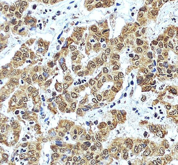
Both anti-DDX17 antibodies list IHC and IF/ICC for human, mouse, and rat (catalog: A01656 and M01656 applications/reactivity). A paraffin-section IHC image documents human liver cancer (A01656 image caption).

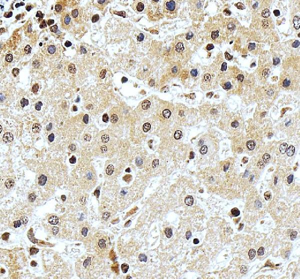
Real IHC data IHC analysis of DDX17 using anti-DDX17 antibody (A01656). DDX17 was detected in a paraffin-embedded section of human liver cancer tissue. Heat mediated antigen retrieval was performed in EDTA buffer (pH 8.0, epitope retrieval solution). The tissue section was blocked with 10% goat serum. The tissue section was then incubated with 2 μg/ml rabbit anti-DDX17 Antibody (A01656) overnight at 4°C. Peroxidase Conjugated Goat Anti-rabbit IgG was used as secondary antibody and incubated for 30 minutes at 37°C. The tissue section was developed using HRP Conjugated Rabbit IgG Super Vision Assay Kit (Catalog # SV0002) with DAB as the chromogen.
Anti-DDX17 Antibody
Cat # A01656

A01656 lists IHC and IF/ICC for human, mouse, and rat, with IHC images from paraffin-embedded human liver cancer and liver sections (catalog: A01656 applications/reactivity and image captions). M01656 lists IHC and IF/ICC for human, mouse, and rat, but has no IHC or IF image in the payload (catalog: M01656 applications/reactivity and image fields).

Which to pick: For tissue IHC, choose A01656 because its own captions document staining in paraffin-embedded human liver sections; the fixative is unreported (A01656 image captions). For IF/ICC, both SKUs list those applications without an IF image; M01656 offers a monoclonal option, while A01656 is polyclonal (catalog: A01656 and M01656 applications, image fields, and antibody types). Both list human, mouse, and rat reactivity for cross-species planning, though the supplied IHC images show human tissue only (catalog: A01656 and M01656 reactivity; A01656 image captions).

Each figure is that product's own IHC / IF validation image from its datasheet.

References

  1. UniProt Consortium. UniProt entry Q92841 (DDX17_HUMAN, Probable ATP-dependent RNA helicase DDX17).
  2. Human Protein Atlas. DDX17 tissue IHC expression (reliability: Supported).
  3. Human Protein Atlas. DDX17 subcellular location (ICC-IF): Localized to the nuclear speckles..
  4. Human Protein Atlas. DDX17 antibody validation summary (2 antibodies).
  5. DDX17 induces epithelial-mesenchymal transition and metastasis through the miR-149-3p/CYBRD1 pathway in colorectal cancer. Cell death & disease 2023 — PMC9807641.
  6. DDX17 promotes hepatocellular carcinoma progression via inhibiting Klf4 transcriptional activity. Cell death & disease 2019 — PMC6814716.
  7. Carfilzomib triggers cardiotoxicity by suppressing SENP1-mediated deSUMOylation of DDX17. Acta biochimica et biophysica Sinica 2025 — PMC13449089.
  8. PLUNC downregulates the expression of PD-L1 by inhibiting the interaction of DDX17/β-catenin in nasopharyngeal carcinoma. Journal of pathology and translational medicine 2025 — PMC11736280.
  9. PubMed PMID:8871553 — UniProt-cited evidence.
  10. PubMed PMID:15461802 — UniProt-cited evidence.
  11. PubMed PMID:17974005 — UniProt-cited evidence.