This website uses cookies to ensure you get the best experience on our website.


- Table of Contents
Plan DDX3Y chromogenic IHC in paraffin sections using the cytoplasmic tissue pattern reported by HPA (HPA tissue IHC). Interpret staining cautiously because HPA reports that its tissue antibody targets proteins from more than one gene (HPA tissue IHC).

Expected localisation, validated protocols, controls and antibodies — the at-a-glance facts below, then the full design guide.
| Expected localisation | Cytoplasmic tissue stain; nuclear shuttling is molecular (HPA tissue IHC; UniProt) | |
| Staining pattern | Cytoplasmic staining in germinal center and hematopoietic cells (HPA tissue IHC) | |
| Antigen retrieval | Tris-EDTA pH 9.0 HIER, 95–98 °C, 20 min (rule: nuclear antigen) | |
| Positive control | Tonsil+4 more · see all | |
| Negative control | Cervix+4 more · see all |
| Fixation | Keep fixation consistent across paraffin sections. (standard IHC practice; not target-specific) | |
| Caveat | Antibody may also detect proteins from other genes (HPA tissue IHC) | |
| Regulation | Testis-specific at protein level (UniProt) | |
| Isoform / epitope | Three isoforms; epitope coverage requires checking (UniProt) |
The catalog antibody’s IHC-P protocol is accompanied by published DDX3Y methods for primate testis and dysgenetic gonadal sections (PMC9142519; PMC10284372).
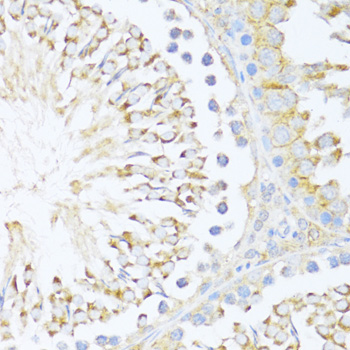
| Sample | Paraffin-embedded rat testis tissue; fixative not specified (datasheet A06062-2) |
| Fixation | Image fixative and duration unreported (datasheet A06062-2); verify before use. |
| Sectioning | 4–5 µm sections on charged slides (standard) |
| Deparaffinisation | Xylene, graded ethanol series to water (standard) |
| Antigen retrieval | Heat-induced epitope retrieval in Tris-EDTA buffer, pH 9.0, 20 min at 95–98 °C (standard rule: nuclear antigen) |
| Peroxidase block | 3% H2O2, 10 min, room temperature (standard) |
| Blocking | 10% normal serum of the secondary host, 30 min, room temperature (standard) |
| Primary antibody | Rabbit anti-DDX3Y, 1:50-1:100 (datasheet A06062-2) |
| Primary incubation | Overnight at 4 °C (standard) |
| Detection | HRP-polymer secondary, DAB chromogen 5–10 min (standard) |
| Counterstain | Hematoxylin, blue, dehydrate and mount (standard) |
| Expected result | DDX3Y-positive staining in germinal center cells of tonsil (HPA tissue IHC: High). HPA tissue profile: Cytoplasmic expression in several different tissue types, highly expressed in testis, lymphoid tissue and bone marrow. No signal in the no-primary control. |
DDX3Y is a soluble protein that shuttles between cytoplasm and nucleus; it has no transmembrane segment (UniProt O15523). In testis, expect staining predominantly in spermatogonia and occasionally in early spermatocytes (UniProt O15523). HPA reports cytoplasmic IHC staining across several tissues, but its Enhanced tissue profile carries a multi-gene targeting and presumed off-target binding caution (HPA tissue IHC).
| Cytoplasmic staining in spermatogonia, with occasional staining in pre-leptotene or leptotene spermatocytes (UniProt O15523). | This cell distribution fits the reported testis protein pattern (UniProt O15523). Assess localization and cell identity together; intensity alone does not establish DDX3Y specificity (HPA tissue IHC: multi-gene targeting caution). |
| Predominantly extracellular, luminal, or sharply membrane-only staining in testis. | This is a poor match for soluble DDX3Y in IHC (UniProt O15523: cytoplasm, nucleus, no transmembrane segment). Review morphology and staining controls before assigning target expression (general IHC practice). Nuclear signal alone needs context because DDX3Y shuttles (UniProt O15523). |
| Strong staining in an unexpected cell population, such as glandular cells reported as undetected in cervix (HPA tissue IHC). | Consider antibody cross-reactivity or endogenous chromogen-generating activity (HPA tissue IHC: presumed off-target binding; general IHC practice). A stained cell is not automatically DDX3Y-positive; compare with a no-primary control and the tissue pattern. |
| Broad haze across tissue and empty spaces, obscuring cell boundaries. | Treat this as background rather than a localized result (general IHC practice). Check whether the no-primary control also stains, then review blocking, washes, detection chemistry, and the catalog antibody dilution before scoring (general IHC practice). |
| No discernible staining in testis spermatogonia (UniProt O15523). | A negative result in the expected cell population is inconclusive until the run controls pass (UniProt O15523; general IHC practice). Check tissue preservation, retrieval, antibody incubation, and chromogen development against the catalog IHC-P instructions; no DDX3Y-specific fixation effect is established here. |
| Tissue choice and cell identity | Testis offers a protein-level expectation centered on spermatogonia (UniProt O15523). HPA also reports high staining in tonsil germinal center cells and medium staining in several other cell populations, but its multi-gene antibody caution limits attribution of those signals to DDX3Y (HPA tissue IHC). |
| Antibody specificity and validation | HPA lists IHC status as Enhanced for HPA059583 and Approved for HPA001648 and HPA005631 (HPA antibodies). Enhanced describes reproducibility or orthogonal support for the observed pattern; it does not remove the tissue profile’s presumed off-target binding warning (HPA antibodies; HPA tissue IHC). |
| Compartment and protein topology | Cytoplasmic signal is consistent with HPA tissue IHC; nuclear signal is biologically possible because DDX3Y shuttles (HPA tissue IHC; UniProt O15523). DDX3Y lacks a transmembrane segment, so a membrane-only IHC pattern needs scrutiny (UniProt O15523). |
| Isoforms and antibody epitope | Three isoforms are annotated, but the supplied record gives no epitope mapping or isoform-specific IHC performance (UniProt O15523). Do not infer which isoform a stained cell contains; check the catalog antibody’s stated immunogen and IHC-P validation when interpreting discordant staining (general IHC practice). |
| Endogenous chromogenic activity | Endogenous enzyme activity can produce chromogenic signal independently of the primary antibody (general IHC practice). A no-primary control helps identify detection-derived staining; assess it alongside morphology before calling a weak or widespread signal positive (general IHC practice). |
| IF/ICC Q&A: should its pattern match IHC? | HPA reports mainly cytosolic ICC-IF localization and an additional approved plasma-membrane localization, with a multi-gene antibody caution (HPA subcellular). UniProt also permits nuclear localization through shuttling (UniProt O15523). Use the separate IF/ICC guide for assay design; these observations do not define an IHC-P membrane pattern. |
| Situation | Likely cause | Next action |
|---|---|---|
| Testis section has no spermatogonial signal (UniProt O15523). | The assay may have failed, or the sampled cells may be absent or poorly preserved (general IHC practice). | Confirm spermatogonia on the counterstained section, inspect run controls, and follow the catalog antibody’s IHC-P retrieval and detection instructions (general IHC practice). |
| Tonsil germinal center cells stain strongly (HPA tissue IHC: High). | HPA reports this pattern, but its tissue profile warns of antibodies targeting multiple genes and presumed off-target binding (HPA tissue IHC). | Record the cell type and compartment; compare with testis and appropriate controls. Do not use tonsil staining alone as proof of DDX3Y specificity (HPA tissue IHC; UniProt O15523). |
| Cervical glandular cells stain despite an HPA ‘Not detected’ entry (HPA tissue IHC). | Possible nonspecific antibody binding or endogenous detection activity (HPA tissue IHC: off-target caution; general IHC practice). | Inspect the no-primary control, staining boundaries, and antibody concentration. Treat the result as unresolved until controls support a cell-associated pattern (general IHC practice). |
| Diffuse brown background masks individual cells. | Excess antibody, inadequate blocking or washing, or endogenous detection activity can obscure chromogenic IHC (general IHC practice). | Compare no-primary and tissue controls, then adjust blocking, washing, or antibody dilution within the catalog IHC-P guidance (general IHC practice). |
| Only nuclei stain in an otherwise negative testis section. | DDX3Y can enter nuclei, but an exclusively nuclear pattern lacks the expected cytoplasmic context (UniProt O15523; HPA tissue IHC). | Check that signal follows the expected spermatogonial distribution and that controls are clean before interpreting nuclear staining as DDX3Y (UniProt O15523; general IHC practice). |
| A membrane-only outline dominates the chromogenic section. | DDX3Y has no transmembrane segment; the HPA additional plasma-membrane observation comes from ICC-IF and carries a multi-gene caution (UniProt O15523; HPA subcellular). | Recheck compartment assignment, no-primary staining, and antibody specificity. Seek a cytoplasmic testis pattern before calling the IHC result consistent with DDX3Y (UniProt O15523; HPA tissue IHC). |
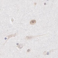


Comprehensive Human Protein Atlas IHC scoring per tissue (reliability: Enhanced — Caution, targets protein from more than one gene. Presumed off target binding observed and disregarded.). Rows are taken directly from the HPA tissue chart — click any row's HPA link to view the source.
| Tissue | Cell type | Level | Evidence | Source |
|---|---|---|---|---|
| Tonsil | Germinal center cells | High | Protein (IHC) | HPA → |
| Appendix | Glandular cells | Medium | Protein (IHC) | HPA → |
| Bone marrow | Hematopoietic cells | Medium | Protein (IHC) | HPA → |
| Breast | Glandular cells | Medium | Protein (IHC) | HPA → |
| Cerebellum | GLUC cells - cytoplasm/membrane | Medium | Protein (IHC) | HPA → |
| Tissue | Cell type | Level | Evidence | Source |
|---|---|---|---|---|
| Cervix | Glandular cells | Not detected | Protein (IHC) | HPA → |
| Endometrium | Cells in endometrial stroma | Not detected | Protein (IHC) | HPA → |
| Heart muscle | Cardiomyocytes | Not detected | Protein (IHC) | HPA → |
| Hippocampus | Glial cells | Not detected | Protein (IHC) | HPA → |
| Liver | Cholangiocytes | Not detected | Protein (IHC) | HPA → |
Troubleshoot DDX3Y chromogenic IHC in paraffin sections by checking retrieval, cell type, compartment, antibody specificity and scoring controls (UniProt O15523; HPA tissue IHC).
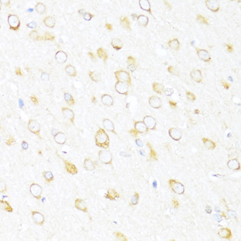
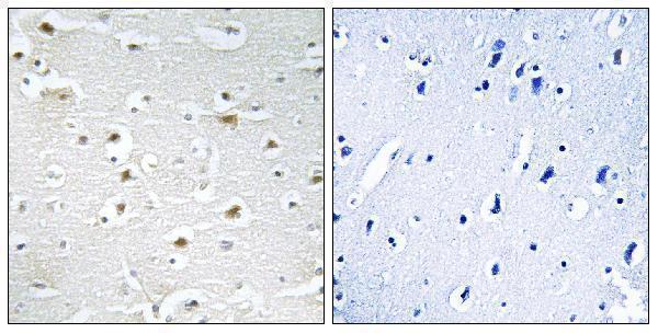
Two anti-DDX3Y antibodies have IHC images from paraffin sections of human brain, rat testis and rat brain (catalog IHC captions). Both list IF/ICC; IF images are unreported (catalog applications; image entries).
A06062-1 lists human and mouse reactivity and IF/ICC, with a peptide-blocked IHC comparison in paraffin-embedded human brain (catalog; A06062-1 IHC caption). A06062-2 lists human, mouse and rat reactivity and IF/ICC, with paraffin-section IHC images from rat testis and rat brain (catalog; A06062-2 IHC captions).
Which to pick: For tissue IHC, choose A06062-1 when the human brain example is most relevant, or A06062-2 for the rat testis or brain examples; the captions identify paraffin sections but do not report the fixative (IHC captions). For IF/ICC, both are listed for those applications, but neither has an IF image in the payload; A06062-1 is identified as polyclonal, while A06062-2 has no reported clonality (catalog). Choose A06062-2 when rat reactivity is needed alongside human and mouse reactivity (catalog reactivity).