This website uses cookies to ensure you get the best experience on our website.


- Table of Contents
Real validated DEFB1 Western blot protocols, expected-band and isoform facts, troubleshooting for weak or shifted signal, and recommended anti-DEFB1 WB antibodies. Everything you need to plan the experiment before you commit precious samples.
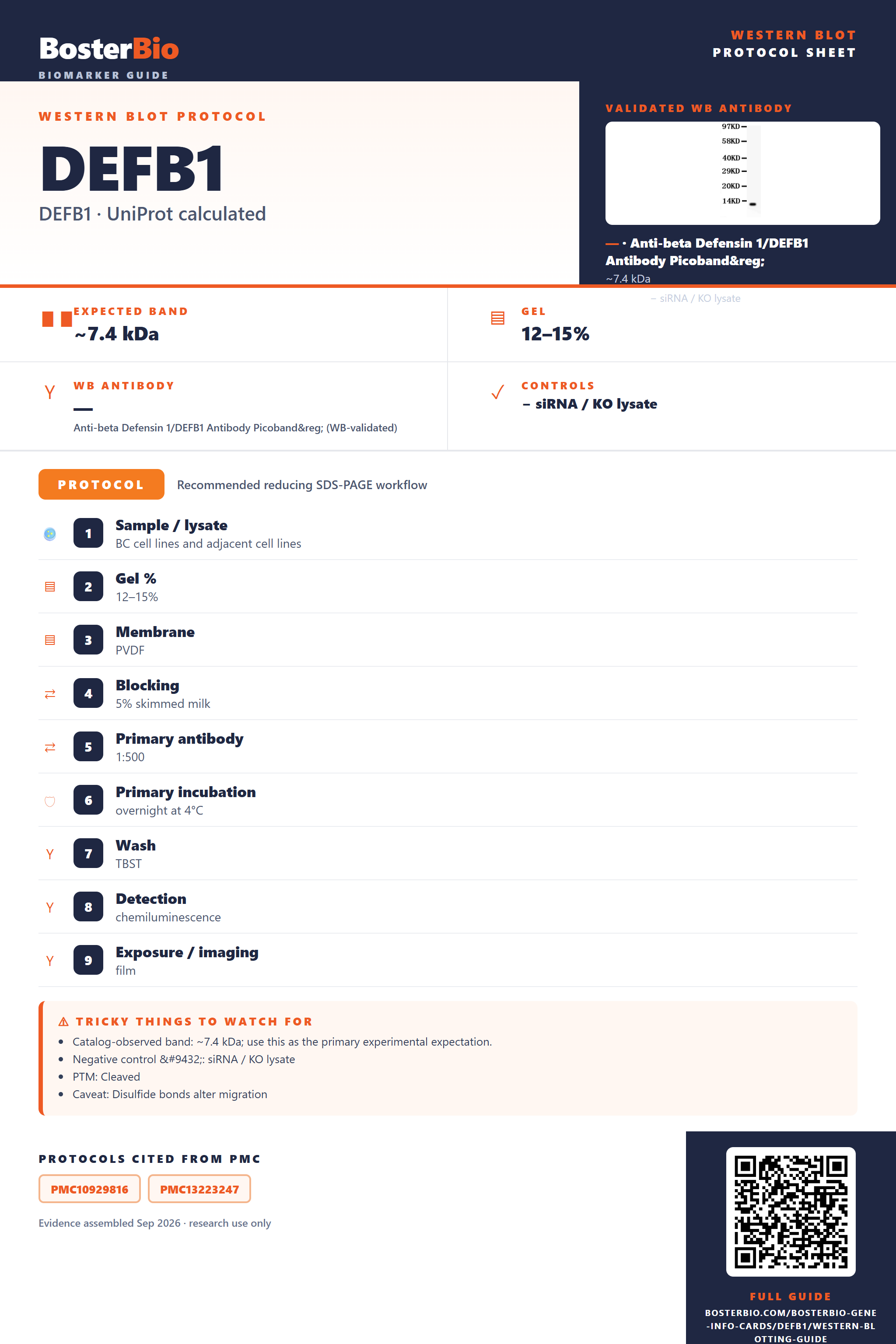
Expected bands, validated protocols, controls and antibodies — the at-a-glance facts below, then the full design guide.
| Expected band | ~7.4 kDa | |
| Observed band | 7 kDa | |
| Gel | 12–15% | |
| Negative control | siRNA / KO lysate |
| PTM | Cleaved | |
| Caveat | Disulfide bonds alter migration | |
| Regulation | LPS-induced | |
| Isoform | 1 isoform(s) |
Literature-validated Western blot parameters for DEFB1 — gel percentage, transfer, blocking, antibody incubation and detection, extracted from published methods.
| Sample / lysate | BC cell lines and adjacent cell lines |
| Gel % | 12–15% |
| Membrane | PVDF |
| Blocking | 5% skimmed milk |
| Primary antibody | 1:500 |
| Primary incubation | overnight at 4°C |
| Wash | TBST |
| Detection | chemiluminescence |
| Exposure / imaging | film |
DEFB1 has a 7.4 kDa predicted precursor mass and runs at an observed ~7 kDa, with homodimerization able to shift the band higher when reduction is incomplete.
| single band near 7 kDa | matches the DEFB1 precursor mass with no glycosylation to add extra weight |
| band smaller than the ~7 kDa precursor | signal peptide and propeptide have been cleaved, leaving the shorter mature secreted peptide |
| faint or absent band in whole-cell lysate | DEFB1 is a secreted peptide, so little of it accumulates intracellularly |
| band near double the monomer mass (~14-15 kDa) under non-reducing conditions | reflects homodimer formation between DEFB1 monomers |
| slightly anomalous migration relative to linear mass | the three intrachain disulfide bonds create a compact folded structure that can run atypically on SDS-PAGE |
| Predicted precursor mass (7.4 kDa) | sets the baseline size before any cleavage or oligomerization, and matches the observed ~7 kDa band |
| Signal peptide (residues 1-21) and propeptide (residues 22-32) cleavage | removes the N-terminal region to generate the shorter mature secreted peptide, which runs below the full-length precursor |
| Three intrachain disulfide bonds (Cys37-Cys66, Cys44-Cys59, Cys49-Cys67) | produce a compact, folded peptide that can migrate slightly anomalously on SDS-PAGE compared to its linear mass, especially if reduction is incomplete |
| Homodimer formation | shifts a portion of the protein to roughly twice the monomer mass under non-reducing or native conditions |
| Secreted subcellular localization | lowers the amount of protein retained in whole-cell lysate relative to secreted fractions |
| Situation | Likely cause | Next action |
|---|---|---|
| No band in lysate | DEFB1 is secreted, so it is exported out of the cell rather than retained intracellularly | test concentrated secreted fractions such as conditioned media or saliva instead of relying solely on whole-cell lysate |
| Band lower than expected | cleavage of the signal peptide and propeptide removes mass to generate the smaller mature secreted peptide | confirm whether the antibody epitope lies in the mature region or the cleaved pro-region, and expect a smaller band in processed samples |
| Band higher than expected | incomplete reduction allows homodimers to persist at roughly twice the monomer mass | use fresh reducing agent and sufficient heat denaturation before loading to fully resolve the monomer |
| Multiple bands | precursor, mature monomer, and homodimer forms can coexist at different apparent masses | run parallel reducing and non-reducing gels to distinguish monomer from dimer and precursor bands |
| Weak or no signal | DEFB1 is a small, low-abundance secreted peptide, limiting the amount present in standard lysate preparations | increase protein loading, use a high-percentage or Tricine gel suited for small peptides, and enrich secreted fractions |

Comprehensive Human Protein Atlas IHC scoring per tissue. Rows are taken directly from the HPA tissue chart — click any row's HPA link to view the source.
| Tissue | Cell type | Level | Evidence | Source |
|---|
| Tissue | Cell type | Level | Evidence | Source |
|---|
Deeper troubleshooting and optimisation questions for DEFB1, answered from its protein features.
BosterBio's DEFB1 antibodies are among the best-performing WB antibodies on the market — well cited, thoroughly validated, and orthogonally cross-validated against negative tissues and complementary methods.
Boster's anti-DEFB1 antibody PA1450 is our top recommendation for Western blot, offering strong citation history and thorough validation, including orthogonal cross-checks against negative tissue controls and complementary methods, ensuring dependable, reproducible detection of DEFB1 in your samples.
Which to pick: Only one Boster anti-DEFB1 antibody is listed, PA1450, which includes an actual Western blot validation image (COLO320 lysate, ~7 kD band matching predicted size)—making it the clear, and only, choice for your DEFB1 WB experiments.