DGKE / Diacylglycerol kinase epsilon · IHC design guide

Design Immunohistochemistry for DGKE

Plan DGKE staining in paraffin sections using cytoplasmic tissue staining and membrane association as guides to interpretation (HPA tissue IHC; UniProt). Compare testis late spermatids with a primary antibody omission control, and interpret staining cautiously because antibody staining has low consistency with RNA expression (HPA tissue IHC).

Evidence assembled Oct 2026 · For research use; verify linked source records and product datasheet before use
Immunohistochemistry protocol sheet for DGKE (IHC for DGKE): expected localisation Cytoplasmic (HPA tissue IHC); membrane-associated (UniProt), antibody A06615S194, validated IHC image, and IHC protocol steps
Printable DGKE IHC protocol sheet — expected localisation Cytoplasmic (HPA tissue IHC); membrane-associated (UniProt), antibody A06615S194, controls and protocol steps. Open the full DGKE IHC guide →

DGKE Immunohistochemistry Experimental Design Guide

Expected localisation, validated protocols, controls and antibodies — the at-a-glance facts below, then the full design guide.

Must know before staining
Expected localisation Cytoplasmic (HPA tissue IHC); membrane-associated (UniProt)
Staining pattern Cytoplasmic staining in several tissues, including testis (HPA tissue IHC)
Antigen retrieval Citrate pH 6.0 HIER, 95–98 °C, 20 min (rule: cytoplasmic / membrane antigen)
Positive control ⓘ Testis+4 more · see all
Negative control ⓘ Adipose tissue+4 more · see all
Important caveats
Reasons your staining may differ from the expected pattern.
Fixation Keep fixation conditions consistent across sections. (standard IHC practice; not target-specific)
Caveat Staining has low consistency with RNA expression (HPA tissue IHC)
Regulation Expression predominates in testis (UniProt)
Isoform / epitope Two isoforms; confirm antibody epitope coverage (UniProt)
Section 1

Recommended DGKE IHC & IF Protocols

The catalog antibody’s IHC-P protocol is accompanied by one published DGKE paraffin-section protocol (PMC3719402).

Recommended immunohistochemistry (IHC-P) protocol parameters
SampleParaffin-embedded human rectum carcinoma tissue; fixative not specified (datasheet A06615S194)
FixationImage fixative and duration unreported (datasheet A06615S194); verify before use.
Sectioning4–5 µm sections on charged slides (standard)
DeparaffinisationXylene, graded ethanol series to water (standard)
Antigen retrievalHeat-induced epitope retrieval in citrate buffer, pH 6.0, 20 min at 95–98 °C (standard rule: cytoplasmic / membrane antigen)
Peroxidase block3% H2O2, 10 min, room temperature (standard)
Blocking10% normal serum of the secondary host, 30 min, room temperature (standard)
Primary antibodyRabbit anti-DGKE, 1:50-1:200 (datasheet A06615S194)
Primary incubationOvernight at 4 °C (standard)
DetectionHRP-polymer secondary, DAB chromogen 5–10 min (standard)
CounterstainHematoxylin, blue, dehydrate and mount (standard)
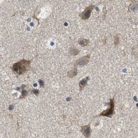
Expected resultDGKE-positive staining in elongated or late spermatids of testis (HPA tissue IHC: High). HPA tissue profile: Cytoplasmic expression in several tissues, including testis. No signal in the no-primary control.
💡Decision noteTry citrate pH 6.0 retrieval first (page retrieval rule; PMC3719402). The paper also reports a Tris-EDTA pH 9.0 variant with a different DGKE antibody (PMC3719402).
Section 2

What Is the Expected DGKE Staining Pattern?

Expect predominantly cytoplasmic DGKE staining in paraffin sections, strongest in elongated or late spermatids in testis (HPA: High; cytoplasmic tissue profile). DGKE is also annotated at membranes, with one transmembrane segment at residues 22–42 (UniProt P52429 topology). Treat the tissue pattern as provisional: HPA rates the IHC data Approved but reports low agreement with RNA expression and pending external verification (HPA: tissue reliability).

What am I looking at on my slide?
Strong cytoplasmic staining in elongated or late spermatids, with weaker staining elsewhere in the section.This matches the clearest supplied positive reference (HPA: High in testis spermatids). Compare staining in the named cells, not whole-section darkness. HPA's Approved rating includes a low-consistency warning, so morphology and controls still matter (HPA: tissue reliability).
A crisp nuclear-only pattern, or staining confined to a compartment other than cytoplasm or membrane.That conflicts with the tissue IHC profile and UniProt localisation (HPA: cytoplasmic tissue profile; UniProt P52429: membrane, cytoplasm). Review tissue morphology and detection controls before assigning such staining to DGKE; the uncertain nucleoplasmic ICC-IF annotation does not establish nuclear IHC staining (HPA: subcellular).
Strong staining in adipocytes or another cell type listed as not detected.HPA reports no detectable staining in adipose adipocytes and several other specified cells (HPA: tissue IHC). Consider cross-reactivity or endogenous detection activity, especially if a reagent-omission control also stains (general IHC practice). A negative reference is cell-specific, not a claim about every cell in that tissue.
Uniform colour across cells, stroma, and empty spaces, with little cell-specific contrast.This does not resemble HPA's cell-resolved cytoplasmic profile (HPA: tissue IHC). Diffuse chromogen can reflect detection background or inadequate blocking and washing (general IHC practice); assess it against a control lacking primary antibody before scoring cells.
No staining in elongated or late spermatids in an otherwise interpretable testis section.That misses the strongest supplied tissue reference (HPA: High in testis spermatids). First confirm those cells are present and identifiable, then check assay controls and reagent performance (general IHC practice). Absence alone cannot establish DGKE loss while HPA's tissue profile awaits external verification (HPA: tissue reliability).
💡Expected DGKE appearanceCall a positive result when elongated or late spermatids show strong, cell-associated cytoplasmic staining (HPA: High in testis spermatids; cytoplasmic tissue profile); uniform background, nuclear-only colour, or equally strong staining in HPA-undetected adipocytes warrants control-based review rather than a DGKE call (HPA: tissue IHC; general IHC practice).
How each factor affects the staining
Cell-specific referenceUse elongated or late spermatids as the strongest supplied IHC comparator (HPA: High). Respiratory epithelial cells, selected neuronal cells, colon and duodenal glandular cells, and cardiomyocytes are Medium references, not equivalent strong-positive controls (HPA: tissue IHC).
Membrane topologyDGKE has one annotated transmembrane segment, residues 22–42 (UniProt P52429 topology). HPA describes tissue staining as cytoplasmic (HPA: tissue profile); topology alone does not require a sharp plasma-membrane outline in chromogenic sections.
Antibody evidenceThe listed antibody HPA017167 is IHC Approved; no IHC Enhanced status is supplied (HPA: antibody validation). HPA also flags low staining–RNA consistency and pending external verification (HPA: tissue reliability). Interpret unexpected patterns with independent controls.
Transcript formsTwo DGKE isoforms are listed (UniProt P52429: isoforms 1 and 2). The payload gives no antibody epitope or isoform-specific IHC pattern, so it cannot explain a particular cell's staining difference.
IF/ICC Q&A: should nuclear fluorescence count as the expected IHC pattern?No. ICC-IF lists supported cytosol and uncertain nucleoplasm (HPA: subcellular), whereas tissue IHC describes cytoplasmic staining (HPA: tissue profile). Assess IF/ICC localisation in its own guide; its nuclear annotation does not validate nuclear chromogenic IHC.
Why is my staining missing, weak or wrong?
SituationLikely causeNext action
Testis positive reference is blank.The expected elongated or late spermatids may be absent from the viewed area, or the staining run may have failed (HPA: High in testis spermatids; general IHC practice).Inspect morphology and another field; verify the run with an appropriate positive control before interpreting DGKE absence (general IHC practice).
All tissues show diffuse brown signal.Non-specific detection background or endogenous enzyme activity may obscure cell-specific staining (general chromogenic IHC practice).Compare a no-primary control, check endogenous-activity blocking where applicable, and review washing and chromogen development (general IHC practice).
Nuclei dominate the signal.A nuclear-only IHC result conflicts with the HPA tissue profile; ICC-IF nucleoplasm is marked uncertain (HPA: tissue IHC; HPA: subcellular).Check counterstain versus chromogen, morphology, and no-primary control; avoid calling nuclear IHC positive without independent validation (general IHC practice).
Adipocytes stain as strongly as the testis reference.HPA lists adipose adipocytes as not detected (HPA: tissue IHC). Cross-reactivity or detection background is possible, but the HPA pattern itself has limited consistency (HPA: tissue reliability).Confirm the stained cells morphologically, inspect a no-primary control, and seek orthogonal support before assigning DGKE positivity (general IHC practice).
Medium-reference tissues vary between sections.HPA's Medium designation applies to named cells, not every cell or field; the tissue profile also awaits external verification (HPA: tissue IHC; tissue reliability).Score the specified cell population and record section quality and controls; avoid using field-wide intensity as the reference (general IHC practice).
A faint, broad cytoplasmic signal is difficult to score.HPA reports low staining in several specified cell populations, including lung alveolar and pancreatic exocrine cells (HPA: tissue IHC); weak colour may approach assay background (general IHC practice).Compare matched negative-control staining and the strong testis reference, then score only cell-associated signal above background (HPA: High in testis spermatids; general IHC practice).

Sample controls for DGKE IHC & IF

🧪Run testis first and expect staining in elongated or late spermatids (HPA: High in elongated or late spermatids). Use adipose tissue adipocytes as the negative tissue (HPA: Not detected in adipocytes); on the testis slide, treat neighboring unstained cells as internal background comparators, without assuming that every other cell type is DGKE-negative (HPA: High in elongated or late spermatids).
Positive control tissue: Testis (Elongated or late spermatids, HPA High)
Negative control tissue: Adipose tissue (HPA Not detected)
ICC-IF cell lines (HPA subcellular resource): HPA ICC-IF images show DGKE in A-431, U-251MG, U2OS, with annotated localisation: Nucleoplasm (uncertain), Cytosol (supported) (HPA subcellular).
Technical controls: Include a no-primary, secondary-only control, a host-species-, immunoglobulin-class-, and clonality-matched isotype control, and a DGKE knockout sample or a peptide-block control if the immunizing peptide is available (standard IHC practice; selected-SKU caption: PBS replaced primary antibody in its negative control). For chromogenic testis sections, block endogenous peroxidase and check for endogenous biotin signal if using avidin–biotin detection (standard IHC practice; selected-SKU caption: avidin–peroxidase detection).
⚠️Feasibility: No target-specific fixation window or fixation effect is reported in the supplied evidence, and the fixative in the selected-SKU paraffin-section caption is unreported (selected-SKU caption: paraffin-embedded rectum carcinoma; fixative not stated). Antigen-retrieval dependence is also unreported, so assess retrieval conditions empirically with the testis positive and adipose negative controls (HPA: High in testis spermatids; Not detected in adipocytes). There is no supplied comparison showing that frozen sections or IF are easier; HPA has ICC-IF images for A-431, U-251MG and U2OS, but those do not establish performance in tissue sections (HPA subcellular: ICC-IF image cell lines).

HPA tissue IHC evidence for DGKE

Comprehensive Human Protein Atlas IHC scoring per tissue (reliability: Approved — Low consistency between antibody staining and RNA expression data. Pending external verification.). Rows are taken directly from the HPA tissue chart — click any row's HPA link to view the source.

Positive expression · recommended positive controls

TissueCell typeLevelEvidenceSource
Testis Elongated or late spermatids High Protein (IHC) HPA →
Bronchus Respiratory epithelial cells Medium Protein (IHC) HPA →
Caudate Neuronal cells Medium Protein (IHC) HPA →
Cerebellum Purkinje cells Medium Protein (IHC) HPA →
Cerebral cortex Neuronal cells Medium Protein (IHC) HPA →

Undetected expression · recommended negative controls

TissueCell typeLevelEvidenceSource
Adipose tissue Adipocytes Not detected Protein (IHC) HPA →
Adrenal gland Glandular cells Not detected Protein (IHC) HPA →
Breast Adipocytes Not detected Protein (IHC) HPA →
Cervix Glandular cells Not detected Protein (IHC) HPA →
Endometrium Cells in endometrial stroma Not detected Protein (IHC) HPA →
Section 3

Advanced DGKE IHC Tips

Troubleshoot DGKE staining in paraffin sections by checking retrieval, cellular context and controls before interpreting chromogenic signal.

How should I retrieve DGKE when paraffin sections stain weakly?
Use citrate buffer at pH 6.0 for heat-induced retrieval at 95–98 °C for 20 minutes (page retrieval rule: cytoplasmic/membrane antigen). Let slides cool consistently, then compare a positive control section processed in the same run; testis with elongated or late spermatids is a documented high-staining reference (HPA tissue IHC: High in elongated or late spermatids). Keep section thickness, detection chemistry and exposure to chromogen constant while assessing retrieval (standard IHC practice). If signal remains weak, check antibody performance and fixation history before changing retrieval conditions, since the supplied evidence does not establish a DGKE-specific alternative buffer (page retrieval rule; supplied caption).
Could fixation explain weak or variable DGKE staining across paraffin blocks?
DGKE-specific sensitivity to fixation is unknown from the supplied evidence; the paraffin-section caption does not report a fixative (selected-SKU tissue-IHC caption). Record each block’s fixative and fixation duration, and compare sections with similar processing histories before attributing differences to DGKE abundance (standard IHC practice). Run a documented positive control alongside the affected section, using the same retrieval at pH 6.0 and 95–98 °C for 20 minutes (HPA tissue IHC: High in elongated or late spermatids; page retrieval rule). Uneven staining between blocks may reflect processing, so interpret it alongside morphology, the no-primary control and repeat staining under matched conditions (standard IHC practice).
What DGKE staining pattern should I expect in chromogenic IHC?
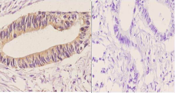
Assess cytoplasmic and membrane-associated staining: DGKE is annotated in the cytoplasm and membrane, with a transmembrane segment at residues 22–42 (UniProt P52429 localisation and topology). The selected paraffin-section image describes cytoplasmic staining in human rectum carcinoma at 1:50 (selected-SKU tissue-IHC caption). HPA also describes cytoplasmic tissue staining, including testis, although its tissue-IHC reliability note reports low consistency with RNA data and pending external verification (HPA tissue IHC). Treat isolated nuclear staining cautiously; the HPA subcellular record calls nucleoplasmic localisation uncertain and cytosolic localisation supported (HPA subcellular). Compare the pattern with a no-primary section before scoring it (standard IHC practice).
How can epitope location change my interpretation of DGKE staining?
DGKE has 2 annotated isoforms and a DAGKc domain at residues 215–356 (UniProt P52429 isoforms and domains). Its single transmembrane segment spans residues 22–42, so epitope position matters when interpreting access after paraffin processing (UniProt P52429 topology; standard IHC practice). Confirm the catalog antibody’s immunogen sequence and stated isoform coverage before assuming that staining represents both isoforms; those details are absent from the supplied caption (selected-SKU tissue-IHC caption). For discordant results, compare independently validated antibodies targeting distinct documented epitopes on adjacent sections, with matched retrieval and no-primary controls (standard IHC practice). Do not infer a modification-specific epitope from the caption’s “S194” designation alone (selected-SKU tissue-IHC caption).
How should I cross-check chromogenic DGKE staining with IF?
Use IF as a separate localisation check: HPA reports supported cytosolic and uncertain nucleoplasmic DGKE localisation in its subcellular record (HPA subcellular). Multiplex DGKE with a validated marker for the expected cell population, such as elongated or late spermatids in testis, and include single-colour controls (HPA tissue IHC: High in elongated or late spermatids; standard IF practice). Choose fluorophores and channels after measuring tissue autofluorescence, and assess bleed-through before calling colocalisation (standard IF practice). Because the antibody’s epitope side of DGKE’s 22–42 transmembrane segment is unspecified, confirm its location before selecting permeabilisation; test mild detergent when cytosolic access is required (UniProt P52429 topology; standard IF practice).
How do I distinguish DGKE signal from chromogenic background?
Run a no-primary control on an adjacent section; the selected paraffin-section image used PBS in place of primary antibody with a biotin-based, avidin-peroxidase detection sequence (selected-SKU tissue-IHC caption). Block endogenous peroxidase before chromogen development, and evaluate endogenous biotin if using the same detection chemistry (standard IHC practice; selected-SKU tissue-IHC caption). Compare the catalog image’s 1:50 antibody dilution only as its reported starting condition, then optimise against both positive and no-primary sections (selected-SKU tissue-IHC caption; standard IHC practice). Diffuse colour in both sections implicates detection or tissue background, while edge-concentrated colour calls for inspection of drying and reagent coverage (standard IHC practice).
What is a defensible way to score DGKE IHC across samples? ⚠ ANSWER MARKED FOR VERIFICATION
Define the relevant cell population and compartment before scoring; DGKE is annotated as cytoplasmic and membrane associated, and HPA reports high staining in elongated or late spermatids (UniProt P52429 localisation; HPA tissue IHC). Score the percentage of positive target cells and intensity using a prespecified H-score, or report positive-cell density per mm² when cell counts vary (standard IHC practice). Normalise to the number or area of the same target cell type, rather than total section area, and apply identical chromogen development and thresholds across sections (standard IHC practice). Record background from no-primary controls and exclude damaged regions before comparing scores (standard IHC practice).
When is a positive DGKE IHC result convincing rather than artefactual?
A convincing result has a reproducible cellular pattern consistent with cytoplasmic or membrane-associated DGKE and exceeds its matched no-primary control (UniProt P52429 localisation; standard IHC practice). Context matters: HPA reports high staining in testis elongated or late spermatids, while its tissue-IHC record also warns of low staining–RNA consistency and pending external verification (HPA tissue IHC). Treat isolated strong nuclear colour cautiously because nucleoplasmic localisation is uncertain in the HPA subcellular record (HPA subcellular). Exclude section edges, necrotic regions and residual endogenous peroxidase signal before interpreting dark chromogen as DGKE (standard IHC practice). Confirm unexpected cell-type patterns with an independent validated assay (standard IHC practice).
Boster reagents

Best DGKE / Diacylglycerol kinase epsilon IHC Antibodies

The IHC-validated DGKE antibody has a paraffin-section image from human rectum carcinoma (catalog image caption); the catalog lists human, mouse, and rat reactivity (catalog applications/reactivity). No IF figure is supplied (catalog images).

Real IHC data Immunohistochemistry (IHC) analyzes of DGK-ε(S194) pAb in paraffin-embedded human rectum carcinoma tissue at 1:50.showing cytoplasmic staining. Negative control (the right)Using PBS instead of primary antibody, secondary antibody is Goat Anti-Rabbit IgG-biotin followed by avidin-peroxidase.
Anti-DGK-Epsilon (S194) DGKE Antibody
Cat # A06615S194

A06615S194 is the only SKU with a card and its own IHC figure: paraffin-embedded human rectum carcinoma shows cytoplasmic staining (catalog image caption). A06615-2 lists IHC and IF applications and human, mouse, and rat reactivity, but has no IHC or IF image in the payload (catalog applications/reactivity/images).

Which to pick: Choose A06615S194 for tissue IHC when a paraffin-section example matters; its caption documents human rectum carcinoma staining, while the fixative is unreported (catalog image caption). For IF, consider A06615-2 because IF is listed at 1:50 (catalog applications/dilution); ICC validation and an IF figure are unreported (catalog applications/images). Both SKUs list human, mouse, and rat reactivity for cross-species planning, though the supplied IHC image documents human tissue only (catalog reactivity; A06615S194 image caption).

Each figure is that product's own IHC / IF validation image from its datasheet.

References

  1. UniProt Consortium. UniProt entry P52429 (DGKE_HUMAN, Diacylglycerol kinase epsilon).
  2. Human Protein Atlas. DGKE tissue IHC expression (reliability: Approved).
  3. Human Protein Atlas. DGKE subcellular location (ICC-IF): Localized to the nucleoplasm and cytosol..
  4. Human Protein Atlas. DGKE antibody validation summary (1 antibodies).
  5. Diacylglycerol kinase epsilon protects against renal ischemia/reperfusion injury in mice through Krüppel-like factor 15/klotho pathway. Renal failure 2022 — PMC9154760.
  6. Recessive mutations in DGKE cause atypical hemolytic-uremic syndrome. Nature genetics 2013 — PMC3719402.
  7. PubMed PMID:8626589 — UniProt-cited evidence.
  8. PubMed PMID:16625196 — UniProt-cited evidence.
  9. PubMed PMID:15489334 — UniProt-cited evidence.