DGKZ / Diacylglycerol kinase zeta · IHC design guide

Design Immunohistochemistry for DGKZ

Plan DGKZ staining in paraffin sections around the cytoplasmic tissue pattern and Purkinje cell membrane staining (HPA tissue IHC). Use the documented catalog antibody conditions (datasheet A06678-2) and score staining by cell type, compartment, and intensity (standard IHC practice).

Evidence assembled Oct 2026 · For research use; verify linked source records and product datasheet before use
Immunohistochemistry protocol sheet for DGKZ (IHC for DGKZ): expected localisation Cytoplasmic staining; Purkinje cell membrane staining (HPA tissue IHC), antibody A06678-2, validated IHC image, and IHC protocol steps
Printable DGKZ IHC protocol sheet — expected localisation Cytoplasmic staining; Purkinje cell membrane staining (HPA tissue IHC), antibody A06678-2, controls and protocol steps. Open the full DGKZ IHC guide →

DGKZ Immunohistochemistry Experimental Design Guide

Expected localisation, validated protocols, controls and antibodies — the at-a-glance facts below, then the full design guide.

Must know before staining
Expected localisation Cytoplasmic staining; Purkinje cell membrane staining (HPA tissue IHC)
Staining pattern Cytoplasmic staining in several tissues, strongest in brain and tonsil (HPA tissue IHC)
Antigen retrieval EDTA pH 8.0 HIER, heat-mediated (datasheet A06678-2)
Positive control ⓘ Cerebellum+4 more · see all
Negative control ⓘ Adipose tissue+4 more · see all
Important caveats
Reasons your staining may differ from the expected pattern.
Fixation Keep fixation consistent across sections (standard IHC practice; not target-specific)
Caveat Antibody staining and RNA have medium consistency (HPA tissue IHC)
Regulation Stimulus regulation not specified (UniProt)
Isoform / epitope 7 isoforms; no transmembrane segment; epitope coverage unknown (UniProt)
Section 1

Recommended DGKZ IHC & IF Protocols

The catalog antibody’s IHC-P protocol (datasheet A06678-2) is accompanied by published DGKZ IHC methods for cervical tissue and osteosarcoma sections (PMC8806244; PMC6328465).

Recommended immunohistochemistry (IHC-P) protocol parameters
SampleParaffin-embedded human tonsil tissue; fixative not specified (datasheet A06678-2)
FixationImage fixative and duration unreported (datasheet A06678-2); verify before use.
Sectioning4–5 µm sections on charged slides (standard)
DeparaffinisationXylene, graded ethanol series to water (standard)
Antigen retrievalHeat retrieval: EDTA pH 8.0 (datasheet A06678-2); 20 min, 95–100 °C (standard)
Peroxidase block3% H2O2, 10 min, room temperature (standard)
Blocking10% goat serum (datasheet A06678-2)
Primary antibodyRabbit anti-DGKZ, 2-5μg/ml (datasheet A06678-2)
Primary incubationOvernight at 4 °C (datasheet A06678-2)
DetectionStreptavidin-biotin complex (SABC), DAB chromogen (datasheet A06678-2)
CounterstainHematoxylin, blue, dehydrate and mount (standard)
Expected resultDGKZ-positive staining in purkinje cells - cytoplasm/membrane of cerebellum (HPA tissue IHC: High). HPA tissue profile: Cytoplasmic expression in several tissues, most abundant in brain and tonsil. No signal in the no-primary control.
💡Decision noteStart with heat-mediated EDTA, pH 8.0 (datasheet A06678-2); microwave citrate, pH 6.0, is a published alternative for osteosarcoma sections (PMC6328465 methods).
Section 2

What Is the Expected DGKZ Staining Pattern?

DGKZ is expected mainly in the cytoplasm of selected cells on paraffin IHC: strongest in cerebellar Purkinje cells, nasopharyngeal ciliated cells and tonsillar germinal center cells (HPA: tissue IHC, Enhanced reliability; medium consistency with RNA). Purkinje staining may include the membrane (HPA: cerebellum). Nuclear, cytosolic, membrane and lamellipodial locations are recorded, while DGKZ has no transmembrane segment (UniProt Q13574: subcellular location and topology).

What am I looking at on my slide?
Strong cell body staining in cerebellar Purkinje cells, with possible membrane accentuation (HPA: cerebellum, High).This fits the most distinctive supplied brain example. Score the identified Purkinje cells and their compartments, rather than treating an entire cerebellar section as uniformly positive (HPA: cerebellum; general IHC practice).
Strong germinal center cell staining in tonsil or strong ciliated cell body staining in nasopharynx (HPA: tonsil and nasopharynx, High).These provide independent, cell-specific positive patterns. Medium neuronal staining in cortex or caudate and medium pancreatic endocrine staining can also fit the atlas record (HPA: tissue IHC).
Staining confined to an unexpected compartment, such as an exclusively nuclear IHC pattern with absent expected cytoplasm (HPA: tissue IHC).Recheck cell identification, morphology and controls before calling it DGKZ: nuclear localization is biologically possible (UniProt Q13574), and nuclear speckles are supported in ICC-IF (HPA: subcellular), but that alone does not establish this IHC pattern.
Prominent staining in HPA-undetected adipocytes or adrenal glandular cells (HPA: adipose tissue and adrenal gland, Not detected).Treat it as discordant with the supplied tissue IHC observations; cross-reactivity or endogenous detection activity are possible explanations, not diagnoses from appearance alone (HPA: tissue IHC; general IHC practice).
Diffuse staining across cells and extracellular areas, or no signal in an expected positive compartment (HPA: tissue IHC).The diffuse pattern is hard to assign to DGKZ-positive cells; absent signal in Purkinje cells, tonsillar germinal center cells or nasopharyngeal ciliated cells calls for a control and workflow check (HPA: those cells, High; general IHC practice).
💡Expected DGKZ appearanceCall a section positive when identifiable Purkinje cell cytoplasm, optionally with membrane staining, or the specified tonsillar or nasopharyngeal cells show strong staining; widespread structure-free color or dominant staining in HPA-undetected cells is suspect (HPA: tissue IHC; general IHC practice).
How each factor affects the staining
Which compartment should guide IHC scoring?Use the cell-specific cytoplasmic pattern as the primary IHC reference; Purkinje cells may also show membrane staining (HPA: tissue IHC). UniProt records nucleus, cytosol, membrane and lamellipodium, so location alone cannot prove antibody specificity (UniProt Q13574).
Does ICC-IF predict a nuclear IHC result?No direct equivalence is established: nuclear speckles are supported in ICC-IF images from A-431 and U-251MG, while the supplied tissue IHC profile emphasizes cytoplasmic expression (HPA: subcellular; HPA: tissue IHC). Interpret each application against its own evidence.
Antibody validation and tissue choiceHPA lists HPA051336 as IHC Enhanced and ICC Supported; its tissue IHC reliability description reports medium consistency with RNA (HPA: antibody validation and tissue IHC). Use a named High cell population alongside a named Not detected population to judge staining specificity (HPA: tissue IHC; general IHC practice).
Isoforms and topologyDGKZ has 7 listed isoforms and no transmembrane segment (UniProt Q13574). The supplied record gives no antibody epitope, so it cannot establish which isoforms this antibody recognizes or whether one compartment should stain preferentially; avoid assigning such differences to isoform recognition.
Why is my staining missing, weak or wrong?
SituationLikely causeNext action
Purkinje cells are unstained or much weaker than expected (HPA: cerebellum, High).The result conflicts with the supplied positive reference; the cause cannot be identified from that section alone (HPA: tissue IHC).Check tissue identity, section integrity, positive-control performance, primary-antibody step and detection reagents; review antigen retrieval as a general IHC workflow variable, without assuming DGKZ-specific fixation sensitivity (general IHC practice).
Tonsillar germinal centers show little or no staining (HPA: tonsil, High).The expected cell population may have been missed during assessment, or the run may have failed (HPA: tonsil; general IHC practice).Locate intact germinal centers on the counterstained section and compare them with a positive control processed in the same run (general IHC practice).
Adipocytes or adrenal glandular cells stain strongly (HPA: both, Not detected).Cross-reactivity or endogenous detection activity could produce a discordant chromogenic signal (general IHC practice).Compare a no-primary control and inspect endogenous-enzyme blocking and detection chemistry; reassess cellular localization before scoring it as DGKZ (general IHC practice).
Color is diffuse or obscures cell boundaries (general IHC practice).Nonspecific background or excessive detection development can impair cell-level interpretation (general IHC practice).Inspect the no-primary control, wash and blocking steps, and development endpoint; score only interpretable cells after background is resolved (general IHC practice).
Signal is exclusively nuclear in paraffin IHC (HPA: tissue IHC emphasizes cytoplasm).The pattern differs from the supplied IHC reference, although nuclear DGKZ is recorded and ICC-IF supports nuclear speckles (UniProt Q13574; HPA: subcellular).Review morphology and controls, then compare with a known-positive IHC tissue; do not transfer the ICC-IF pattern directly into an IHC scoring rule (HPA: tissue IHC and subcellular; general IHC practice).
A medium or low atlas population is scored negative (HPA: cortical neurons, Medium; hippocampal neurons, Low).Weak signal may fall below the run's practical detection threshold; this does not overturn the atlas observation (HPA: tissue IHC; general IHC practice).First confirm strong staining in a High reference cell population, then assess the weaker population against background and matched run controls (HPA: tissue IHC; general IHC practice).

Sample controls for DGKZ IHC & IF

🧪Run tonsil first; germinal center cells should stain (HPA: High in tonsil germinal center cells). Use adipose tissue as the negative comparator, where adipocytes are not detected (HPA: Not detected in adipocytes); on the tonsil slide, cells without specific staining should show counterstain without DAB deposition, but HPA does not identify a particular tonsil cell type as negative.
Positive control tissue: Cerebellum (Purkinje cells - cytoplasm/membrane, HPA High)
Negative control tissue: Adipose tissue (HPA Not detected)
ICC-IF cell lines (HPA subcellular resource): HPA ICC-IF images show DGKZ in A-431, U-251MG, with annotated localisation: Nuclear speckles (supported) (HPA subcellular).
Technical controls: Include a no-primary (secondary-only) slide, an isotype control matched to the primary antibody’s host species and clonality, and a DGKZ knockout specimen or validated peptide-block control (standard IHC practice; caption: rabbit primary antibody). Check tonsil sections for endogenous peroxidase and endogenous biotin signal because the caption uses biotinylated secondary antibody, streptavidin–biotin detection, and DAB (caption: biotinylated secondary, SABC, DAB).
⚠️Feasibility: The selected A06678-2 paraffin-section caption does not report a fixative, and no target-specific fixation window or fixation effect is reported in the supplied evidence (caption: fixative unreported). The demonstrated tonsil procedure uses heat-mediated EDTA retrieval at pH 8.0 before chromogenic detection; the evidence does not establish whether retrieval is essential (caption: EDTA retrieval, DAB). Frozen-section or IF ease cannot be ranked against this IHC procedure from the supplied evidence; in tonsil, assess endogenous peroxidase and biotin as possible sources of background with the caption’s detection method (caption: biotinylated secondary, SABC, DAB).

HPA tissue IHC evidence for DGKZ

Comprehensive Human Protein Atlas IHC scoring per tissue (reliability: Enhanced — Medium consistency between antibody staining and RNA expression data.). Rows are taken directly from the HPA tissue chart — click any row's HPA link to view the source.

Positive expression · recommended positive controls

TissueCell typeLevelEvidenceSource
Cerebellum Purkinje cells - cytoplasm/membrane High Protein (IHC) HPA →
Nasopharynx Ciliated cells (cell body) High Protein (IHC) HPA →
Tonsil Germinal center cells High Protein (IHC) HPA →
Bronchus Ciliated cells (cell body) Medium Protein (IHC) HPA →
Caudate Neuronal cells Medium Protein (IHC) HPA →

Undetected expression · recommended negative controls

TissueCell typeLevelEvidenceSource
Adipose tissue Adipocytes Not detected Protein (IHC) HPA →
Adrenal gland Glandular cells Not detected Protein (IHC) HPA →
Bone marrow Hematopoietic cells Not detected Protein (IHC) HPA →
Breast Adipocytes Not detected Protein (IHC) HPA →
Cervix Glandular cells Not detected Protein (IHC) HPA →
Section 3

Advanced DGKZ IHC Tips

Use compartment, cell type and matched controls to troubleshoot DGKZ staining in paraffin sections; the catalog image provides a starting retrieval and detection workflow.

How should I optimise retrieval when DGKZ staining is weak in paraffin sections?
Start with heat-mediated EDTA retrieval at pH 8.0 (datasheet A06678-2). The catalog tonsil image pairs that retrieval with 2 µg/ml primary antibody overnight at 4°C, so retain those conditions while assessing retrieval (datasheet A06678-2). Compare adjacent sections processed with shorter and longer heating under the same EDTA conditions, recording tissue integrity and staining in germinal center cells, where DGKZ is reported high (HPA: tonsil). Include a section without primary antibody to distinguish retrieval-associated background from specific signal (general IHC practice). Change one variable per run, and judge improvement by cellular staining and preserved morphology rather than stronger DAB alone (general IHC practice).
Could fixation explain weak or uneven DGKZ staining?
Target-specific sensitivity to fixation is unknown: the selected paraffin-section caption does not state a fixative (datasheet A06678-2). Record each specimen’s fixative, fixation duration and processing history, then compare sections with matched retrieval and detection conditions (general IHC practice). Use heat-mediated EDTA at pH 8.0 and the reported 2 µg/ml primary concentration as a common starting point (datasheet A06678-2). If staining differs, repeat the comparison on adjacent sections and check morphology before assigning the difference to fixation (general IHC practice). HPA tissue staining describes expression patterns; it cannot establish this antibody’s fixation sensitivity (HPA: tissue IHC).
Is nuclear staining credible when my DGKZ IHC is mainly cytoplasmic?
DGKZ has reported nuclear, cytosolic, cell-membrane and lamellipodial locations (UniProt Q13574: subcellular location). HPA tissue IHC describes mainly cytoplasmic expression, including high cytoplasm/membrane staining in cerebellar Purkinje cells, whereas HPA ICC/IF supports nuclear speckles (HPA: tissue IHC; HPA: subcellular). Score nuclear and cytoplasmic DAB separately in morphologically intact cells rather than treating every nuclear signal as nonspecific (general IHC practice). Compare matched sections using EDTA at pH 8.0, and include a no-primary control to assess nuclear background (datasheet A06678-2; general IHC practice). Interpret compartment differences in light of assay and cell type; the ICC/IF pattern alone does not validate nuclear staining in paraffin sections (HPA: subcellular).
Could isoform choice or epitope accessibility explain a discrepant DGKZ pattern?
DGKZ has 7 listed isoforms, so establish which sequence the antibody’s immunogen covers before interpreting absent staining as absent protein (UniProt Q13574: isoforms). Its catalytic DAGKc domain spans residues 291–425, and reported phosphoserines include residues 705 and 781 (UniProt Q13574: domains; modified residues). These annotations do not identify the catalog antibody’s epitope or show that phosphorylation changes binding (UniProt Q13574: protein record; datasheet A06678-2). If sequence information becomes available, map it against the isoforms and repeat staining with a separately validated epitope where practical (general IHC practice). Hold EDTA retrieval at pH 8.0 constant during antibody comparisons to make staining differences interpretable (datasheet A06678-2; general IHC practice).
How can I investigate DGKZ staining by multiplex IF without mistaking autofluorescence for signal?
For the separate IF/ICC workflow, pair DGKZ with a validated marker for the expected cell type, such as germinal center cells when examining tonsil (HPA: tonsil; general IF practice). Choose spectrally separated fluorophores and consider a far-red DGKZ channel if the tissue shows strong shorter-wavelength autofluorescence; inspect single-stain and no-primary controls (general IF practice). DGKZ lacks a transmembrane segment yet has cytosolic and nuclear locations, so optimise permeabilisation for intracellular epitope access without assuming a membrane-exposed epitope (UniProt Q13574: topology; subcellular location). HPA reports supported nuclear-speckle localisation in ICC/IF, which offers a compartment check for that assay (HPA: subcellular). Record permeabilisation time and compare matched conditions; the IHC caption’s pH 8.0 retrieval is evidence for paraffin IHC, not an IF setting (datasheet A06678-2; general IF practice).
What should I change when DAB obscures DGKZ-positive cells?
The catalog tonsil image used 10% goat-serum block, 2 µg/ml primary overnight at 4°C, and biotinylated secondary for 30 minutes at 37°C (datasheet A06678-2). Start by checking no-primary and secondary-only sections, then adjust primary concentration or wash stringency one variable at a time (general IHC practice). Use a peroxidase block and monitor DAB development as general chromogenic IHC steps, recording their conditions for each run (general IHC practice). Because the illustrated detection uses a biotin-based complex, assess endogenous biotin if background persists in that workflow (datasheet A06678-2; general IHC practice). Judge improvement against cellular staining in tonsil germinal centers and preserved negative areas, rather than overall section darkness (HPA: tonsil; general IHC practice).
How should I score DGKZ IHC across heterogeneous tissue sections? ⚠ ANSWER MARKED FOR VERIFICATION
Define the cell population and compartment before scoring: HPA reports high DGKZ staining in tonsil germinal center cells and cytoplasm/membrane staining in cerebellar Purkinje cells (HPA: tissue IHC). For a defined population, record the percentage of positive cells and an H-score based on intensity categories; use positive cells per mm² when cell density is the question (general IHC practice). Normalise counts to viable, evaluable cells or sampled tissue area, and report nuclear and cytoplasmic scores separately when both occur (general IHC practice). Apply the same retrieval, including EDTA at pH 8.0, and identical imaging or scoring thresholds across comparison sections (datasheet A06678-2; general IHC practice). Exclude folds, necrosis and section edges from the denominator according to a prespecified rule (general IHC practice).
How do I distinguish genuine DGKZ signal from a convincing staining artefact?
Look for cell-associated staining in expected populations: HPA reports high signal in tonsil germinal center cells and cerebellar Purkinje cells, while adipocytes in adipose tissue are not detected (HPA: tissue IHC). Cytoplasmic or membrane staining is plausible, and nuclear signal can also warrant evaluation because DGKZ has reported nuclear localisation (UniProt Q13574: subcellular location). Recheck isolated staining in an unexpected cell or compartment against morphology, adjacent sections and a no-primary control (general IHC practice). Treat edge-only staining, necrotic deposits and signal present after omitting primary antibody as artefact candidates; assess endogenous peroxidase in the DAB workflow (general IHC practice). Reproduce the pattern with matched EDTA retrieval at pH 8.0 before making a biological claim (datasheet A06678-2; general IHC practice).
Boster reagents

Best DGKZ / Diacylglycerol kinase zeta IHC Antibodies

Two anti-DGKZ antibodies have paraffin-section IHC images from human tissue; one also has a mouse brain image (catalog image captions). IF/ICC is listed for A06678, without an IF image (catalog applications and images).

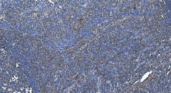
Real IHC data IHC analysis of DGKZ/DGK-zeta using anti-DGKZ/DGK-zeta antibody (A06678-2). DGKZ/DGK-zeta was detected in paraffin-embedded section of human tonsil tissue. Heat mediated antigen retrieval was performed in EDTA buffer (pH8.0, epitope retrieval solution). The tissue section was blocked with 10% goat serum. The tissue section was then incubated with 2μg/ml rabbit anti-DGKZ/DGK-zeta Antibody (A06678-2) overnight at 4°C. Biotinylated goat anti-rabbit IgG was used as secondary antibody and incubated for 30 minutes at 37°C. The tissue section was developed using Strepavidin-Biotin-Complex (SABC) (Catalog # SA1022) with DAB as the chromogen.
Anti-DGKZ/DGK-zeta Antibody ®
Cat # A06678-2
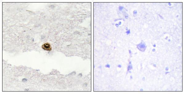
Real IHC data Immunohistochemistry analysis of paraffin-embedded human brain, using DGKZ Antibody. The picture on the right is blocked with the synthesized peptide.
Anti-DGKZ/Dgk Zeta Antibody
Cat # A06678

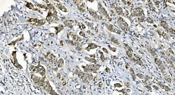
A06678 has paraffin-section IHC images of human brain, including a peptide-blocked comparison, and is listed for IF/ICC in human, mouse and rat (catalog image caption and applications/reactivity). A06678-2 has paraffin-section IHC images of human tonsil, breast cancer and liver cancer, plus mouse brain; its listed applications exclude IF/ICC (catalog image captions and applications).

Which to pick: For tissue IHC, choose A06678-2 when a documented retrieval and staining workflow is useful: its captions specify EDTA pH 8.0 retrieval and 2 μg/ml primary antibody on paraffin sections (catalog image captions). Choose polyclonal A06678 for IF/ICC (1:50 IF dilution; catalog dilution and applications); both list human, mouse and rat reactivity, but the shown IHC examples cover human and mouse only (catalog reactivity and image captions). The IHC captions identify paraffin sections but do not report the fixative (catalog image captions).

Each figure is that product's own IHC / IF validation image from its datasheet.

References

  1. UniProt Consortium. UniProt entry Q13574 (DGKZ_HUMAN, Diacylglycerol kinase zeta).
  2. Human Protein Atlas. DGKZ tissue IHC expression (reliability: Enhanced).
  3. Human Protein Atlas. DGKZ subcellular location (ICC-IF): Localized to the nuclear speckles..
  4. Human Protein Atlas. DGKZ antibody validation summary (1 antibodies).
  5. Downregulation of Diacylglycerol kinase zeta (DGKZ) suppresses tumorigenesis and progression of cervical cancer by facilitating cell apoptosis and cell cycle arrest. Bioengineered 2021 — PMC8806244.
  6. DGKZ Acts as a Potential Oncogene in Osteosarcoma Proliferation Through Its Possible Interaction With ERK1/2 and MYC Pathway. Frontiers in oncology 2018 — PMC6328465.
  7. PubMed PMID:8626588 — UniProt-cited evidence.
  8. PubMed PMID:9159104 — UniProt-cited evidence.
  9. PubMed PMID:14702039 — UniProt-cited evidence.