This website uses cookies to ensure you get the best experience on our website.


- Table of Contents
Plan DHX15 chromogenic IHC on paraffin sections using the nuclear and nucleolar tissue pattern (HPA tissue IHC). The guide covers catalog antibody conditions and tissue controls, with caution about low agreement between staining and RNA expression (datasheet A08140-1; HPA tissue IHC).

Expected localisation, validated protocols, controls and antibodies — the at-a-glance facts below, then the full design guide.
| Expected localisation | Nuclear and nucleolar staining in several tissues (HPA tissue IHC) | |
| Staining pattern | Nuclear and nucleolar signal in several tissue cell types (HPA tissue IHC) | |
| Antigen retrieval | EDTA pH 8.0 HIER, heat-mediated (datasheet A08140-1) | |
| Positive control | Cerebellum+4 more · see all | |
| Negative control | Adipose tissue+4 more · see all |
| Fixation | Keep fixation consistent across paraffin sections (standard IHC practice; not target-specific) | |
| Caveat | Cytoplasmic staining was observed and disregarded (HPA tissue IHC) | |
| Regulation | No expression regulator is specified (UniProt) | |
| Isoform / epitope | No annotated isoforms; one full-length chain (UniProt) |
The catalog antibody’s IHC-P protocol (datasheet A08140-1) is followed by published DHX15 IHC methods from three articles (PMC6861564; PMC10894276; PMC8862312).
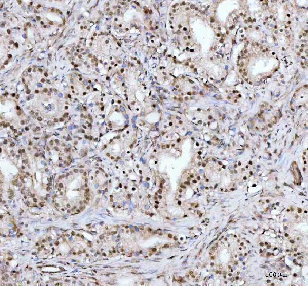
| Sample | Paraffin-embedded human prostate cancer tissue; fixative not specified (datasheet A08140-1) |
| Fixation | Image fixative and duration unreported (datasheet A08140-1); verify before use. |
| Sectioning | 4–5 µm sections on charged slides (standard) |
| Deparaffinisation | Xylene, graded ethanol series to water (standard) |
| Antigen retrieval | Heat retrieval: EDTA pH 8.0 (datasheet A08140-1); 20 min, 95–100 °C (standard) |
| Peroxidase block | 3% H2O2, 10 min, room temperature (standard) |
| Blocking | 10% goat serum (datasheet A08140-1) |
| Primary antibody | Rabbit anti-DHX15, 2-5μg/ml (datasheet A08140-1) |
| Primary incubation | Overnight at 4 °C (datasheet A08140-1) |
| Detection | HRP-conjugated secondary, DAB chromogen (datasheet A08140-1) |
| Counterstain | Hematoxylin, blue, dehydrate and mount (standard) |
| Expected result | DHX15-positive staining in purkinje cells of cerebellum (HPA tissue IHC: Medium). HPA tissue profile: Nuclear and nucleolar expression in several tissues. No signal in the no-primary control. |
DHX15 is reported in the nucleus and nucleolus (UniProt O43143: subcellular location), with nuclear and nucleolar staining in several tissues (HPA: tissue IHC). Expect staining in selected glandular cells, pancreatic endocrine cells, ovarian follicle cells and cerebellar Purkinje cells (HPA: medium staining). Interpret this pattern cautiously: HPA rates the IHC antibody Approved, reports low consistency with RNA data and notes that external verification is pending (HPA: reliability). DHX15 has no transmembrane segment (UniProt O43143: topology).
| Nuclear or nucleolar signal in the listed positive cell populations. | This fits the reported tissue pattern; HPA records medium staining in those populations, without establishing identical intensity in every tissue (HPA: tissue IHC). |
| Predominantly cytoplasmic signal, with little nuclear staining. | Treat this as suspect: HPA observed and disregarded cytoplasmic staining, while UniProt places DHX15 in the nucleus and nucleolus (HPA: reliability; UniProt O43143: subcellular location). |
| Strong staining in cells listed as undetected, such as adipocytes or cardiomyocytes. | Check for cross-reactivity or endogenous detection activity before assigning DHX15 positivity; those particular cell populations were not detected by HPA IHC (HPA: tissue IHC; general IHC practice). |
| Diffuse chromogen across cells and surrounding tissue, without clear nuclear boundaries. | A compartment-free deposit does not match the reported nuclear and nucleolar pattern. Review background controls and the detection step before scoring cells (HPA: tissue IHC; general IHC practice). |
| No nuclear signal in a section containing a listed medium-staining population. | First check tissue identity, section quality and assay controls. A negative run in one section cannot alone overturn an HPA observation, especially given its stated validation caveat (HPA: tissue IHC and reliability; general IHC practice). |
| Choice of comparison tissue | Use a listed medium-staining population to judge whether the assay can show the expected compartment; HPA lists Purkinje cells and several glandular populations, but reports low staining in skin keratinocytes (HPA: tissue IHC). |
| Cell identity within a section | Score the named cells rather than an entire organ: HPA reports pancreatic endocrine cells as medium, for example, and reports liver cholangiocytes as not detected (HPA: tissue IHC). |
| Evidence strength | Approved is the HPA IHC rating for HPA047047; HPA also reports low agreement between antibody staining and RNA expression, with external verification pending (HPA: antibody validation and reliability). |
| DHX15 distribution and structure | UniProt calls tissue expression ubiquitous and reports no transmembrane segment. These facts support broad biological presence but do not predict equal visible IHC staining in every cell (UniProt O43143: tissue specificity and topology; HPA: tissue IHC). |
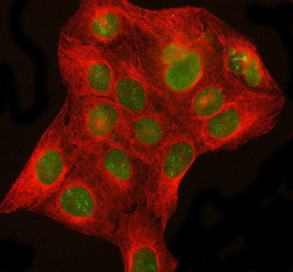
| IF/ICC question: where should signal appear? | HPA reports enhanced nuclear-speckle localization in ICC-IF; UniProt also lists nuclear and nucleolar locations. Compare each image with its assay's own controls rather than requiring identical IHC and IF detail (HPA: subcellular ICC-IF; UniProt O43143: subcellular location; general microscopy practice). |
| Situation | Likely cause | Next action |
|---|---|---|
| The listed positive cells show no nuclear signal. | The section may lack the expected cells, or the staining run may have failed; absence alone does not identify the cause (HPA: tissue IHC; general IHC practice). | Confirm the named cell population and inspect a suitable positive control, then review the general IHC retrieval and detection workflow before interpreting the negative section (HPA: tissue IHC; general IHC practice). |
| Signal is mainly cytoplasmic. | HPA explicitly disregarded observed cytoplasmic staining, so it is weak evidence for DHX15 localization (HPA: reliability). | Check the negative control and antibody-dependent staining; score DHX15 only where a credible nuclear or nucleolar pattern is present (HPA: tissue IHC; general IHC practice). |
| An HPA-listed undetected cell type stains strongly. | Cross-reactivity or endogenous detection activity is possible; an HPA undetected call is also limited to its observed assay and sample (HPA: tissue IHC; general IHC practice). | Compare with a control lacking primary antibody and check whether staining follows cell boundaries or the reported nuclear compartment (HPA: tissue IHC; general IHC practice). |
| Chromogen appears broadly across the section. | Diffuse background can obscure compartment assignment and may reflect nonspecific binding or detection background (general IHC practice). | Inspect a control lacking primary antibody, blocking and detection conditions; score only resolved nuclear or nucleolar signal in the relevant cells (HPA: tissue IHC; general IHC practice). |
| Staining varies between named positive tissues. | HPA reports selected cells at medium intensity, other cells at low intensity or undetected, and low staining-to-RNA consistency (HPA: tissue IHC and reliability). | Record tissue, cell type, compartment and intensity separately; use the listed HPA observations as comparisons rather than assuming uniform staining (HPA: tissue IHC). |
| Nuclear puncta appear in IF/ICC but are less distinct in chromogenic IHC. | HPA describes enhanced nuclear speckles in ICC-IF and nuclear and nucleolar expression in tissue IHC; these are assay-specific observations (HPA: subcellular ICC-IF and tissue IHC). | Evaluate each assay against its reported pattern and controls; do not reject tissue IHC solely because it lacks the speckle detail reported for ICC-IF (HPA: subcellular ICC-IF and tissue IHC; general microscopy practice). |


Comprehensive Human Protein Atlas IHC scoring per tissue (reliability: Approved — Low consistency between antibody staining and RNA expression data. Pending external verification. Cytoplasmic staining observed and disregarded.). Rows are taken directly from the HPA tissue chart — click any row's HPA link to view the source.
| Tissue | Cell type | Level | Evidence | Source |
|---|---|---|---|---|
| Cerebellum | Purkinje cells | Medium | Protein (IHC) | HPA → |
| Fallopian tube | Glandular cells | Medium | Protein (IHC) | HPA → |
| Gallbladder | Glandular cells | Medium | Protein (IHC) | HPA → |
| Ovary | Follicle cells | Medium | Protein (IHC) | HPA → |
| Pancreas | Pancreatic endocrine cells | Medium | Protein (IHC) | HPA → |
| Tissue | Cell type | Level | Evidence | Source |
|---|---|---|---|---|
| Adipose tissue | Adipocytes | Not detected | Protein (IHC) | HPA → |
| Appendix | Glandular cells | Not detected | Protein (IHC) | HPA → |
| Bone marrow | Hematopoietic cells | Not detected | Protein (IHC) | HPA → |
| Bronchus | Respiratory epithelial cells | Not detected | Protein (IHC) | HPA → |
| Cerebral cortex | Endothelial cells | Not detected | Protein (IHC) | HPA → |
Troubleshoot DHX15 staining in paraffin-section chromogenic IHC using the catalog antibody’s tissue image, reported localisation, and matched controls.
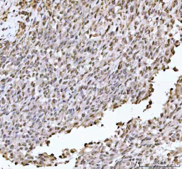
A08140-1 has paraffin-section IHC images from human tumors and an IF/ICC image from U2OS cells (catalog: IHC and IF image captions). Listed reactivity includes human, mouse and rat (catalog: reactivity).
A08140-1 will render with its human prostate cancer paraffin-section IHC figure (catalog: IHC image caption). Its additional IHC captions cover human testicular germ cell tumor, colorectal adenocarcinoma and liver cancer; its IF caption shows U2OS cells (catalog: IHC and IF image captions).
Which to pick: Choose A08140-1 for paraffin-section tissue IHC: its figure documents EDTA pH 8.0 retrieval, 2 μg/ml primary antibody and DAB detection; the fixative is unreported (catalog: IHC image caption). Choose the same SKU for IF/ICC based on its U2OS cell image and 5 μg/ml IF concentration (catalog: IF image caption). Human, mouse and rat reactivity is listed, but the supplied IHC and IF images show human samples only; the antibody is rabbit-hosted and its clone is unreported (catalog: reactivity, host and clone; IHC and IF image captions).