This website uses cookies to ensure you get the best experience on our website.


- Table of Contents
Plan paraffin-section DHX9 IHC around ubiquitous nuclear staining (HPA tissue IHC). This guide covers fixation consistency, the catalog antibody’s 0.5–1 μg/ml IHC dilution (datasheet A02550-1), and nuclear scoring, including the possibility of absent nuclear staining in mitotic cells (UniProt).

Expected localisation, validated protocols, controls and antibodies — the at-a-glance facts below, then the full design guide.
| Expected localisation | Ubiquitous nuclear tissue staining (HPA tissue IHC) | |
| Staining pattern | Nuclear staining across tissue cell types (HPA tissue IHC) | |
| Antigen retrieval | EDTA pH 8.0 HIER, heat-mediated (datasheet A02550-1) | |
| Positive control | Adrenal gland+4 more · see all | |
| Negative control | None in HPA (detected in all 45 tissues); use no-primary + isotype controls |
| Fixation | Keep fixation consistent across sections (standard IHC practice; not target-specific) | |
| Caveat | Mitotic cells may lack nuclear staining (UniProt) | |
| Regulation | No expression regulator annotated (UniProt) | |
| Isoform / epitope | 2 isoforms; epitope coverage is unspecified (UniProt) |
The catalog antibody has a datasheet IHC protocol (datasheet A02550-1). The published options below report DHX9 staining in liver cancer tissue arrays and mouse tissue (PMC11816161; PMC8649379; PMC5023453).
| Sample | Paraffin-embedded human gastric cancer tissue; fixative not specified (datasheet A02550-1) |
| Fixation | Image fixative and duration unreported (datasheet A02550-1); verify before use. |
| Sectioning | 4–5 µm sections on charged slides (standard) |
| Deparaffinisation | Xylene, graded ethanol series to water (standard) |
| Antigen retrieval | Heat retrieval: EDTA pH 8.0 (datasheet A02550-1); 20 min, 95–100 °C (standard) |
| Peroxidase block | 3% H2O2, 10 min, room temperature (standard) |
| Blocking | 10% goat serum (datasheet A02550-1) |
| Primary antibody | Rabbit anti-DHX9, 0.5-1μg/ml (datasheet A02550-1) |
| Primary incubation | Overnight at 4 °C (datasheet A02550-1) |
| Detection | Streptavidin-biotin complex (SABC), DAB chromogen (datasheet A02550-1) |
| Counterstain | Hematoxylin, blue, dehydrate and mount (standard) |
| Expected result | DHX9-positive staining in glandular cells of adrenal gland (HPA tissue IHC: High). HPA tissue profile: Ubiquitous nuclear expression. No signal in the no-primary control. |
DHX9 should show predominantly nuclear staining in paraffin sections, consistent with HPA’s supported, ubiquitous nuclear tissue pattern and UniProt’s nucleoplasmic and nucleolar localization (HPA: tissue IHC; UniProt Q08211: subcellular location). Strong signal is reported in glandular cells of several tissues, bone marrow hematopoietic cells, and selected brain cell populations (HPA: High in listed cells). DHX9 has no transmembrane segment (UniProt Q08211: topology).
| Distinct nuclear staining in glandular cells of adrenal gland, appendix, breast, or cervix. | This matches reported high staining in those glandular cells (HPA: tissue IHC). Judge the nuclear pattern within the named cells; HPA’s high category does not specify a chromogen intensity threshold for an individual slide. |
| Nuclear staining in bone marrow hematopoietic cells, caudate or cortical glia, or cerebellar granular-layer cells. | These are additional high-staining cell populations (HPA: tissue IHC). Compare signal with the local tissue architecture and counterstain; variation between cells does not, by itself, overturn the reported ubiquitous nuclear pattern. |
| Cytoplasmic or membrane staining dominates while nuclei lack a clear signal. | That conflicts with the expected tissue IHC pattern (HPA: ubiquitous nuclear expression). UniProt also lists cytoplasmic localization and nuclear shuttling, so a minor cytoplasmic component alone is not proof of artefact (UniProt Q08211: subcellular location). |
| Strong staining appears in a cell population outside the named high-staining populations. | Investigate cross-reactivity or endogenous detection activity before assigning that signal to DHX9 (general IHC practice). The supplied HPA record lists no negative cell populations, so an unlisted cell type cannot be declared DHX9-negative from this evidence. |
| Diffuse haze obscures nuclei, or a known high-staining population has no detectable signal. | Haze makes localization unreliable; absent signal in a listed high-staining population warrants a run-quality check (general IHC practice; HPA: High in listed cells). Neither appearance alone establishes altered DHX9 expression. |
| Compartment and tissue context | HPA reports ubiquitous nuclear tissue expression and high staining in specific listed cell populations (HPA: tissue IHC). Score nuclei in their identified cells rather than treating all chromogen in a section as target signal. |
| Nucleolar and cytoplasmic localization | UniProt lists nucleoplasm, nucleolus, cytoplasm, and nuclear shuttling (UniProt Q08211: subcellular location). HPA’s ICC-IF summary emphasizes nucleoplasm with additional nucleoli; it does not establish an equally prominent cytoplasmic IHC pattern (HPA: subcellular ICC-IF). |
| Antibody validation | Two listed antibodies have Supported IHC status, while the third has no IHC status in this payload (HPA: HPA028050, CAB011819, HPA055684). Apply the validation status to the antibody actually used; ICC support alone is not an IHC validation claim. |
| Isoform recognition | UniProt lists two DHX9 isoforms (UniProt Q08211: isoforms 1 and 2). Their relative tissue staining and the epitopes recognized by these antibodies are not supplied, so an isoform-specific interpretation requires separate antibody information. |
| Detection chemistry | Endogenous activity and nonspecific chromogen deposition can imitate cellular staining (general IHC practice). Detection controls can help distinguish these contributions from primary-antibody-dependent nuclear signal; this is a general workflow consideration, not a DHX9-specific HPA finding. |
| Situation | Likely cause | Next action |
|---|---|---|
| No nuclear signal in a section containing a listed high-staining cell population. | The run may have failed, or the tissue and antibody conditions may be unsuitable (general IHC practice). HPA’s high category predicts a useful comparison population, not guaranteed staining in every preparation (HPA: tissue IHC). | Check tissue integrity and counterstain, then review the IHC-validated antibody’s stated dilution, retrieval, and detection instructions. Repeat with a run control before interpreting the absence as biological. |
| Uniform brown haze makes cell boundaries and nuclei hard to distinguish. | Excess background from antibody, detection chemistry, or inadequate blocking or washing can obscure compartment assignment (general IHC practice). HPA’s reported nuclear pattern cannot validate a diffuse result (HPA: tissue IHC). | Compare the primary-omission control, inspect washing and blocking, and adjust antibody and detection conditions within the antibody’s instructions. Reassess nuclear localization only after background clears. |
| Most apparent signal lies outside nuclei. | The result departs from HPA’s ubiquitous nuclear tissue pattern (HPA: tissue IHC). UniProt permits some cytoplasmic localization, so compartment alone cannot identify the source of the discrepancy (UniProt Q08211: subcellular location). | Review morphology, counterstain alignment, and primary-omission staining. Confirm that the antibody has IHC support; avoid scoring a predominantly membrane or diffuse pattern as the expected positive result. |
| A cell population not listed as high shows strong staining. | Cross-reactivity or endogenous detection activity is possible (general IHC practice). The supplied HPA list has no negative populations, so unexpected staining is a validation question rather than proof of a false positive (HPA: tissue IHC). | Check the cell identity against morphology and compare a primary-omission control. If the signal persists as primary-dependent staining, seek independent antibody or orthogonal evidence before assigning it to DHX9. |
| Punctate deposits or staining at section edges dominate the image. | Localized reagent deposits or uneven staining can resemble discrete signal (general IHC practice). UniProt reports intranuclear foci in particular contexts, but that annotation does not identify edge deposits as DHX9 (UniProt Q08211: subcellular location). | Inspect adjacent tissue and the primary-omission control for the same deposits. Evaluate any true intranuclear puncta only in intact cells with interpretable nuclear morphology. |
| Q: Should an IF/ICC image be expected to match the paraffin IHC slide exactly? | HPA describes mainly nucleoplasmic IF signal with additional nucleolar localization (HPA: subcellular ICC-IF). Its tissue IHC summary reports ubiquitous nuclear expression; the records describe different assay readouts (HPA: tissue IHC; subcellular ICC-IF). | A: Use nucleoplasmic enrichment with possible nucleolar signal as the IF/ICC localization reference (HPA: subcellular ICC-IF). For paraffin IHC, judge nuclear staining in the reported cell populations and assess assay controls separately. |


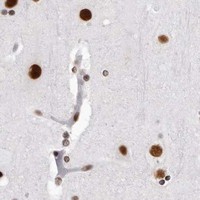
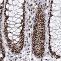


Comprehensive Human Protein Atlas IHC scoring per tissue (reliability: Supported — High consistency between antibody staining and RNA expression data.). Rows are taken directly from the HPA tissue chart — click any row's HPA link to view the source.
| Tissue | Cell type | Level | Evidence | Source |
|---|---|---|---|---|
| Adrenal gland | Glandular cells | High | Protein (IHC) | HPA → |
| Appendix | Glandular cells | High | Protein (IHC) | HPA → |
| Bone marrow | Hematopoietic cells | High | Protein (IHC) | HPA → |
| Breast | Glandular cells | High | Protein (IHC) | HPA → |
| Caudate | Glial cells | High | Protein (IHC) | HPA → |
| Tissue | Cell type | Level | Evidence | Source |
|---|---|---|---|---|
| None in HPA: DHX9 is detected in all 45 scored tissues. Use a no-primary (secondary-only) and an isotype control instead. | ||||
Troubleshoot DHX9 chromogenic IHC by checking retrieval, nuclear localisation, staining controls and cell-level scoring before interpreting tissue differences.
A02550-1 has DHX9 IHC images from human gastric and lung cancer paraffin sections and IF images from human gastric cancer sections and U20S cells (catalog image captions).
A02550-1 lists IHC and IF for human, mouse and rat, with images documenting human paraffin-section IHC and human tissue and cell IF (catalog applications; catalog reactivity; catalog image captions). M02550 lists IHC and IF and human, mouse and rat reactivity, but has no IHC or IF images in the payload (catalog applications; catalog reactivity; catalog image captions).
Which to pick: Choose A02550-1 for tissue IHC: its own caption documents heat retrieval in EDTA at pH 8.0 and staining at 1 μg/ml on human paraffin sections; the fixative is unreported (A02550-1 IHC image caption). Choose A02550-1 for IF/ICC when an image example matters; its captions document human gastric cancer sections and U20S cells at 2 μg/ml (A02550-1 IF image captions). For mouse or rat work, both SKUs list reactivity, while M02550 is the monoclonal option with no supplied IHC or IF image; validate staining in the intended sample (catalog reactivity; M02550 catalog clone; catalog image captions).