This website uses cookies to ensure you get the best experience on our website.


- Table of Contents
Plan paraffin IHC for DIABLO using its granular cytoplasmic tissue pattern (HPA tissue IHC). This guide covers fixation, controls and scoring, with testis pachytene spermatocytes as a high staining reference (HPA tissue IHC).

Expected localisation, validated protocols, controls and antibodies — the at-a-glance facts below, then the full design guide.
| Expected localisation | Granular cytoplasm in tissue (HPA tissue IHC); mitochondria (UniProt) | |
| Staining pattern | Granular cytoplasm; high in testis pachytene spermatocytes (HPA tissue IHC) | |
| Antigen retrieval | Citrate pH 6.0 HIER, 95–98 °C, 20 min (rule: cytoplasmic / membrane antigen) | |
| Positive control | Gallbladder+4 more · see all | |
| Negative control | Adipose tissue+1 more · see all |
| Fixation | Keep fixation consistent across paraffin sections (standard IHC practice; not target-specific) | |
| Caveat | Apoptosis can release DIABLO into the cytosol (UniProt) | |
| Regulation | Highest expression in testis (UniProt) | |
| Isoform / epitope | 3 isoforms; 22–239 and 56–239 forms warrant epitope review (UniProt) |
The catalog antibody’s IHC-P protocol is accompanied by published DIABLO methods for gastrointestinal carcinoma (PMC4214500), rat endometrium (PMC194660), and cervical cancer (PMC3225885).
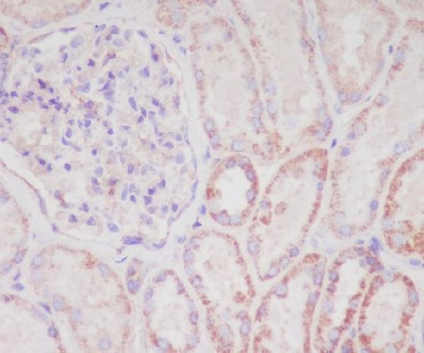
| Sample | Paraffin-embedded human kidney tissue; fixative not specified (datasheet M03790) |
| Fixation | Image fixative and duration unreported (datasheet M03790); verify before use. |
| Sectioning | 4–5 µm sections on charged slides (standard) |
| Deparaffinisation | Xylene, graded ethanol series to water (standard) |
| Antigen retrieval | Heat-induced epitope retrieval in citrate buffer, pH 6.0, 20 min at 95–98 °C (standard rule: cytoplasmic / membrane antigen) |
| Peroxidase block | 3% H2O2, 10 min, room temperature (standard) |
| Blocking | 10% normal serum of the secondary host, 30 min, room temperature (standard) |
| Primary antibody | Rabbit monoclonal (clone FGG-4) anti-DIABLO, 1:50 (datasheet M03790) |
| Primary incubation | Overnight at 4 °C (standard) |
| Detection | HRP-polymer secondary, DAB chromogen 5–10 min (standard) |
| Counterstain | Hematoxylin, blue, dehydrate and mount (standard) |
| Expected result | DIABLO-positive staining in glandular cells of gallbladder (HPA tissue IHC: High). HPA tissue profile: General cytoplasmic expression with a granular pattern. No signal in the no-primary control. |
DIABLO should appear mainly as granular cytoplasmic staining consistent with mitochondria in expressing cells (HPA tissue IHC; HPA subcellular: supported mitochondrial location). Strong examples include gallbladder and stomach glandular cells, pancreatic endocrine cells, and testis pachytene spermatocytes (HPA: High). Cytosolic DIABLO can occur during apoptosis (UniProt Q9NR28: apoptosis-dependent release). DIABLO has no transmembrane segment (UniProt Q9NR28 topology). The HPA tissue IHC pattern is Supported, with high consistency between staining and RNA data (HPA tissue IHC).
| Granular cytoplasmic staining in gallbladder or stomach glandular cells, pancreatic endocrine cells, or pachytene spermatocytes (HPA: High). | This matches the reported tissue pattern and predominant mitochondrial location (HPA tissue IHC; HPA subcellular: supported). Judge intensity within the identified cell population: neighboring cells need not share its staining level (HPA: cell-specific tissue observations). |
| Predominantly nuclear staining, with little granular cytoplasmic signal, in an otherwise positive region. | Nuclear localization is outside the reported principal pattern (HPA tissue IHC; UniProt Q9NR28 subcellular location). Treat it as suspect and compare controls before scoring it as DIABLO; nonspecific antibody binding or detection artefact is possible (general IHC practice). |
| Strong staining in adipocytes or soft-tissue fibroblasts while expected positive cells are also present. | HPA reports DIABLO as Not detected in those respective cell populations (HPA: adipocytes; soft-tissue fibroblasts). Check cell identification and negative controls: cross-reactivity or endogenous detection activity may explain discordant staining (general IHC practice). |
| Diffuse color over tissue, stroma, and blank areas, obscuring cell boundaries and granules. | This does not resolve the reported granular cytoplasmic pattern (HPA tissue IHC). Broad deposition can reflect background from blocking, detection, or washing steps; inspect a reagent control and distinguish precipitate from cellular staining (general IHC practice). |
| No convincing signal in a gallbladder, stomach, pancreas, or testis cell population reported as High (HPA tissue IHC). | An absent result conflicts with those HPA observations but does not alone prove biological absence. Confirm the sampled cell population, then assess retrieval, primary antibody, detection, and counterstain settings against a working control (general IHC practice). |
| Cell population and tissue selection | HPA reports High staining in selected glandular cells, pancreatic endocrine cells, and pachytene spermatocytes, but Not detected in adipocytes and soft-tissue fibroblasts (HPA tissue IHC). Use the named cells when judging a control; a whole-section average can hide a focal positive population (general IHC practice). |
| Compartment and apoptosis | The main location is mitochondrial, with a granular cytoplasmic tissue pattern (HPA subcellular: supported; HPA tissue IHC). DIABLO may enter the cytosol when cells undergo apoptosis (UniProt Q9NR28). A less punctate cytoplasmic pattern therefore needs tissue context; it is not, by itself, evidence of failed staining. |
| Protein forms and antibody recognition | UniProt lists three isoforms and chains beginning at residues 22 and 56 (UniProt Q9NR28 processing). Antibody epitope information is absent from this payload, so these annotations cannot establish which forms the IHC-validated antibody detects. Interpret a negative section using a positive tissue control rather than assuming loss of one form (general IHC practice). |
| Antibody validation and IF/ICC Q&A | Q: Should IF/ICC show the same main compartment? A: Mitochondria are the supported main location; cytosol is an uncertain additional ICC-IF location (HPA subcellular). HPA001825 is Supported for both IHC and ICC; CAB003857 is Supported for IHC, while CAB016688 is Supported for ICC (HPA antibodies). IF/ICC protocol choices belong to its own guide. |
| Situation | Likely cause | Next action |
|---|---|---|
| A reported High cell population is unstained, although the section has intact morphology (HPA tissue IHC). | The expected cells may be absent from the sampled area, or a routine retrieval, antibody, or detection step may have failed (general IHC practice). | Verify the cell population and run a known positive section through the same IHC workflow; review retrieval and reagent records before interpreting the negative result (general IHC practice). |
| Signal is pale and lacks the expected granules in otherwise positive cells (HPA tissue IHC). | Weak detection or excessive counterstain can obscure fine cytoplasmic detail (general IHC practice). | Compare a working positive control and counterstain balance, then adjust one routine IHC variable at a time; no DIABLO-specific dilution or retrieval condition is supplied here (general IHC practice). |
| Nuclei dominate the stain while cytoplasm is weak. | A nuclear dominant pattern conflicts with the recorded mitochondrial and cytosolic locations (HPA subcellular; UniProt Q9NR28); nonspecific signal is possible (general IHC practice). | Inspect the no-primary control and repeat with an IHC-supported antibody if available (HPA antibodies; general IHC practice). Score DIABLO only where the cellular pattern is credible. |
| Adipocytes or soft-tissue fibroblasts stain strongly (HPA: Not detected). | Cell misidentification, antibody cross-reactivity, or endogenous detection activity could create a misleading positive (general IHC practice). | Confirm the cell type on the counterstained section and compare appropriate negative controls; check detection blocking if color persists without primary antibody (general IHC practice). |
| Brown color coats many cell types and extracellular areas without discrete granules. | This departs from the granular cytoplasmic HPA profile (HPA tissue IHC) and may reflect chromogen deposit or broad background (general IHC practice). | Inspect blank areas and a no-primary control, then review washing, blocking, and chromogen handling; avoid assigning diffuse deposit to DIABLO-positive cells (general IHC practice). |
| Staining becomes more diffuse within cytoplasm in a region with apoptotic morphology. | DIABLO can be released from mitochondria into cytosol during apoptosis (UniProt Q9NR28); HPA lists cytosol as an uncertain additional ICC-IF location (HPA subcellular). | Record the morphology and compare adjacent viable cells and controls. Do not call apoptosis from DIABLO staining alone; assess the pattern alongside independent morphological evidence (general IHC practice). |


Comprehensive Human Protein Atlas IHC scoring per tissue (reliability: Supported — High consistency between antibody staining and RNA expression data.). Rows are taken directly from the HPA tissue chart — click any row's HPA link to view the source.
| Tissue | Cell type | Level | Evidence | Source |
|---|---|---|---|---|
| Gallbladder | Glandular cells | High | Protein (IHC) | HPA → |
| Pancreas | Pancreatic endocrine cells | High | Protein (IHC) | HPA → |
| Stomach | Glandular cells | High | Protein (IHC) | HPA → |
| Testis | Pachytene spermatocytes | High | Protein (IHC) | HPA → |
| Adrenal gland | Glandular cells | Medium | Protein (IHC) | HPA → |
Use these checks to optimise DIABLO staining in paraffin sections and distinguish its expected granular cytoplasmic signal from artefact.
Anti-DIABLO IHC images cover paraffin-embedded human kidney and human, mouse, and rat testis; an IF image shows U20S cells (M03790 IHC image caption; PB9860 IHC and IF image captions).
M03790 is the sole card that will render, with an IHC image of paraffin-embedded human kidney (M03790 IHC image caption). Its catalog lists IHC and IF/ICC applications and human, mouse, and rat reactivity; no IF image is supplied for this SKU (M03790 catalog).
Which to pick: For tissue IHC, M03790 offers a rabbit monoclonal with a human kidney paraffin-section example; the caption does not report the fixative (M03790 catalog; M03790 IHC image caption). For IF/ICC, PB9860 has an image of U20S cells, while M03790 lists IF/ICC without a supplied IF image (PB9860 IF image caption; M03790 catalog). For cross-species tissue IHC, PB9860 has paraffin-section images from human, mouse, and rat testis, though those captions do not report fixation (PB9860 IHC image captions).