This website uses cookies to ensure you get the best experience on our website.


- Table of Contents
Plan chromogenic DIDO1 IHC in paraffin sections using the catalog antibody’s 1:100–1:300 dilution range (datasheet A06019). Assess the mainly nuclear tissue pattern (HPA tissue IHC) with consistent fixation and cell-type scoring (standard IHC practice).

Expected localisation, validated protocols, controls and antibodies — the at-a-glance facts below, then the full design guide.
| Expected localisation | Mainly nuclear in tissue (HPA tissue IHC); cytoplasmic location annotated (UniProt) | |
| Staining pattern | Mainly nuclear staining in most tissues (HPA tissue IHC) | |
| Antigen retrieval | Tris-EDTA pH 9.0 HIER, heat-mediated (datasheet A06019) | |
| Positive control | Adrenal gland+4 more · see all | |
| Negative control | None in HPA (detected in all 45 tissues); use no-primary + isotype controls |
| Fixation | Keep fixation consistent across sections (standard IHC practice; not target-specific) | |
| Caveat | Antibody staining and RNA show medium consistency (HPA tissue IHC) | |
| Regulation | No specific induction reported (UniProt) | |
| Isoform / epitope | Four isoforms; verify antibody epitope coverage (UniProt) |
The catalog antibody protocol and two published DIDO1 IHC methods cover paraffin sections (datasheet A06019; PMC12994943; PMC8188684).
| Sample | Paraffin-embedded human tonsil tissue; fixative not specified (datasheet A06019) |
| Fixation | Image fixative and duration unreported (datasheet A06019); verify before use. |
| Sectioning | 4–5 µm sections on charged slides (standard) |
| Deparaffinisation | Xylene, graded ethanol series to water (standard) |
| Antigen retrieval | Heat retrieval: Tris-EDTA pH 9.0 (datasheet A06019); 20 min, 95–100 °C (standard) |
| Peroxidase block | 3% H2O2, 10 min, room temperature (standard) |
| Blocking | 10% normal serum of the secondary host, 30 min, room temperature (standard) |
| Primary antibody | Rabbit anti-DIDO1, 1:100-1:300 (datasheet A06019) |
| Primary incubation | Overnight at 4 °C (standard) |
| Detection | HRP-polymer secondary, DAB chromogen 5–10 min (standard) |
| Counterstain | Hematoxylin, blue, dehydrate and mount (standard) |
| Expected result | DIDO1-positive staining in glandular cells of adrenal gland (HPA tissue IHC: High). HPA tissue profile: Mainly nuclear expression in most tissues. No signal in the no-primary control. |
DIDO1 should stain mainly nuclei across many tissues (HPA tissue IHC: mainly nuclear expression; UniProt Q9BTC0: ubiquitous). Examples of cells with high staining include bone marrow hematopoietic cells, adrenal glandular cells and bronchial respiratory epithelial cells (HPA tissue IHC). HPA rates the tissue pattern Supported, with medium antibody–RNA consistency and external verification pending (HPA tissue IHC). DIDO1 has no transmembrane segment (UniProt Q9BTC0 topology).
| Predominant nuclear chromogenic signal in expected cells, including adrenal glandular or bone marrow hematopoietic cells. | This matches the reported pattern and high staining in those cell populations (HPA tissue IHC). Assess compartment and cell identity together; a dark deposit alone does not establish specific DIDO1 staining (general IHC practice). |
| Signal is exclusively membranous, or strongly cytoplasmic while nuclei remain unstained. | A purely membranous pattern conflicts with DIDO1 topology (UniProt Q9BTC0 topology). Cytoplasmic DIDO1 is possible (UniProt Q9BTC0 subcellular location), but a cytoplasm-only tissue pattern conflicts with HPA’s mainly nuclear IHC profile; investigate artefact or nonspecific binding (HPA tissue IHC; general IHC practice). |
| Prominent staining appears in cells outside the reported positive populations within a reference tissue. | HPA identifies specific strongly stained cell populations, but supplies no negative-cell list (HPA tissue IHC). Treat the finding as unresolved: cross-reactivity or endogenous chromogenic activity is possible, and an unlisted cell type cannot automatically be called DIDO1-negative (general IHC practice). |
| Chromogen forms a diffuse haze over cells or extracellular areas, obscuring nuclei. | The distribution cannot be scored confidently against the mainly nuclear reference pattern (HPA tissue IHC). Review background controls, blocking, antibody concentration and detection steps to locate nonspecific signal (general IHC practice). |
| No convincing signal appears in an expected positive population, such as bone marrow hematopoietic cells. | This disagrees with the reported high level for that population (HPA tissue IHC). First check section quality, the staining run and positive control; a failed stain alone does not establish biological absence (general IHC practice). |
| Compartment and cell identity | HPA reports mainly nuclear tissue staining and high levels in selected cell populations (HPA tissue IHC). Score nuclei in identified cells rather than treating all tissue staining as equivalent (general IHC practice). |
| Context-dependent localization | UniProt places DIDO1 in nucleus and cytoplasm and describes spindle translocation in early mitosis after loss of H3K4me3 interaction (UniProt Q9BTC0). This allows a context-dependent exception; it does not redefine the predominant tissue IHC pattern (HPA tissue IHC). |
| Tissue evidence limits | HPA lists no negative or low-staining tissues in the supplied record and rates the pattern Supported, with external verification pending (HPA tissue IHC). Use listed positives as references without declaring unlisted tissues negative. |
| Antibody evidence | HPA049904 and CAB004374 each have IHC status Supported; each has ICC status Uncertain (HPA antibodies). Their IHC status supports comparison with the tissue pattern, but does not establish identical staining for every antibody or assay. |
| Isoforms and processing | UniProt lists 4 isoforms and a single 1–2240 chain, with no signal peptide or propeptide (UniProt Q9BTC0). The supplied record does not map the IHC antibody epitope, so isoform-specific staining cannot be predicted. |
| IF/ICC Q: what localization is reported? | A: HPA approves nucleoplasm as the main ICC-IF location, with vesicles and cytosol also reported (HPA subcellular). This is a localization cross-check; the listed antibodies’ ICC validation is Uncertain (HPA antibodies). |
| Situation | Likely cause | Next action |
|---|---|---|
| Known-positive tissue is blank. | The result conflicts with HPA’s high staining in the specified positive cells (HPA tissue IHC); an unsuccessful staining run is possible (general IHC practice). | Verify the positive-control section, reagent sequence, retrieval and detection controls before interpreting absence of DIDO1 (general IHC practice). |
| Nuclei are obscured by widespread background. | Diffuse deposit prevents comparison with the mainly nuclear pattern (HPA tissue IHC); excess antibody or incomplete blocking may contribute (general IHC practice). | Inspect a negative reagent control, then review blocking, washes, dilution and chromogen development (general IHC practice). |
| Only cell borders stain. | An exclusively membranous pattern is inconsistent with the lack of a transmembrane segment (UniProt Q9BTC0 topology). | Compare with a known-positive nuclear pattern and check negative reagent controls for nonspecific detection (HPA tissue IHC; general IHC practice). |
| Unexpected cells stain strongly. | HPA supplies selected positive populations but no negative-cell list (HPA tissue IHC); cross-reactivity or endogenous detection activity may mimic signal (general IHC practice). | Identify the stained cells, check reagent controls and compare the compartment with a listed positive population before assigning specificity (general IHC practice; HPA tissue IHC). |
| A few dividing cells show signal near spindle structures. | DIDO1 can move to the mitotic spindle during early mitosis under the stated chromatin-binding condition (UniProt Q9BTC0). | Assess whether staining is confined to plausible mitotic cells; keep predominant nuclear staining as the tissue-level reference (UniProt Q9BTC0; HPA tissue IHC; general IHC practice). |
| IF/ICC appears more cytosolic than the tissue IHC section. | HPA reports additional cytosol and vesicle localization in ICC-IF, while tissue IHC is mainly nuclear (HPA subcellular; HPA tissue IHC). | Compare compartment patterns within each assay and note that the listed antibodies have ICC status Uncertain (HPA antibodies). |


Comprehensive Human Protein Atlas IHC scoring per tissue (reliability: Supported — Medium consistency between antibody staining and RNA expression data. Pending external verification.). Rows are taken directly from the HPA tissue chart — click any row's HPA link to view the source.
| Tissue | Cell type | Level | Evidence | Source |
|---|---|---|---|---|
| Adrenal gland | Glandular cells | High | Protein (IHC) | HPA → |
| Appendix | Glandular cells | High | Protein (IHC) | HPA → |
| Bone marrow | Hematopoietic cells | High | Protein (IHC) | HPA → |
| Breast | Glandular cells | High | Protein (IHC) | HPA → |
| Bronchus | Respiratory epithelial cells | High | Protein (IHC) | HPA → |
| Tissue | Cell type | Level | Evidence | Source |
|---|---|---|---|---|
| None in HPA: DIDO1 is detected in all 45 scored tissues. Use a no-primary (secondary-only) and an isotype control instead. | ||||
Troubleshoot DIDO1 staining in paraffin-section chromogenic IHC using the catalog antibody’s tonsil example and compartment-specific controls.
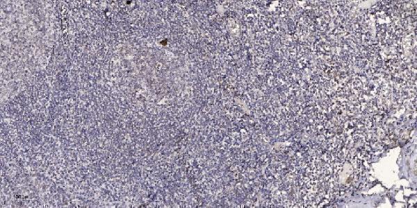
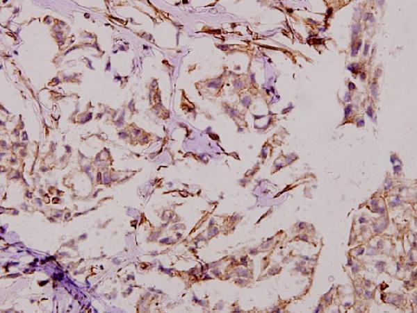
IHC images document paraffin-embedded human tonsil and breast carcinoma (image captions: A06019, A06019S186); catalog reactivity also lists mouse and rat (catalog: reactivity). No IF image is supplied (catalog: IF image captions).
A06019 will render with human tonsil paraffin-section IHC at 1:200; its listed applications include IF, and its reactivity is human and mouse (A06019 image caption; catalog: applications and reactivity). A06019S186 will render with human breast carcinoma paraffin-section IHC at 1:50; its listed reactivity is human, mouse and rat (A06019S186 image caption; catalog: reactivity).
Which to pick: For tissue IHC, choose A06019 for the documented human tonsil example or A06019S186 for the documented human breast carcinoma example; both captions specify paraffin-embedded tissue, and neither reports the fixative (respective IHC image captions). For IF/ICC, A06019-1 lists both applications and is described as polyclonal, but has no supplied IF image; A06019 lists IF and 1:50 without an IF image (catalog: applications, dilution, clonality and IF image captions). For rat tissue IHC, A06019S186 lists rat reactivity and IHC, while its own IHC image shows human tissue only (catalog: reactivity and applications; A06019S186 image caption).