DKC1 / H/ACA ribonucleoprotein complex subunit DKC1 · IHC design guide

Design Immunohistochemistry for DKC1

Plan DKC1 staining in paraffin sections around its nuclear and nucleolar tissue pattern (HPA tissue IHC). The catalog antibody is listed for IHC at 2–5 μg/ml (datasheet A01535-2).

Evidence assembled Oct 2026 · For research use; verify linked source records and product datasheet before use
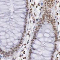
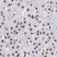
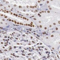
Immunohistochemistry protocol sheet for DKC1 (IHC for DKC1): expected localisation Nuclear and nucleolar tissue staining (HPA tissue IHC), antibody A01535-2, validated IHC image, and IHC protocol steps
Printable DKC1 IHC protocol sheet — expected localisation Nuclear and nucleolar tissue staining (HPA tissue IHC), antibody A01535-2, controls and protocol steps. Open the full DKC1 IHC guide →

DKC1 Immunohistochemistry Experimental Design Guide

Expected localisation, validated protocols, controls and antibodies — the at-a-glance facts below, then the full design guide.

Must know before staining
Expected localisation Nuclear and nucleolar tissue staining (HPA tissue IHC)
Staining pattern Ubiquitous nuclear and nucleolar staining (HPA tissue IHC)
Antigen retrieval EDTA pH 8.0 HIER, heat-mediated (datasheet A01535-2)
Positive control ⓘ Adrenal gland+4 more · see all
Negative control ⓘ None in HPA (detected in all 45 tissues); use no-primary + isotype controls
Important caveats
Reasons your staining may differ from the expected pattern.
Fixation Keep fixation consistent across tissue sections. (standard IHC practice; not target-specific)
Caveat Chondrocytes show low staining (HPA tissue IHC)
Regulation Broad tissue expression (UniProt)
Isoform / epitope 2 isoforms; epitope differences are unspecified (UniProt)
Section 1

Recommended DKC1 IHC & IF Protocols

The catalog antibody’s IHC-P protocol uses EDTA retrieval (datasheet A01535-2). Four published DKC1 IHC protocols provide tissue-specific alternatives (PMC7054532; PMC8056518; PMC7653035; PMC12302887).

Recommended immunohistochemistry (IHC-P) protocol parameters
SampleParaffin-embedded human breast cancer tissue; fixative not specified (datasheet A01535-2)
FixationImage fixative and duration unreported (datasheet A01535-2); verify before use.
Sectioning4–5 µm sections on charged slides (standard)
DeparaffinisationXylene, graded ethanol series to water (standard)
Antigen retrievalHeat retrieval: EDTA pH 8.0 (datasheet A01535-2); 20 min, 95–100 °C (standard)
Peroxidase block3% H2O2, 10 min, room temperature (standard)
Blocking10% goat serum (datasheet A01535-2)
Primary antibodyRabbit anti-DKC1, 2-5 μg/ml (datasheet A01535-2)
Primary incubationOvernight at 4 °C (datasheet A01535-2)
DetectionHRP-conjugated secondary, DAB chromogen (datasheet A01535-2)
CounterstainHematoxylin, blue, dehydrate and mount (standard)
Expected resultDKC1-positive staining in glandular cells of adrenal gland (HPA tissue IHC: High). HPA tissue profile: Ubiquitous nuclear and nucleolar expression. No signal in the no-primary control.
💡Decision noteTry EDTA pH 8.0 first with the catalog antibody (datasheet A01535-2). Published colorectal and breast protocols used citrate pH 6.0 (PMC7054532; PMC7653035).
Section 2

What Is the Expected DKC1 Staining Pattern?

DKC1 should stain chiefly in nuclei and nucleoli across many cell types (HPA: ubiquitous nuclear and nucleolar IHC expression; UniProt O60832: ubiquitously expressed). HPA rates the tissue pattern Supported, with medium consistency between staining and RNA data (HPA: tissue IHC). DKC1 has no transmembrane segment (UniProt O60832: topology), so a membrane rim is unexpected.

What am I looking at on my slide?
Distinct nuclear staining with darker nucleolar foci in several cell types.This matches the observed tissue pattern (HPA: ubiquitous nuclear and nucleolar IHC expression). High staining is reported in bone marrow hematopoietic cells and bronchial respiratory epithelial cells (HPA: tissue IHC). Compare compartments within cells, rather than calling a whole tissue positive from scattered dark deposits (general IHC interpretation).
Predominantly membrane, extracellular, or diffuse cytoplasmic staining without nuclear enrichment.Treat this as suspect localisation and check controls and antibody conditions (HPA: nuclear and nucleolar IHC pattern; UniProt O60832: no transmembrane segment). UniProt also lists cytoplasm, so a cytoplasmic component alone cannot prove artefact (UniProt O60832: subcellular location).
Strong staining appears in cells expected to be faint, while adjacent nuclei lack the expected pattern.For example, HPA reports low staining in soft-tissue chondrocytes (HPA: tissue IHC). Check cell identification and background before considering cross-reactivity or endogenous detection activity (general IHC practice). DKC1 is broadly expressed, so this is not an absolute negative-cell criterion (UniProt O60832: tissue specificity).
Haze or chromogen deposit spreads across tissue and obscures nuclear boundaries.The distribution cannot be scored confidently against the nuclear and nucleolar reference pattern (HPA: tissue IHC). Review negative controls, washing, blocking, and chromogen development for general background or endogenous detection activity (general IHC practice).
No nuclear signal in a section containing a reported high-staining cell population.Bone marrow hematopoietic cells and adrenal glandular cells are reported High (HPA: tissue IHC). Check tissue preservation, retrieval, antibody application, and detection with controls (general IHC practice). A failed run is possible; HPA's Supported rating does not guarantee every section will stain (HPA: tissue IHC reliability).
💡Expected DKC1 appearanceCall positive when cells show clear nuclear staining with nucleolar enrichment, including High signal in a documented positive population such as bronchial respiratory epithelium (HPA: tissue IHC); isolated membrane rims or diffuse deposit without nuclear enrichment are suspect (HPA: nuclear and nucleolar pattern; UniProt O60832: topology).
How each factor affects the staining
Tissue and cell population (HPA: tissue IHC)HPA reports ubiquitous nuclear and nucleolar expression, with High staining in several listed populations and Low staining in soft-tissue chondrocytes (HPA: tissue IHC). Score the identified cells and compartments; do not assume every cell has equal intensity.
Antibody validation (HPA: antibody records)IHC is Supported for HPA000166, HPA000447, and CAB033086 (HPA: antibody records). Their listed status does not establish an Enhanced rating or validate a different antibody. Interpret a new reagent against its own controls (general IHC practice).
Protein location and processing (UniProt O60832)DKC1 is reported in nucleolus, Cajal body, and cytoplasm, with no transmembrane segment or signal peptide (UniProt O60832: subcellular location, topology, processing). A membrane-only pattern lacks support; these annotations do not predict an IHC fixation effect.
IF/ICC Q: Where should DKC1 appear? (HPA: subcellular ICC-IF)A: Mainly at the nucleoli fibrillar center, with additional nucleoplasmic signal (HPA: supported ICC-IF locations). This is a localisation cross-check; the IF/ICC workflow belongs on its own guide page.
Why is my staining missing, weak or wrong?
SituationLikely causeNext action
Nuclear staining is absent in a documented High population (HPA: tissue IHC).Possible section or detection failure, or an unsuitable antibody condition (general IHC practice). HPA staining levels do not identify which step failed.Run a documented positive tissue alongside the sample; verify antibody application, retrieval, detection reagents, and counterstain (general IHC practice). Do not infer DKC1-specific fixation sensitivity from this result.
Only diffuse cytoplasmic colour is visible.The pattern does not match HPA's dominant nuclear and nucleolar IHC result (HPA: tissue IHC), although UniProt also lists cytoplasm (UniProt O60832: subcellular location).Inspect nuclear detail and a negative control; adjust general background controls and repeat with an IHC-supported reagent if needed (general IHC practice; HPA: antibody records).
Membrane outlines dominate the slide.Membrane-only staining is inconsistent with the reported nuclear and nucleolar pattern and absence of a transmembrane segment (HPA: tissue IHC; UniProt O60832: topology).Check whether the deposit follows cell edges in the negative control, then review blocking, washing, and detection conditions (general IHC practice). Avoid scoring membrane rims as DKC1 positive.
Many cells appear uniformly dark, with nucleoli indistinguishable.Excess background or overdeveloped chromogen may hide the expected compartment contrast (HPA: nuclear and nucleolar IHC pattern; general IHC practice).Compare a negative control and shorten chromogen development or optimize antibody conditions as appropriate (general IHC practice). Score only cells whose nuclear pattern remains readable.
A presumed negative cell population stains strongly.A cell-type assignment or nonspecific signal may be at issue; HPA lists soft-tissue chondrocytes as Low, not negative (HPA: tissue IHC).Verify morphology and controls, then compare intensity within the same run (general IHC practice). Do not designate an unlisted tissue as DKC1 negative (UniProt O60832: ubiquitous expression).
A particular antibody yields a pattern unlike the HPA reference.HPA reports IHC Supported for three specified antibodies; that status cannot be transferred to an unlisted reagent (HPA: antibody records).Check the reagent's IHC validation and controls, and compare a documented positive population with the HPA nuclear and nucleolar pattern (HPA: tissue IHC; general IHC practice).

Sample controls for DKC1 IHC & IF

🧪Run bronchus first: respiratory epithelial cells should stain strongly (HPA: High in bronchus respiratory epithelial cells). HPA detects DKC1 in all 45 scored tissues, so there is no supported negative tissue; no cell type on the positive slide is a validated internal negative, and cells without nucleolar staining should be treated cautiously rather than called target-negative (HPA: no negative tissue rows; HPA: nucleoli fibrillar center localization).
Positive control tissue: Adrenal gland (Glandular cells, HPA High)
Negative control tissue: None in HPA: DKC1 is detected in all 45 scored tissues. Use a no-primary (secondary-only) and an isotype control instead.
ICC-IF cell lines (HPA subcellular resource): HPA ICC-IF images show DKC1 in A-431, U-251MG, U2OS, with annotated localisation: Nucleoli fibrillar center (supported) (HPA subcellular).
Technical controls: Run no-primary (secondary-only) and rabbit IgG isotype controls matched to the primary antibody’s format and concentration; a matched DKC1-knockout specimen, if available, provides a biological negative (datasheet: rabbit primary; standard IHC practice). For bronchus, quench endogenous peroxidase and check staining in inflammatory cells against the negative controls (standard chromogenic IHC practice).
⚠️Feasibility: The selected A01535-2 paraffin-section caption reports heat retrieval in EDTA at pH 8.0, but reports no fixative; a DKC1-specific fixation window or fixation effect is unreported (datasheet: A01535-2 tissue-IHC caption). Paraffin IHC is demonstrated in the caption, while ICC-IF images exist for A-431, U-251MG and U2OS; the supplied evidence does not establish that frozen sections or IF are easier (datasheet: A01535-2 tissue-IHC caption; HPA: subcellular ICC-IF images). In bronchus, inflammatory-cell peroxidase can create chromogenic background, so interpret epithelial staining against the no-primary control (standard chromogenic IHC practice).

HPA tissue IHC evidence for DKC1

Comprehensive Human Protein Atlas IHC scoring per tissue (reliability: Supported — Medium consistency between antibody staining and RNA expression data.). Rows are taken directly from the HPA tissue chart — click any row's HPA link to view the source.

Positive expression · recommended positive controls

TissueCell typeLevelEvidenceSource
Adrenal gland Glandular cells High Protein (IHC) HPA →
Appendix Non-germinal center cells High Protein (IHC) HPA →
Bone marrow Hematopoietic cells High Protein (IHC) HPA →
Bronchus Respiratory epithelial cells High Protein (IHC) HPA →
Caudate Glial cells High Protein (IHC) HPA →

Undetected expression · recommended negative controls

TissueCell typeLevelEvidenceSource
None in HPA: DKC1 is detected in all 45 scored tissues. Use a no-primary (secondary-only) and an isotype control instead.
Section 3

Advanced DKC1 IHC Tips

Troubleshoot DKC1 staining in paraffin sections by checking retrieval, compartment-specific signal, controls, and consistent scoring (datasheet A01535-2; HPA tissue IHC).

What retrieval should I try when DKC1 staining is weak in paraffin sections?
Start with heat-mediated retrieval in EDTA at pH 8.0 (datasheet A01535-2). The selected paraffin-section example used that retrieval before 2 μg/ml primary antibody overnight at 4°C, so keep those conditions together for the initial comparison (datasheet A01535-2). If staining remains weak, vary heating time on matched sections while holding antibody concentration, detection, and development constant; record tissue preservation alongside signal (standard IHC practice). Check whether recovered staining is predominantly nucleolar or nuclear, rather than judging intensity alone, because those are expected DKC1 locations (HPA subcellular; UniProt O60832 subcellular). Include a no-primary section to reveal detection-related staining (standard IHC practice).
Could fixation explain variable DKC1 staining between paraffin blocks?
DKC1-specific fixation sensitivity is unknown here: the selected paraffin-section caption does not state its fixative (datasheet A01535-2). Record each block’s fixative and fixation duration, and compare sections processed with the same retrieval and detection conditions before attributing an intensity difference to biology (standard IHC practice). If processing histories differ, test comparable material under one staining run and assess morphology as well as nucleolar signal (standard IHC practice; HPA subcellular). Do not infer a fixation effect from DKC1’s reported modifications or intracellular location, which do not establish how this antibody performs after fixation (UniProt O60832 modified residues and subcellular; datasheet A01535-2).
How should I assess DKC1 staining outside the nucleolus?
Expect a prominent nucleolar pattern, with possible nucleoplasmic staining, when assessing chromogenic sections (HPA subcellular). UniProt also lists the nucleus, Cajal body, and cytoplasm, so a non-nucleolar signal warrants examination rather than automatic rejection (UniProt O60832 subcellular). Compare its distribution with intact cell morphology and the nucleolar pattern in the same staining run, and check a no-primary control for diffuse detection signal (standard IHC practice; HPA subcellular). Strong, uniform cytoplasmic DAB with little nuclear staining should prompt review of blocking, antibody concentration, and section preservation before assigning it to DKC1 (standard IHC practice; HPA tissue IHC).
Can this stain distinguish DKC1 isoforms or modified epitopes?
The record lists DKC1 isoforms 1 and 3, but the selected staining caption does not identify the antibody epitope or establish isoform-specific detection (UniProt O60832 isoforms; datasheet A01535-2). DKC1 contains a PUA domain at residues 296–371 and reported modified residues, including phosphoserines at 21, 387, 451, and 453 (UniProt O60832 domains and modified residues). Those features alone cannot identify what the antibody recognizes in paraffin sections (UniProt O60832; datasheet A01535-2). Interpret intensity as staining with this antibody, and obtain epitope mapping or an independently validated reagent before making isoform- or modification-specific claims (standard IHC practice).
How can I compare DKC1 immunofluorescence with this IHC result?
Treat IF as a separate assay: the selected antibody evidence documents chromogenic staining in a paraffin section, while HPA reports DKC1 mainly in nucleoli with additional nucleoplasmic localization (datasheet A01535-2; HPA subcellular). For multiplex IF, pair DKC1 with a marker for the cell population being assessed and choose well-separated fluorophores, favoring a far-red channel when tissue autofluorescence is troublesome (standard IF practice). Because DKC1 is intracellular and has no transmembrane segment, permeabilisation must allow antibody access to nuclear and nucleolar epitopes; titrate it against preserved morphology (UniProt O60832 topology and subcellular; standard IF practice). Compare compartment patterns, using no-primary and single-stain controls to assess background and channel bleed-through (standard IF practice).
What should I check if DAB obscures nucleolar DKC1 staining?
First compare the stained section with a no-primary control to locate signal caused by detection reagents or endogenous enzyme activity (standard IHC practice). The selected paraffin-section example used 10% goat serum blocking, a peroxidase-linked secondary, and DAB development; it does not establish that those steps remove every source of background (datasheet A01535-2). Apply an endogenous peroxidase block and titrate primary concentration or DAB development on matched sections while retaining the documented 2 μg/ml condition as a reference (standard IHC practice; datasheet A01535-2). Evaluate background in the context of the expected nucleolar and nuclear pattern, especially where dense precipitate hides individual nuclei (HPA subcellular; standard IHC practice).
How should I quantify DKC1 staining across paraffin sections? ⚠ ANSWER MARKED FOR VERIFICATION
Define the scored compartment before analysis: DKC1 is reported predominantly in nucleoli, with additional nucleoplasmic staining (HPA subcellular). Record the percentage of positive cells and, if intensity is reproducible, calculate an H-score as the sum of intensity grade multiplied by its cell percentage, giving a 0–300 scale for grades 0–3 (standard IHC practice). Alternatively, count positive cells per mm² when cell density is the question, normalising to viable tissue area and reporting the cell population assessed (standard IHC practice). Use the same staining run, image settings, thresholds, and compartment rules across compared sections; exclude folds and necrotic areas (standard IHC practice).
How can I distinguish genuine DKC1 staining from artefact?
A convincing result shows staining in intact nuclei, especially nucleoli, in a pattern consistent across comparable cells and sections (HPA tissue IHC; HPA subcellular; standard IHC practice). DKC1 is reported as ubiquitously expressed, so cell identity and compartment carry more weight than a presumed positive-versus-negative tissue contrast (UniProt O60832 tissue specificity; HPA tissue IHC). Recheck signal confined to section edges, folds, or necrotic regions, where processing and DAB deposition can distort interpretation (standard IHC practice). If a no-primary section stains similarly, investigate endogenous peroxidase or detection background before assigning that signal to DKC1 (standard IHC practice). Treat isolated cytoplasmic staining cautiously when nucleolar staining is absent (HPA subcellular; standard IHC practice).
Boster reagents

Best DKC1 / H/ACA ribonucleoprotein complex subunit DKC1 IHC Antibodies

A01535-2 has human paraffin-section IHC images and U2OS ICC/IF data (catalog image captions). Both antibodies list human, mouse and rat reactivity (catalog reactivity).

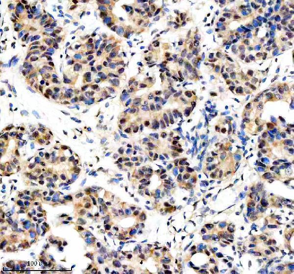
Real IHC data IHC analysis of DKC1 using anti-DKC1 antibody (A01535-2). DKC1 was detected in a paraffin-embedded section of human breast cancer tissue. Heat mediated antigen retrieval was performed in EDTA buffer (pH 8.0, epitope retrieval solution). The tissue section was blocked with 10% goat serum. The tissue section was then incubated with 2 μg/ml rabbit anti-DKC1 Antibody (A01535-2) overnight at 4°C. Peroxidase Conjugated Goat Anti-rabbit IgG was used as secondary antibody and incubated for 30 minutes at 37°C. The tissue section was developed using HRP Conjugated Rabbit IgG Super Vision Assay Kit (Catalog # SV0002) with DAB as the chromogen.
Anti-DKC1 Antibody ®
Cat # A01535-2

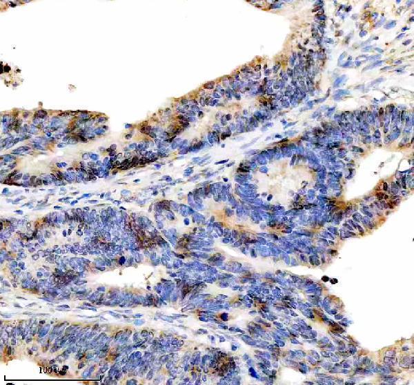
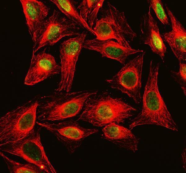
A01535-2 has IHC images from paraffin-embedded human breast cancer, colon cancer and colon tissue, plus an ICC/IF image from U2OS cells (catalog image captions). M01535-1 lists IHC and ICC/IF applications, but no IHC or IF image is supplied for it (catalog applications and image alts).

Which to pick: For tissue IHC, start with A01535-2: its caption documents a human paraffin section, EDTA retrieval at pH 8.0 and 2 μg/ml primary antibody; the fixative is unreported (A01535-2 IHC caption). For IF/ICC, A01535-2 has a U2OS image at 5 μg/ml (A01535-2 IF caption); M01535-1 is a rabbit monoclonal listed for IF/ICC, without an image in this payload (catalog host, clone, applications and image alts). For mouse or rat samples, either SKU lists reactivity, but the supplied images do not establish performance in those species (catalog reactivity and image captions).

Each figure is that product's own IHC / IF validation image from its datasheet.

References

  1. UniProt Consortium. UniProt entry O60832 (DKC1_HUMAN, H/ACA ribonucleoprotein complex subunit DKC1).
  2. Human Protein Atlas. DKC1 tissue IHC expression (reliability: Supported).
  3. Human Protein Atlas. DKC1 subcellular location (ICC-IF): Mainly localized to the nucleoli fibrillar center. In addition localized to the nucleoplasm..
  4. Human Protein Atlas. DKC1 antibody validation summary (4 antibodies).
  5. DKC1 enhances angiogenesis by promoting HIF-1α transcription and facilitates metastasis in colorectal cancer. British journal of cancer 2020 — PMC7054532.
  6. Inhibition of DKC1 induces telomere-related senescence and apoptosis in lung adenocarcinoma. Journal of translational medicine 2021 — PMC8056518.
  7. The nucleolar-related protein Dyskerin pseudouridine synthase 1 (DKC1) predicts poor prognosis in breast cancer. British journal of cancer 2020 — PMC7653035.
  8. Targeting DKC1/NF-κB axis suppresses tumorigenesis and enhances 5-FU sensitivity in gastric cancer. Cancer cell international 2025 — PMC12302887.
  9. PubMed PMID:9590285 — UniProt-cited evidence.
  10. PubMed PMID:10364516 — UniProt-cited evidence.
  11. PubMed PMID:9888995 — UniProt-cited evidence.