This website uses cookies to ensure you get the best experience on our website.


- Table of Contents
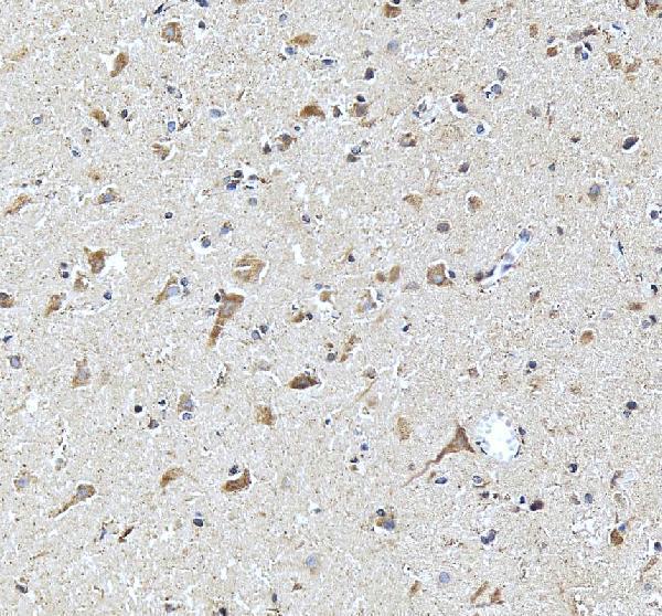
Plan DKK3 chromogenic IHC in paraffin sections using the catalog antibody’s 1:50 starting dilution (datasheet A02248-2). Compare staining with the generally cytoplasmic tissue pattern and high neuronal staining in caudate and hippocampus (HPA tissue IHC).

Expected localisation, validated protocols, controls and antibodies — the at-a-glance facts below, then the full design guide.
| Expected localisation | Cytoplasmic staining (HPA tissue IHC); secreted (UniProt) | |
| Staining pattern | High neuronal staining; generally cytoplasmic (HPA tissue IHC) | |
| Antigen retrieval | EDTA pH 8.0 HIER, heat-mediated (datasheet A02248-2) | |
| Positive control | Caudate+4 more · see all | |
| Negative control | Lymph node+1 more · see all |
| Fixation | Keep fixation consistent across paraffin sections (standard IHC practice; not target-specific) | |
| Caveat | Secreted DKK3 may stain away from RNA-positive cells (HPA tissue IHC) | |
| Regulation | Expression regulation is not specified (UniProt record) | |
| Isoform / epitope | No isoforms annotated; mature chain starts at 22 (UniProt) |
The catalog antibody protocol uses EDTA pH 8.0 retrieval (datasheet A02248-2). The four published DKK3 IHC protocols below cover cartilage, glioblastoma, oral biopsies, and bladder cancer tissue (PMC12645725; PMC11012478; PMC4933828; PMC7150467).
| Sample | Paraffin-embedded human brain tissue; fixative not specified (datasheet A02248-2) |
| Fixation | Image fixative and duration unreported (datasheet A02248-2); verify before use. |
| Sectioning | 4–5 µm sections on charged slides (standard) |
| Deparaffinisation | Xylene, graded ethanol series to water (standard) |
| Antigen retrieval | Heat retrieval: EDTA pH 8.0 (datasheet A02248-2); 20 min, 95–100 °C (standard) |
| Peroxidase block | 3% H2O2, 10 min, room temperature (standard) |
| Blocking | 10% goat serum (datasheet A02248-2) |
| Primary antibody | Rabbit anti-DKK3, 1:50 recommended; image 2 μg/ml (datasheet A02248-2) |
| Primary incubation | Overnight at 4 °C (datasheet A02248-2) |
| Detection | HRP-conjugated secondary, DAB chromogen (datasheet A02248-2) |
| Counterstain | Hematoxylin, blue, dehydrate and mount (standard) |
| Expected result | DKK3-positive staining in neuronal cells of caudate (HPA tissue IHC: High). HPA tissue profile: General cytoplasmic expression. No signal in the no-primary control. |
DKK3 is secreted, has a signal peptide and lacks a transmembrane segment (UniProt Q9UBP4 topology). In tissue IHC, expect mainly cytoplasmic staining, especially in caudate and hippocampal neuronal cells, where HPA reports high staining (HPA tissue IHC). HPA rates its tissue IHC profile Approved but reports low consistency with RNA; secretion can separate the sites of RNA expression and protein staining (HPA tissue IHC).
| Strong cytoplasmic staining in caudate or hippocampal neurons. | This matches HPA's high neuronal staining and general cytoplasmic tissue profile (HPA tissue IHC). Judge the signal by cell identity and compartment as well as intensity; a positive control should reproduce that distribution. |
| Predominant nuclear staining, with little cytoplasmic signal. | This departs from the general tissue IHC pattern (HPA tissue IHC). HPA describes nuclear bodies only as an uncertain additional ICC-IF location (HPA subcellular); dominant nuclear IHC staining therefore needs artifact and antibody-specificity checks before interpretation. |
| Strong staining in lymph-node germinal-center cells. | HPA reports these cells as not detected (HPA tissue IHC). Recheck cell identification, the no-primary control and detection background; unexpected staining may reflect cross-reactivity or endogenous detection activity (standard IHC practice). |
| Diffuse chromogen across cells, stroma and the section edge. | A distribution unrelated to the HPA cell pattern is difficult to score as DKK3 (HPA tissue IHC). Section-wide signal may arise from nonspecific binding or detection background; a secreted target alone does not explain uniform staining (UniProt Q9UBP4; standard IHC practice). |
| No signal in caudate or hippocampal neurons. | These are HPA high-staining cells, so absent signal calls the staining run into question (HPA tissue IHC). Check tissue preservation, retrieval, antibody dilution and detection controls before treating the result as biological absence (standard IHC practice). |
| Secretion and topology | DKK3 has a signal peptide at residues 1–21, a mature chain at 22–350, and no transmembrane segment (UniProt Q9UBP4 topology). Interpret tissue signal in its cell context; protein staining need not coincide with the cells showing the most RNA (HPA tissue IHC). |
| Glycosylation and processing | UniProt lists 6 glycosylation sites and no propeptide (UniProt Q9UBP4). These annotations describe DKK3 processing; they do not establish an IHC epitope, retrieval requirement or fixation sensitivity. |
| Tissue and cell choice | Caudate and hippocampal neurons are high-staining reference cells; adrenal and appendix glandular cells show medium staining (HPA tissue IHC). Germinal-center cells are a reported not-detected comparator, but their absence of staining does not validate every positive signal (HPA tissue IHC). |
| Strength of tissue evidence | The HPA tissue profile is Approved, with low consistency between antibody staining and RNA expression (HPA tissue IHC). HPA lists HPA011868 and CAB024949 as IHC Approved; this payload does not report an Enhanced IHC classification (HPA antibodies). |
| IF/ICC comparison | Q: Should IF/ICC look identical to tissue IHC? A: HPA supports vesicles as the main ICC-IF location; nuclear bodies and cytosol are uncertain additional locations (HPA subcellular). Compare each application with its own observed pattern. |
| Situation | Likely cause | Next action |
|---|---|---|
| Positive-control neurons are blank. | The observed result conflicts with HPA's high caudate and hippocampal neuronal staining (HPA tissue IHC); the failed step is undetermined. | Review section quality, retrieval, antibody dilution, detection reagents and a run control systematically (standard IHC practice). Do not assign DKK3 absence from this run alone. |
| The whole section is brown. | Uniform signal does not follow HPA's cell-specific tissue observations (HPA tissue IHC). Nonspecific binding or detection background is possible (standard IHC practice). | Inspect a no-primary control; review blocking, washes, detection chemistry and development time (standard IHC practice). Reassess the pattern only after background is controlled. |
| Germinal-center cells stain strongly. | HPA reports no detected staining in these cells (HPA tissue IHC). Cross-reactivity or endogenous detection activity is possible, depending on the detection system (standard IHC practice). | Confirm cell identity and examine no-primary and detection-only controls (standard IHC practice). Treat the signal as unresolved until its source is established. |
| Nuclei dominate the signal. | General cytoplasmic tissue staining is expected (HPA tissue IHC); nuclear bodies are an uncertain ICC-IF observation, not an established dominant IHC pattern (HPA subcellular). | Compare a known-positive section and controls, then review antibody and detection conditions (standard IHC practice). Avoid scoring isolated nuclear signal as confirmed DKK3. |
| Heart staining seems inconsistent with an RNA-based expectation. | UniProt reports high heart expression, while HPA reports enhanced heart-muscle RNA and warns that secreted protein staining may differ from RNA location (UniProt Q9UBP4; HPA tissue IHC). | Record the actual stained cell type and compartment. Use the observed IHC pattern and matched controls for interpretation rather than treating RNA abundance as a staining map (HPA tissue IHC). |
| A proposed retrieval or fixation change is described as DKK3-specific. | Target-specific fixation effects are not established by the supplied assay evidence. Verify with a matched IHC source before attributing a result to fixation. | Treat retrieval adjustment as general paraffin-IHC optimization and compare controlled sections (standard IHC practice). Do not claim a target-specific effect without direct evidence. |
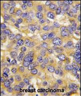


Comprehensive Human Protein Atlas IHC scoring per tissue (reliability: Approved — Low consistency between antibody staining and RNA expression data. Secreted protein, tissue location of RNA and protein is expected to differ.). Rows are taken directly from the HPA tissue chart — click any row's HPA link to view the source.
| Tissue | Cell type | Level | Evidence | Source |
|---|---|---|---|---|
| Caudate | Neuronal cells | High | Protein (IHC) | HPA → |
| Hippocampus | Neuronal cells | High | Protein (IHC) | HPA → |
| Adrenal gland | Glandular cells | Medium | Protein (IHC) | HPA → |
| Appendix | Glandular cells | Medium | Protein (IHC) | HPA → |
| Breast | Glandular cells | Medium | Protein (IHC) | HPA → |
Troubleshoot DKK3 staining in paraffin sections using the catalog antibody’s tissue example, protein biology, and tissue staining patterns as separate lines of evidence.
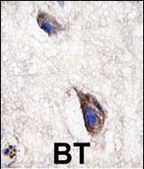
Both anti-DKK3 antibodies have human brain IHC images (catalog image captions); A02248-2 also lists IF/ICC applications and human, mouse, and rat reactivity (catalog applications/reactivity).
A02248-1 shows DAB staining in formalin-fixed, paraffin-embedded human brain (catalog image caption) and lists human IHC-P use (catalog applications/reactivity). A02248-2 shows DAB staining in paraffin-embedded human brain after EDTA retrieval (catalog image caption) and lists IHC, IF, and ICC use with human, mouse, and rat reactivity (catalog applications/reactivity).
Which to pick: For human tissue IHC with paraffin-section paraffin sections, A02248-1 matches the preparation in its own image; its listed IHC-P dilution is 1:50–1:100 (catalog image caption; datasheet: IHC-P dilution). For IF/ICC or cross-species work, choose A02248-2 because it lists those applications and human, mouse, and rat reactivity; its listed IF dilution is 1:50 (catalog applications/reactivity; datasheet: IF dilution). A02248-2’s own IHC image supports paraffin-embedded human brain, but does not report the fixative (catalog image caption); both antibodies are polyclonal (catalog dilution_raw). The selected A02248-2 tissue-IHC caption documents paraffin sections, but does not specify the fixative (selected-SKU IHC image A02248-2).