This website uses cookies to ensure you get the best experience on our website.


- Table of Contents
Plan chromogenic IHC on paraffin sections with the catalog antibody at 1:100–1:300 (datasheet). Assess membranous and luminal staining in glandular cells, keeping in mind the uncertain tissue staining reliability (HPA tissue IHC).

Expected localisation, validated protocols, controls and antibodies — the at-a-glance facts below, then the full design guide.
| Expected localisation | Membranous and luminal tissue staining (HPA tissue IHC) | |
| Staining pattern | Membranous and luminal staining in glandular cells (HPA tissue IHC) | |
| Antigen retrieval | Citrate pH 6.0 HIER, 95–98 °C, 20 min (rule: cytoplasmic / membrane antigen) | |
| Positive control | Gallbladder+4 more · see all | |
| Negative control | Adipose tissue+4 more · see all |
| Fixation | Keep paraffin-section fixation consistent (standard IHC practice; not target-specific) | |
| Caveat | Staining has low agreement with RNA; verification is pending (HPA tissue IHC) | |
| Regulation | Expression regulation is not established (UniProt) | |
| Isoform / epitope | 6 isoforms; antibody epitope coverage is unknown (UniProt) |
Compare the catalog antibody’s IHC-P protocol with 3 published DLC1 staining protocols for paraffin sections.
| Sample | Paraffin-embedded human prostate carcinoma tissue; fixative not specified (datasheet A30552) |
| Fixation | Image fixative and duration unreported (datasheet A30552); verify before use. |
| Sectioning | 4–5 µm sections on charged slides (standard) |
| Deparaffinisation | Xylene, graded ethanol series to water (standard) |
| Antigen retrieval | Heat-induced epitope retrieval in citrate buffer, pH 6.0, 20 min at 95–98 °C (standard rule: cytoplasmic / membrane antigen) |
| Peroxidase block | 3% H2O2, 10 min, room temperature (standard) |
| Blocking | 10% normal serum of the secondary host, 30 min, room temperature (standard) |
| Primary antibody | Rabbit anti-DLC1, 1:100 - 1:300 (datasheet A30552) |
| Primary incubation | Overnight at 4 °C (standard) |
| Detection | HRP-polymer secondary, DAB chromogen 5–10 min (standard) |
| Counterstain | Hematoxylin, blue, dehydrate and mount (standard) |
| Expected result | DLC1-positive staining in glandular cells of gallbladder (HPA tissue IHC: High). HPA tissue profile: Membranous and luminal expression in several tissues. No signal in the no-primary control. |
DLC1 is a cytoplasmic protein associated with focal adhesions and membranes, with no transmembrane segment (UniProt Q96QB1). In paraffin-section IHC, look for signal in gallbladder glandular cells and, at lower reported intensity, colon and small-intestine glandular cells or kidney tubule cells (HPA tissue IHC). HPA describes membranous and luminal staining across several tissues, but rates its tissue IHC profile Uncertain because staining and RNA data have low consistency (HPA tissue IHC).
| Glandular cells show localized staining in gallbladder, with weaker staining in colon or small intestine. | This follows HPA's High gallbladder and Medium colon and small-intestine observations (HPA tissue IHC). Assess cell identity and localization together; those observations remain provisional because the tissue IHC profile is Uncertain (HPA tissue IHC). |
| Signal appears at cell edges or in cytoplasm, sometimes alongside luminal staining. | Cytoplasmic and focal-adhesion-associated localization is consistent with UniProt Q96QB1; HPA reports membranous and luminal tissue staining (HPA tissue IHC). Luminal signal alone deserves scrutiny: HPA's pattern has not received strong tissue-level validation (HPA tissue IHC). |
| Nuclear staining dominates, with little signal in the reported-positive cells. | A dominant nuclear pattern conflicts with the supplied cytoplasmic, focal-adhesion and membrane annotations (UniProt Q96QB1). Treat it as a suspected nonspecific or technical signal and check controls before assigning it to DLC1 (general IHC practice). |
| Reported-negative cells stain as strongly as the expected glandular cells. | HPA reports DLC1 as Not detected in adipocytes of adipose tissue and in adrenal glandular cells (HPA tissue IHC). Strong staining there raises concern about cross-reactivity or endogenous detection activity; it does not alone disprove expression, given the Uncertain HPA profile (HPA tissue IHC; general IHC practice). |
| The whole section is diffusely colored, or a reported-positive tissue has no discernible cellular signal. | Uniform color obscures compartment and cell-type assessment, while absent signal cannot distinguish a technical failure from an antibody or specimen difference (general IHC practice). Compare controls and the complete cellular pattern before interpreting either result (general IHC practice). |
| How firm is the tissue reference pattern? | HPA calls tissue IHC Uncertain because antibody staining and RNA expression have low consistency; the supplied IHC antibody entry HPA017753 is also Uncertain (HPA tissue IHC; HPA antibodies). Use reported intensities as comparison points, not absolute pass or fail criteria (general IHC practice). |
| Which compartment should guide scoring? | UniProt places DLC1 in cytoplasm, focal adhesions and membrane-associated regions, without a transmembrane segment (UniProt Q96QB1). HPA tissue IHC reports membranous and luminal expression (HPA tissue IHC). Record the exact compartment seen rather than treating all edge or luminal color as equivalent (general IHC practice). |
| Can one stain represent every isoform? | UniProt lists 6 DLC1 isoforms (UniProt Q96QB1). Antibody epitope coverage is not supplied, so an isoform-specific staining prediction is unavailable; interpret an absent signal cautiously when comparing reagents (general IHC practice). |
| What does IF/ICC add? | HPA ICC-IF places DLC1 mainly in supported cytosol, with approved additional endoplasmic-reticulum and vesicle locations (HPA subcellular). This answers the localization question for IF/ICC; its cell images and supported ICC antibody entries do not validate the uncertain tissue IHC pattern (HPA subcellular; HPA antibodies; HPA tissue IHC). |
| Situation | Likely cause | Next action |
|---|---|---|
| Gallbladder glandular cells show no signal. | HPA reports High staining there, but its tissue IHC assessment is Uncertain; a technical failure or reagent-dependent result is possible (HPA tissue IHC; general IHC practice). | Check tissue preservation, the detection control and the antibody's supplied IHC-P instructions; repeat with a working control before calling the sample negative (general IHC practice). |
| Only nuclei stain strongly. | Nuclear dominance does not fit the supplied DLC1 localization (UniProt Q96QB1). Nonspecific antibody or detection signal is possible (general IHC practice). | Review the no-primary control and compare nuclear color with cytoplasmic and cell-edge signal in reported-positive cells (general IHC practice). |
| Adipocytes or adrenal glandular cells stain strongly. | Those cells are reported Not detected by HPA tissue IHC, although the overall profile is Uncertain (HPA tissue IHC). Cross-reactivity or endogenous detection activity may contribute (general IHC practice). | Run a no-primary control and check detection blocking; compare the same run with reported-positive glandular tissue before interpreting specificity (general IHC practice). |
| Diffuse color obscures cell boundaries. | Background from antibody or detection reagents can make a cellular pattern unreadable (general IHC practice). | Check the no-primary control, blocking, washes and reagent concentration under the chosen IHC-P workflow; score only interpretable cellular signal (general IHC practice). |
| Luminal color is prominent but glandular cells are faint. | HPA reports luminal staining, yet the tissue IHC profile has low consistency with RNA data (HPA tissue IHC). Luminal color alone is therefore weak evidence for DLC1-positive cells (HPA tissue IHC; general IHC practice). | Document luminal and cellular staining separately; compare reported-positive and reported-negative tissues plus the no-primary control (HPA tissue IHC; general IHC practice). |
| Two antibodies give different tissue patterns. | The supplied HPA tissue IHC entry is Uncertain, and the listed antibodies have different IHC and ICC validation entries (HPA tissue IHC; HPA antibodies). Epitope coverage is unspecified (HPA antibodies). | Keep each antibody's observations separate, compare localization and controls, and avoid declaring either pattern validated from agreement with HPA intensity alone (general IHC practice; HPA tissue IHC). |


![HPA ICC-IF BJ [Human fibroblast] — DLC1 (HPA061655)](https://images.proteinatlas.org/61655/1801_B4_4_cr59ca0d3dc6143_blue_red_green.jpg)
![HPA ICC-IF BJ [Human fibroblast] — DLC1 (HPA061655)](https://images.proteinatlas.org/61655/1801_B4_6_cr59c4f4e721d1a_blue_red_green.jpg)
Comprehensive Human Protein Atlas IHC scoring per tissue (reliability: Uncertain — Low consistency between antibody staining and RNA expression data. Pending external verification.). Rows are taken directly from the HPA tissue chart — click any row's HPA link to view the source.
| Tissue | Cell type | Level | Evidence | Source |
|---|---|---|---|---|
| Gallbladder | Glandular cells | High | Protein (IHC) | HPA → |
| Caudate | Glial cells | Medium | Protein (IHC) | HPA → |
| Cerebellum | Cells in granular layer | Medium | Protein (IHC) | HPA → |
| Colon | Glandular cells | Medium | Protein (IHC) | HPA → |
| Duodenum | Glandular cells | Medium | Protein (IHC) | HPA → |
| Tissue | Cell type | Level | Evidence | Source |
|---|---|---|---|---|
| Adipose tissue | Adipocytes | Not detected | Protein (IHC) | HPA → |
| Adrenal gland | Glandular cells | Not detected | Protein (IHC) | HPA → |
| Appendix | Glandular cells | Not detected | Protein (IHC) | HPA → |
| Bone marrow | Hematopoietic cells | Not detected | Protein (IHC) | HPA → |
| Breast | Adipocytes | Not detected | Protein (IHC) | HPA → |
Troubleshoot DLC1 staining in paraffin sections by checking retrieval, compartment, cell type and controls before interpreting chromogenic signal.
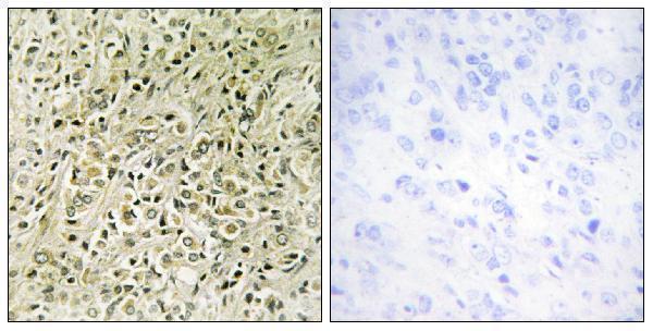
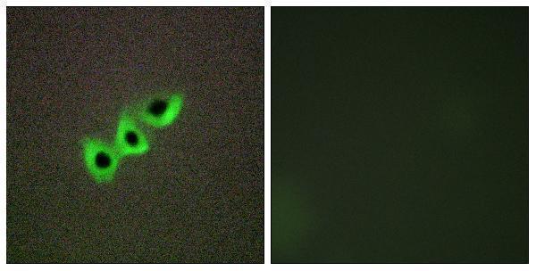
A30552 has IHC data from paraffin-embedded human prostate carcinoma tissue and IF data from A549 cells (catalog image captions). Human, Mouse, and Rat reactivity is listed (catalog reactivity).
A30552 shows IHC staining of paraffin-embedded human prostate carcinoma tissue (catalog IHC image caption). It also shows IF staining of A549 cells and is listed for IHC, IF, and ICC (catalog IF image caption; catalog applications).
Which to pick: Choose A30552 for tissue IHC: its IHC image uses paraffin-embedded human prostate carcinoma tissue, but the fixative is unreported (catalog IHC image caption). Choose A30552 for IF/ICC because those applications are listed and its IF image shows A549 cells (catalog applications; catalog IF image caption). For cross-species work, A30552 is a rabbit polyclonal antibody with Human, Mouse, and Rat reactivity listed; the supplied IHC image documents human tissue only (catalog host; catalog dilution_raw; catalog reactivity; catalog IHC image caption).