This website uses cookies to ensure you get the best experience on our website.


- Table of Contents
Real validated DLX2 Western blot protocols, expected-band and isoform facts, troubleshooting for weak or shifted signal, and recommended anti-DLX2 WB antibodies. Everything you need to plan the experiment before you commit precious samples.
Expected bands, validated protocols, controls and antibodies — the at-a-glance facts below, then the full design guide.
| Expected band | ~34.2 kDa | |
| Observed band | 37 kDa | |
| Gel | 10–12% | |
| Negative control | siRNA / KO lysate |
| PTM | Phosphorylated | |
| Caveat | Phosphorylation-induced mobility shift | |
| Regulation | LPS-induced | |
| Isoform | 2 isoform(s) |
Literature-validated Western blot parameters for DLX2 — gel percentage, transfer, blocking, antibody incubation and detection, extracted from published methods.
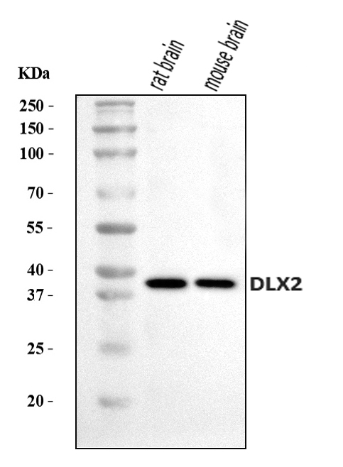
| Sample / lysate | rat brain , Lane 2: mouse brain . After electrophoresis, proteins were transferred to a nitrocellulose membrane at 150 mA for 50-90 minutes. Blocked the membrane with 5% non-fat milk/TBS for 1.5 hour at RT. The membrane was incubated with rabbit anti-DLX2 antigen affinity purified polyclonal antibody (Catalog # A03617-2) at 0.5 μg/mL overnight at 4°C, then washed with TBS-0.1%Tween 3 times with 5 minutes each and probed with a goat anti-rabbit IgG-HRP secondary antibody at a dilution of 1:5000 for 1.5 hour at RT. The signal is developed using an Enhanced Chemiluminescent detection (ECL) kit (Catalog # EK1002) with Tanon 5200 system. A specific band was detected for DLX2 at approximately 37 kDa. The expected band size for DLX2 is at 37 kDa |
| Gel % | 10–12% |
| Load | 30 ug |
| Transfer | nitrocellulose membrane, 150 mA, 50–90 min |
| Membrane | nitrocellulose |
| Blocking | 5% non-fat milk / TBS, 1.5 h RT |
| Primary antibody | 0.5 µg/mL |
| Primary incubation | overnight at 4 °C |
| Secondary antibody | goat anti-rabbit IgG-HRP, 1:5000 |
| Wash | TBS-0.1% Tween, 3 × 5 min |
| Detection | ECL |
| Exposure / imaging | Tanon 5200 |
| Observed band | 37 kDa |
DLX2 has a 34.2 kDa predicted backbone but typically runs around 37 kDa on Western blots, largely attributable to phosphorylation at Ser232.
| single band around 37 kDa | mature DLX2 running modestly above its 34.2 kDa predicted mass, consistent with phosphorylation at Ser232 |
| doublet or two closely spaced bands | reflects the two annotated DLX2 isoforms (1 and 2) generated by alternative splicing |
| band shifts down toward ~34 kDa after phosphatase treatment | loss of the Ser232 phosphate removes the mass contribution that pushes the native band above the predicted backbone |
| band strong in nuclear fraction but weak in cytoplasmic fraction | DLX2 is annotated as a nuclear protein, so it partitions into the nuclear extract rather than cytoplasmic lysate |
| no smearing and no higher-molecular-weight band under non-reducing conditions | DLX2 has no annotated glycosylation and no annotated disulfide bonds or homodimer formation |
| Predicted mass (UniProt, 328 aa) | sets the 34.2 kDa theoretical baseline for unmodified DLX2 |
| Phosphorylation at Ser232 | adds apparent mass, contributing to the observed ~37 kDa band rather than the bare 34.2 kDa prediction |
| Alternative splicing (isoforms 1 and 2) | can produce two bands of differing apparent size, or a single band whose size depends on which isoform predominates, without a defined per-isoform kDa |
| No glycosylation annotated | rules out a smeared or diffusely higher band from carbohydrate modification |
| No disulfide bonds or homodimer annotated | rules out a band at roughly twice the monomer mass under non-reducing or incompletely reduced conditions |
| Nuclear subcellular localization | band strength depends on efficient nuclear lysis or fractionation, rather than whole-cell or cytoplasmic-only extraction |
| Situation | Likely cause | Next action |
|---|---|---|
| No band in lysate | DLX2 is a nuclear developmental transcription factor with restricted tissue expression, and standard whole-cell lysis may not efficiently release nuclear protein | use a nuclear extraction protocol and confirm sample source expresses DLX2, such as developing neural or branchial arch tissue |
| Band higher than expected | phosphorylation at Ser232 raises the apparent mass above the 34.2 kDa predicted mass to around 37 kDa | treat a lysate aliquot with phosphatase and compare mobility, and use 37 kDa as the reference size rather than the unmodified prediction |
| Band lower than expected | loss of the Ser232 phosphate during sample handling can shift the band down toward or below the 34.2 kDa predicted mass | include phosphatase inhibitors in the lysis buffer to preserve the native phosphorylated form |
| Multiple bands | the two annotated splice isoforms (1 and 2) can migrate as separate bands | confirm isoform identity with isoform-specific detection or transcript analysis rather than assuming nonspecific binding |
| Weak or no signal | DLX2 is a low-abundance nuclear transcription factor confined to developing tissues, so signal can be faint outside those contexts | increase protein loading, enrich for the nuclear fraction, and include a known DLX2-expressing tissue as a positive control |
Comprehensive Human Protein Atlas IHC scoring per tissue. Rows are taken directly from the HPA tissue chart — click any row's HPA link to view the source.
| Tissue | Cell type | Level | Evidence | Source |
|---|
| Tissue | Cell type | Level | Evidence | Source |
|---|
Deeper troubleshooting and optimisation questions for DLX2, answered from its protein features.
BosterBio's DLX2 antibodies are among the best-performing WB antibodies on the market — well cited, thoroughly validated, and orthogonally cross-validated against negative tissues and complementary methods.
Our recommended anti-DLX2 antibodies are top-performing reagents, extensively cited across the literature and rigorously validated through orthogonal methods, including comparison against negative-control tissues and complementary techniques, ensuring reliable, reproducible specificity for confident Western blot detection of DLX2.
Which to pick: Only one DLX2 antibody is catalogued here, A03617-2, which includes an actual Western blot validation image showing detection of DLX2 by SDS-PAGE, making it the clear, and only, choice for your Western blot experiments.