This website uses cookies to ensure you get the best experience on our website.


- Table of Contents
Plan chromogenic IHC for DMRT1 in paraffin sections using the catalog antibody’s validated conditions. Compare nuclear staining in a subset of spermatogonia with HPA tissue IHC observations, and assess unexpected staining cautiously because presumed off-target binding was reported (HPA tissue IHC).

Expected localisation, validated protocols, controls and antibodies — the at-a-glance facts below, then the full design guide.
| Expected localisation | Nuclear in a subset of spermatogonia (HPA tissue IHC) | |
| Staining pattern | A subset of spermatogonia shows nuclear staining (HPA tissue IHC) | |
| Antigen retrieval | Citrate pH 6 HIER, heat-mediated (datasheet RP1083) | |
| Positive control | Testis | |
| Negative control | Adipose tissue+4 more · see all |
| Fixation | Keep fixation conditions consistent across sections. (standard IHC practice; not target-specific) | |
| Caveat | Presumed off-target staining was disregarded (HPA tissue IHC) | |
| Regulation | Testis-specific expression (UniProt) | |
| Isoform / epitope | 3 isoforms; epitope coverage is unknown (UniProt) |
The catalog antibody’s paraffin IHC protocol is paired with a published chromogenic IHC protocol for orange-spotted grouper sections (PMC6529513).
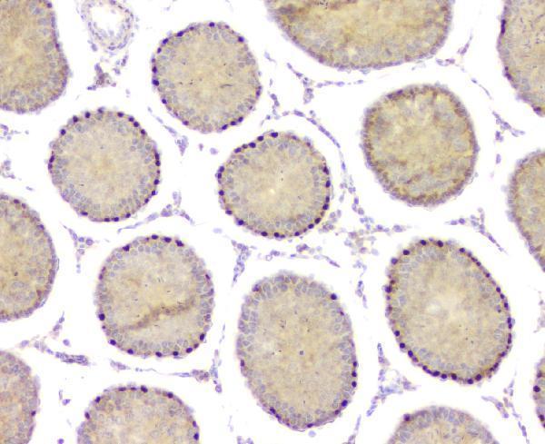
| Sample | Paraffin-embedded rat testis tissue; fixative not specified (datasheet RP1083) |
| Fixation | Image fixative and duration unreported (datasheet RP1083); verify before use. |
| Sectioning | 4–5 µm sections on charged slides (standard) |
| Deparaffinisation | Xylene, graded ethanol series to water (standard) |
| Antigen retrieval | Heat retrieval: Citrate pH 6, 20 min (datasheet RP1083) |
| Peroxidase block | 3% H2O2, 10 min, room temperature (standard) |
| Blocking | 10% goat serum (datasheet RP1083) |
| Primary antibody | Rabbit anti-DMRT1, 0.5-1μg/ml (datasheet RP1083) |
| Primary incubation | Overnight at 4 °C (datasheet RP1083) |
| Detection | Streptavidin-biotin complex (SABC), DAB chromogen (datasheet RP1083) |
| Counterstain | Hematoxylin, blue, dehydrate and mount (standard) |
| Expected result | DMRT1-positive staining in preleptotene spermatocytes of testis (HPA tissue IHC: High). HPA tissue profile: Nuclear expression in a subset spermatogonia. No signal in the no-primary control. |
DMRT1 is a nuclear transcription factor with no transmembrane segment (UniProt Q9Y5R6). In testis IHC, expect nuclear staining in a subset of spermatogonia (HPA tissue IHC profile); HPA also lists high staining in preleptotene spermatocytes (HPA tissue IHC). HPA rates the tissue pattern Enhanced, while noting medium agreement with RNA expression and presumed off-target staining that was disregarded (HPA tissue IHC).
| Distinct nuclear stain in some germ cells within testis, with neighboring cells unstained. | This fits the reported nuclear pattern in a subset of spermatogonia (HPA tissue IHC profile; UniProt Q9Y5R6). Do not require every germ cell to stain: HPA describes a subset and separately lists high staining in preleptotene spermatocytes (HPA tissue IHC). |
| Cytoplasmic or membrane-dominant stain, without convincing nuclear signal. | The dominant compartment conflicts with the annotated nuclear location (UniProt Q9Y5R6) and observed testis pattern (HPA tissue IHC). Treat it as suspect, then compare morphology and a no-primary control; background or detection artefact cannot be identified from location alone (general IHC practice). |
| Strong stain in adipocytes or other cells listed as unstained by HPA. | HPA reports no detection in adipocytes from adipose tissue and breast, among its negative examples (HPA tissue IHC). Such staining raises concern for cross-reactivity or endogenous detection activity, especially if a no-primary control also stains; it does not establish a new DMRT1 expression site (general IHC practice). |
| Broad, hazy stain obscures nuclei across the section. | Diffuse colour makes nuclear localisation and cell-level scoring unreliable (general IHC practice). Assess a no-primary control and the distribution of precipitate; excessive detection signal or insufficient washing are possible general workflow causes, not DMRT1-specific effects (general IHC practice). |
| No nuclear signal in an otherwise interpretable testis section. | Target-specific fixation effects are not established by the supplied assay evidence. Verify with a matched IHC source before attributing a result to fixation. |
| Cell population and scoring | Score nuclear signal by cell type rather than averaging the whole section: HPA describes a subset of spermatogonia and lists preleptotene spermatocytes as high (HPA tissue IHC). The two HPA descriptions should be reported as given, without assuming they identify the same cells. |
| IHC evidence and antibody choice | HPA rates its tissue IHC reliability Enhanced and lists antibody HPA027850 as IHC Enhanced (HPA tissue IHC; HPA antibodies). HPA also notes medium agreement with RNA and disregarded presumed off-target binding; an unexpected pattern still needs controls (HPA tissue IHC; general IHC practice). |
| Intracellular location and processing | A nuclear result is biologically consistent with DMRT1's annotated location and transcription-factor function (UniProt Q9Y5R6). UniProt lists no transmembrane segment, signal peptide or propeptide, and one chain spanning residues 1–373; these annotations do not predict a fixation or retrieval response (UniProt Q9Y5R6). |
| Molecular variants | UniProt lists 3 isoforms and phosphoserine at residue 339 (UniProt Q9Y5R6). The supplied records do not map an antibody epitope or establish isoform-specific staining, phosphorylation-dependent staining, or a preferred retrieval condition; avoid explaining a slide pattern by those mechanisms. |
| Situation | Likely cause | Next action |
|---|---|---|
| Expected testis nuclei are unstained. | The assay may have failed, or the sampled area may lack the reported positive population (HPA tissue IHC; general IHC practice). | Locate appropriate germ cells on the counterstained section; compare a known-positive testis control and follow the IHC-validated antibody procedure before interpreting absence (general IHC practice). |
| Only cytoplasmic or membrane-like colour appears. | That distribution conflicts with nuclear DMRT1 (UniProt Q9Y5R6); nonspecific or detection-derived colour remains possible (general IHC practice). | Compare the stained section with a no-primary control, inspect nuclear boundaries, and score only reproducible nuclear signal in the expected cells (general IHC practice; HPA tissue IHC). |
| HPA-negative cell types stain strongly. | Cross-reactivity or endogenous detection activity is possible; HPA reports several unstained cell types, including adipocytes (HPA tissue IHC; general IHC practice). | Check a no-primary control and tissue morphology, then confirm any proposed new positive pattern independently before assigning it to DMRT1 (general IHC practice). |
| Diffuse chromogen obscures the nuclei. | Excess detection signal or inadequate washing can obscure cell-level localisation (general IHC practice). | Review reagent exposure and washes, compare the no-primary control, and adjust the general IHC workflow until nuclear boundaries can be judged (general IHC practice). |
| Testis stains, but nearly every cell appears positive. | HPA reports a subset pattern, so widespread staining exceeds the reported distribution (HPA tissue IHC). | Recheck cell identification and background controls; record the observed distribution separately from the HPA pattern rather than scoring all colour as specific (general IHC practice; HPA tissue IHC). |
| IF/ICC shows a pattern different from IHC. | HPA provides no DMRT1 ICC-IF images or validated main subcellular location in this payload (HPA subcellular); UniProt annotates nuclear location (UniProt Q9Y5R6). | Treat the IF/ICC result as unconfirmed here and use its dedicated guide for assay design; this section establishes no IF/ICC protocol or image-based validation (HPA subcellular). |


Comprehensive Human Protein Atlas IHC scoring per tissue (reliability: Enhanced — Medium consistency between antibody staining and RNA expression data. Presumed off target binding observed and disregarded.). Rows are taken directly from the HPA tissue chart — click any row's HPA link to view the source.
| Tissue | Cell type | Level | Evidence | Source |
|---|---|---|---|---|
| Testis | Preleptotene spermatocytes | High | Protein (IHC) | HPA → |
| Tissue | Cell type | Level | Evidence | Source |
|---|---|---|---|---|
| Adipose tissue | Adipocytes | Not detected | Protein (IHC) | HPA → |
| Adrenal gland | Glandular cells | Not detected | Protein (IHC) | HPA → |
| Appendix | Glandular cells | Not detected | Protein (IHC) | HPA → |
| Bone marrow | Hematopoietic cells | Not detected | Protein (IHC) | HPA → |
| Breast | Adipocytes | Not detected | Protein (IHC) | HPA → |
Troubleshoot DMRT1 staining in paraffin sections by checking retrieval, nuclear localisation, cell identity and controls before interpreting chromogenic signal.
RP1083 has real IHC images of paraffin testis sections from human, mouse, and rat (RP1083 IHC captions). No IF/ICC image or application is listed (catalog applications; image alts).
RP1083 is listed for IHC in human, mouse, and rat (catalog applications and reactivity). Its IHC captions show paraffin testis sections from all three species; no IF/ICC validation is shown (RP1083 IHC and IF image alts).
Which to pick: For tissue IHC, choose RP1083: its captions document staining of paraffin testis sections, while the fixative is unreported (RP1083 IHC captions). For IF/ICC, there is no validated choice in this catalog because RP1083 lists IHC and WB only, has no IF image, and reports no clone (catalog applications, IF image alts, clone). For cross-species IHC, RP1083 covers human, mouse, and rat (catalog reactivity; RP1083 IHC captions).