This website uses cookies to ensure you get the best experience on our website.


- Table of Contents
Use nuclear and cytoplasmic staining as the DNAJB1 tissue IHC benchmark (HPA tissue IHC). Adrenal glandular cells offer a high-staining reference (HPA tissue IHC); score nuclear and cytoplasmic signal separately because heat shock can shift DNAJB1 toward nucleoli (UniProt).

Expected localisation, validated protocols, controls and antibodies — the at-a-glance facts below, then the full design guide.
| Expected localisation | Nuclear and cytoplasmic in tissue (HPA tissue IHC) | |
| Staining pattern | General nuclear and cytoplasmic staining (HPA tissue IHC) | |
| Antigen retrieval | EDTA pH 8.0 HIER, heat-mediated (datasheet A03100-1) | |
| Positive control | Adrenal gland+4 more · see all | |
| Negative control | None in HPA (detected in all 45 tissues); use no-primary + isotype controls |
| Fixation | Keep tissue fixation consistent across samples (standard IHC practice; not target-specific) | |
| Caveat | Heat shock can shift DNAJB1 toward nucleoli (UniProt) | |
| Regulation | Heat shock shifts nuclear signal (UniProt) | |
| Isoform / epitope | 2 isoforms; epitope coverage unspecified (UniProt) |
The catalog antibody’s IHC-P protocol (datasheet A03100-1) appears alongside published DNAJB1 IHC protocols for fish ovarian frozen sections (PMC6485077) and liver sections (PMC11278829).
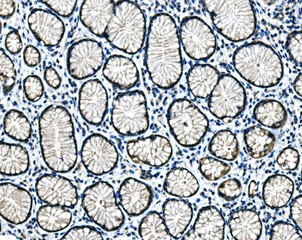
| Sample | Paraffin-embedded human gastric signet ring cell carcinoma tissue; fixative not specified (datasheet A03100-1) |
| Fixation | Image fixative and duration unreported (datasheet A03100-1); verify before use. |
| Sectioning | 4–5 µm sections on charged slides (standard) |
| Deparaffinisation | Xylene, graded ethanol series to water (standard) |
| Antigen retrieval | Heat retrieval: EDTA pH 8.0 (datasheet A03100-1); 20 min, 95–100 °C (standard) |
| Peroxidase block | 3% H2O2, 10 min, room temperature (standard) |
| Blocking | 10% goat serum (datasheet A03100-1) |
| Primary antibody | Rabbit anti-DNAJB1, 2-5 μg/ml (datasheet A03100-1) |
| Primary incubation | Overnight at 4 °C (datasheet A03100-1) |
| Detection | Streptavidin-biotin complex (SABC), DAB chromogen (datasheet A03100-1) |
| Counterstain | Hematoxylin, blue, dehydrate and mount (standard) |
| Expected result | DNAJB1-positive staining in glandular cells of adrenal gland (HPA tissue IHC: High). HPA tissue profile: General nuclear and cytoplasmic expression. No signal in the no-primary control. |
DNAJB1 is a soluble protein found in the cytoplasm and nucleus; it has no transmembrane segment (UniProt P25685 topology and subcellular location). Expect nuclear and cytoplasmic staining across several cell types, with high staining in selected epithelial, glandular and follicle cells (HPA tissue IHC). The tissue IHC profile has Enhanced reliability, reflecting high consistency between antibody staining and RNA expression (HPA tissue IHC).
| Nuclear and cytoplasmic staining in bronchial respiratory epithelial cells or ovarian follicle cells. | This fits the general tissue pattern and the High level reported for those cells (HPA tissue IHC). Judge intensity against matched controls; a positive stain alone does not establish a particular cellular stress state (general IHC interpretation). |
| Predominantly membrane outlining or extracellular deposits, with little cellular staining. | That distribution conflicts with the reported cytoplasmic and nuclear locations and lack of a transmembrane segment (UniProt P25685; HPA tissue IHC). Review morphology, detection controls and antibody specificity before assigning it to DNAJB1 (general IHC practice). |
| Strong staining in cardiomyocytes or smooth muscle cells while expected positive cells stain weakly. | Those cell types are listed as Low, so this contrast warrants review; Low does not mean absent (HPA tissue IHC). Check cell identification and controls for cross-reactivity or endogenous chromogenic activity (general IHC practice). |
| Diffuse color over tissue, empty spaces and the slide, without clear cell boundaries. | Noncellular background cannot be read as the nuclear and cytoplasmic pattern (HPA tissue IHC). Compare a no-primary control and assess blocking, washes and detection chemistry (general IHC practice). |
| No staining in a selected High reference cell population, such as adrenal glandular cells. | This fails the expected reference pattern (HPA tissue IHC). Check section quality, retrieval and reagent performance with a known-positive control before interpreting other tissues as negative (general IHC practice). |
| Compartment and stress state | DNAJB1 can move rapidly from cytoplasm to nucleus, especially nucleoli, after heat shock (UniProt P25685). Record handling and compare like conditions before treating a nuclear shift as an assay failure (general IHC interpretation). |
| Tissue and cell selection | HPA reports Low tissue RNA specificity and a general nuclear and cytoplasmic IHC profile; its named High and Low cell populations provide useful references, not universal positive and negative tissues (HPA tissue IHC). |
| Antibody validation | HPA063247 is IHC Enhanced and CAB017450 is IHC Supported (HPA antibodies). These are antibody-specific validation labels; apply the label only to the antibody actually used (HPA antibodies). |
| Isoforms and antigen design | Two isoforms are listed, but the supplied record does not map an IHC epitope to either one (UniProt P25685). Do not infer isoform-specific staining from this record; consult the selected antibody's documented immunogen before making that claim. |
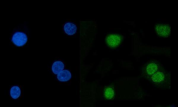
| IF/ICC comparison | What should IF/ICC show? Mainly nucleoplasm, with additional cytosol, in the HPA cell images (HPA subcellular ICC-IF). Treat this as a localisation cross-check; the HPA tissue IHC levels describe sections, not IF/ICC intensity. |
| Situation | Likely cause | Next action |
|---|---|---|
| Known High reference cells are blank. | The assay may have failed, or the selected cells may be missing from the section (general IHC practice). | Verify the cell population and run a known-positive section; review the selected antibody's documented retrieval and detection conditions (general IHC practice). High reference examples include bronchial respiratory epithelial cells (HPA tissue IHC). |
| Only a crisp membrane rim appears. | The pattern disagrees with DNAJB1's nuclear and cytoplasmic locations and absent transmembrane segment (UniProt P25685). | Compare a no-primary control, inspect morphology and confirm the antibody's IHC validation before scoring the rim as target staining (general IHC practice; HPA antibodies). |
| Strong color appears in a Low reference population. | Cross-reactivity or endogenous detection activity is possible, but Low is not equivalent to negative (HPA tissue IHC; general IHC practice). | Confirm cell identity; compare no-primary and appropriate detection controls alongside a High reference population before assigning specificity (general IHC practice; HPA tissue IHC). |
| Haze obscures nuclei and cytoplasm. | Background from blocking, washes or detection may prevent compartment scoring (general IHC practice). | Compare a no-primary control, review blocking and washes, and adjust detection conditions according to the selected assay's instructions (general IHC practice). |
| Staining is much more nuclear in one sample. | Stress-dependent nuclear and nucleolar translocation is possible; the stain alone cannot establish its cause (UniProt P25685). | Compare sample handling and matched controls, then score nuclear and cytoplasmic compartments separately (general IHC practice). |
| IF/ICC localisation seems different from tissue IHC. | The HPA ICC-IF summary emphasizes nucleoplasm with additional cytosol, whereas tissue IHC reports general nuclear and cytoplasmic expression (HPA subcellular ICC-IF; HPA tissue IHC). | Compare compartments within each application and check the antibody's application-specific validation; do not transfer tissue IHC intensity categories to IF/ICC (HPA antibodies; HPA tissue IHC). |


Comprehensive Human Protein Atlas IHC scoring per tissue (reliability: Enhanced — High consistency between antibody staining and RNA expression data.). Rows are taken directly from the HPA tissue chart — click any row's HPA link to view the source.
| Tissue | Cell type | Level | Evidence | Source |
|---|---|---|---|---|
| Adrenal gland | Glandular cells | High | Protein (IHC) | HPA → |
| Bronchus | Respiratory epithelial cells | High | Protein (IHC) | HPA → |
| Cervix | Squamous epithelial cells | High | Protein (IHC) | HPA → |
| Esophagus | Squamous epithelial cells | High | Protein (IHC) | HPA → |
| Gallbladder | Glandular cells | High | Protein (IHC) | HPA → |
| Tissue | Cell type | Level | Evidence | Source |
|---|---|---|---|---|
| None in HPA: DNAJB1 is detected in all 45 scored tissues. Use a no-primary (secondary-only) and an isotype control instead. | ||||
Troubleshoot DNAJB1 staining in paraffin sections by checking retrieval, compartment, controls, and scoring against its documented expression pattern (UniProt P25685; HPA tissue IHC).
A03100-1 has IHC data from human gastric signet ring cell carcinoma paraffin sections and IF data from A549 cells; listed reactivity covers human, mouse, and rat (catalog image captions and reactivity).
The card for A03100-1 will show chromogenic IHC in a human gastric signet ring cell carcinoma paraffin section (A03100-1 IHC image caption). Its IF image shows staining in A549 cells, and the catalog lists both IF and ICC applications (A03100-1 IF image caption; catalog applications).
Which to pick: Choose A03100-1 for tissue IHC: its own image shows a paraffin section with EDTA pH 8.0 retrieval, 2 μg/ml primary antibody, and DAB detection; the fixative is unreported (A03100-1 IHC image caption). For IF/ICC, choose A03100-1 based on its A549 cell IF image and listed IF and ICC applications (A03100-1 IF image caption; catalog applications). For cross-species work, A03100-1 lists human, mouse, and rat reactivity, though its supplied IHC image and IHC dilution listing cover human samples; its host is rabbit and clonality is unreported (catalog reactivity, host, and dilution; A03100-1 IHC image caption).