DNAJC10 / Endoplasmic reticulum disulfide reductase DNAJC10 · IHC design guide

Design Immunohistochemistry for DNAJC10

Plan DNAJC10 IHC in paraffin sections using the cytoplasmic tissue staining pattern and high glandular staining in endometrium and epididymis (HPA tissue IHC). Start with the catalog antibody at 2–5 μg/mL and compare staining with appropriate controls (datasheet A07954-1; standard IHC practice).

Evidence assembled Oct 2026 · For research use; verify linked source records and product datasheet before use
Immunohistochemistry protocol sheet for DNAJC10 (IHC for DNAJC10): expected localisation Cytoplasmic tissue staining (HPA tissue IHC); ER lumen (UniProt), antibody A07954-1, validated IHC image, and IHC protocol steps
Printable DNAJC10 IHC protocol sheet — expected localisation Cytoplasmic tissue staining (HPA tissue IHC); ER lumen (UniProt), antibody A07954-1, controls and protocol steps. Open the full DNAJC10 IHC guide →

DNAJC10 Immunohistochemistry Experimental Design Guide

Expected localisation, validated protocols, controls and antibodies — the at-a-glance facts below, then the full design guide.

Must know before staining
Expected localisation Cytoplasmic tissue staining (HPA tissue IHC); ER lumen (UniProt)
Staining pattern Cytoplasmic across tissues; high in glandular cells (HPA tissue IHC)
Antigen retrieval EDTA pH 8.0 HIER, heat-mediated (datasheet A07954-1)
Positive control ⓘ Endometrium+4 more · see all
Negative control ⓘ None in HPA (detected in all 45 tissues); use no-primary + isotype controls
Important caveats
Reasons your staining may differ from the expected pattern.
Fixation Keep fixation consistent across sections (standard IHC practice; not target-specific)
Caveat Staining and RNA expression show medium consistency (HPA tissue IHC)
Regulation Expression regulation not specified (UniProt)
Isoform / epitope 3 isoforms; epitope coverage unknown; mature chain 33–793 (UniProt)
Section 1

Recommended DNAJC10 IHC & IF Protocols

The catalog antibody’s IHC-P protocol (datasheet A07954-1) is paired with one published DNAJC10 tissue-staining protocol (PMC11190951).

Recommended immunohistochemistry (IHC-P) protocol parameters
SampleParaffin-embedded human breast cancer tissue; fixative not specified (datasheet A07954-1)
FixationImage fixative and duration unreported (datasheet A07954-1); verify before use.
Sectioning4–5 µm sections on charged slides (standard)
DeparaffinisationXylene, graded ethanol series to water (standard)
Antigen retrievalHeat retrieval: EDTA pH 8.0 (datasheet A07954-1); 20 min, 95–100 °C (standard)
Peroxidase block3% H2O2, 10 min, room temperature (standard)
Blocking10% goat serum (datasheet A07954-1)
Primary antibodyRabbit anti-DNAJC10, 2-5μg/ml (datasheet A07954-1)
Primary incubationOvernight at 4 °C (datasheet A07954-1)
DetectionStreptavidin-biotin complex (SABC), DAB chromogen (datasheet A07954-1)
CounterstainHematoxylin, blue, dehydrate and mount (standard)
Expected resultDNAJC10-positive staining in glandular cells of endometrium (HPA tissue IHC: High). HPA tissue profile: Ubiquitous cytoplasmic expression. No signal in the no-primary control.
💡Decision noteStart with heat-mediated EDTA pH 8.0 retrieval (datasheet A07954-1); compare the published microwave treatment in 5 mM Tris-HCl for 10 min (PMC11190951).
Section 2

What Is the Expected DNAJC10 Staining Pattern?

DNAJC10 is a soluble protein of the endoplasmic reticulum lumen, so positive staining should appear within the cytoplasm rather than on the cell surface (UniProt Q8IXB1 topology). In paraffin sections, expect the strongest reported staining in glandular cells of endometrium and epididymis (HPA: High). HPA describes the overall tissue pattern as ubiquitous cytoplasmic expression, with Approved IHC reliability and medium consistency between staining and RNA data (HPA tissue IHC).

What am I looking at on my slide?
Cytoplasmic staining in endometrial or epididymal glandular cells.This matches the reported High staining in these cells (HPA: High) and the expected intracellular location (UniProt Q8IXB1: ER lumen). Assess cell identity and background alongside intensity; the pattern alone does not establish antibody specificity (HPA: IHC Approved).
Predominantly nuclear, surface, or extracellular signal with little cytoplasmic staining.These compartments conflict with the annotated ER lumen location and absence of a transmembrane segment (UniProt Q8IXB1 topology). Treat an isolated signal there as suspect; review morphology and controls before calling it DNAJC10 staining (general IHC practice).
Strong signal in a cell population reported as Low, especially when adjacent expected cells lack signal.Consider cross-reactivity or endogenous chromogenic detection activity (general IHC practice). Low HPA staining is a relative observation, not proof that those cells cannot express DNAJC10; compare the full cellular and compartment pattern before rejecting the result (HPA tissue IHC: Low).
Diffuse staining across tissue spaces, cell types, or the entire section.A nonselective haze obscures the expected cytoplasmic pattern (HPA tissue IHC: ubiquitous cytoplasmic expression). Excess detection reagent, incomplete blocking, or inadequate washing can contribute to diffuse background (general IHC practice).
No detectable cytoplasmic signal in endometrial or epididymal glandular cells.This disagrees with reported High staining in those cells (HPA: High). Check the run controls and section quality before interpreting the tissue as negative; HPA reports medium consistency between antibody staining and RNA data, so its pattern is a guide rather than a guarantee for every specimen (HPA tissue IHC).
💡Expected DNAJC10 appearanceCall a positive result when glandular cells show clear cytoplasmic staining, strongest in the reported High endometrium or epididymis examples (HPA: High; UniProt Q8IXB1: ER lumen); isolated nuclear, surface, or extracellular color is suspect (UniProt Q8IXB1 topology).
How each factor affects the staining
Tissue and cell contextHPA reports High glandular staining in endometrium and epididymis, Medium staining in several other listed cell populations, and Low staining in others (HPA tissue IHC). Compare like cell types rather than expecting equal intensity throughout a section (general IHC practice).
Compartment and protein processingDNAJC10 has a signal sequence at residues 1–32 and a mature chain at 33–793; it is annotated in the ER lumen without a transmembrane segment (UniProt Q8IXB1). Use these annotations to assess location. The payload does not map the catalog antibody's epitope, so cleavage cannot predict its staining sensitivity.
Isoforms and glycosylationUniProt lists three isoforms and one glycosylation site at residue 530 (UniProt Q8IXB1). Their effects on this antibody's IHC signal are unreported in the supplied evidence; do not assign different staining patterns to them.
IHC evidence strengthThe listed antibody HPA031111 has Approved IHC status; the supplied record does not mark it Enhanced (HPA antibodies). HPA describes medium consistency between tissue staining and RNA expression (HPA tissue IHC). Interpret unexpected staining with appropriate controls (general IHC practice).
IF/ICC: what location should be expected?An intracellular, ER-associated pattern is the expectation from UniProt's ER lumen annotation (UniProt Q8IXB1). HPA provides no main ICC-IF location or cell-line images here, so this is a prediction rather than an HPA-observed IF pattern (HPA subcellular).
Chromogenic detection backgroundEndogenous enzyme activity can produce color unrelated to primary-antibody binding, depending on the detection chemistry (general IHC practice). Evaluate a suitable negative detection control before assigning unusual cellular staining to DNAJC10 (general IHC practice).
Why is my staining missing, weak or wrong?
SituationLikely causeNext action
Expected glandular cells show no stain, and the run control is also blank.A failed staining or detection step is possible (general IHC practice).Check the antibody and detection workflow against the catalog antibody's IHC-P instructions, then repeat with a working positive run control (general IHC practice). HPA reports High glandular staining in endometrium and epididymis (HPA: High).
Expected glandular cells are blank, but the run control stains.Specimen quality, cell sampling, or variable expression may explain a local negative result (general IHC practice; HPA: medium staining–RNA consistency).Confirm glandular cells are present and assess another suitable section or documented positive tissue (general IHC practice). Avoid claiming a DNAJC10-specific fixation effect; none is reported in the supplied UniProt or HPA evidence.
Color is concentrated in nuclei or along cell surfaces.The distribution conflicts with DNAJC10's annotated ER lumen location and lack of a transmembrane segment (UniProt Q8IXB1 topology).Review cell morphology, compare a negative detection control, and reassess antibody and detection specificity before scoring this as positive (general IHC practice).
Broad brown haze obscures cell boundaries.Nonselective background may follow insufficient blocking or washing, or excess detection signal (general IHC practice).Check the negative detection control and follow the IHC-P workflow for blocking, washing, and detection conditions (general IHC practice). Score DNAJC10 only where cellular cytoplasmic staining can be distinguished (HPA tissue IHC: cytoplasmic expression).
Low-listed cells stain more strongly than nearby High-listed glandular cells.Cross-reactivity, endogenous detection activity, or specimen variation may contribute (general IHC practice). HPA levels summarize observed patterns and do not define absolute negatives (HPA tissue IHC).Verify cell identity and compare compartment, negative control, and documented positive tissue before changing the interpretation (general IHC practice; HPA: High endometrial and epididymal glandular cells).
IF/ICC appears diffuse or yields an unexpected nuclear pattern.HPA supplies no ICC-IF images or assigned subcellular location for this record; the ER lumen expectation comes from UniProt (HPA subcellular; UniProt Q8IXB1).Treat the IF pattern as unverified here and consult the separate IF/ICC guide for assay controls (HPA subcellular: no ICC-IF images). Do not use an IF observation to override the documented IHC tissue pattern without further evidence (HPA tissue IHC).

Sample controls for DNAJC10 IHC & IF

🧪Run endometrium first and expect staining in its glandular cells (HPA: High in endometrial glandular cells). HPA detects DNAJC10 in all 45 scored tissues, so there is no HPA negative tissue; use no-primary and isotype slides for negative controls, and treat cells without visible staining on the positive slide as background references rather than confirmed DNAJC10-negative cells (HPA: no negative tissue rows; detected in all 45 scored tissues).
Positive control tissue: Endometrium (Glandular cells, HPA High)
Negative control tissue: None in HPA: DNAJC10 is detected in all 45 scored tissues. Use a no-primary (secondary-only) and an isotype control instead.
ICC-IF cell lines (HPA subcellular resource): HPA carries no ICC-IF cell line for DNAJC10; derive a cell-line control from the positive tissue's cell type (Glandular cells) and confirm it by RNA or western blot first.
Technical controls: Include a no-primary, secondary-only slide and a concentration-matched rabbit IgG isotype slide alongside a DNAJC10 knockout specimen, if available, to assess staining specificity (selected-SKU caption: rabbit primary antibody; standard IHC control practice). With the caption’s biotin-based, DAB detection, check endogenous biotin and peroxidase background in the positive tissue (selected-SKU caption: biotinylated secondary, SABC and DAB).
⚠️Feasibility: A target-specific fixation window or fixation effect is unreported, and the selected-SKU paraffin-section caption does not state a fixative (selected-SKU caption: fixative not stated). The caption used heat-mediated retrieval in EDTA at pH 8.0, but whether DNAJC10 staining depends on that retrieval is unreported (selected-SKU caption: EDTA retrieval at pH 8.0). The supplied evidence does not establish whether frozen sections or IF are easier, and it reports no tissue-specific artefact (HPA subcellular: no ICC-IF image cell lines; selected-SKU caption: paraffin-section IHC only).

HPA tissue IHC evidence for DNAJC10

Comprehensive Human Protein Atlas IHC scoring per tissue (reliability: Approved — Medium consistency between antibody staining and RNA expression data.). Rows are taken directly from the HPA tissue chart — click any row's HPA link to view the source.

Positive expression · recommended positive controls

TissueCell typeLevelEvidenceSource
Endometrium Glandular cells High Protein (IHC) HPA →
Epididymis Glandular cells High Protein (IHC) HPA →
Adipose tissue Adipocytes Medium Protein (IHC) HPA →
Adrenal gland Glandular cells Medium Protein (IHC) HPA →
Appendix Glandular cells Medium Protein (IHC) HPA →

Undetected expression · recommended negative controls

TissueCell typeLevelEvidenceSource
None in HPA: DNAJC10 is detected in all 45 scored tissues. Use a no-primary (secondary-only) and an isotype control instead.
Section 3

Advanced DNAJC10 IHC Tips

Troubleshoot DNAJC10 staining in paraffin sections by checking retrieval, cellular localisation, controls, and scoring against the available tissue evidence.

Which retrieval conditions should I try first for weak DNAJC10 staining?
Start with heat-mediated antigen retrieval in EDTA at pH 8.0 for paraffin sections (datasheet A07954-1). The catalog antibody produced staining in a human breast cancer section after this retrieval, followed by 2 μg/ml primary antibody overnight at 4°C (datasheet A07954-1). If signal remains weak, compare a carefully controlled alternative retrieval condition on adjacent sections, keeping antibody concentration and detection constant (standard IHC practice). Include a no-primary control and inspect tissue morphology: excessive heating can damage sections, while a stronger signal alone does not establish DNAJC10 specificity (standard IHC practice).
How should I troubleshoot fixation-related loss of DNAJC10 staining?
The selected tissue caption identifies a paraffin section but does not report its fixative, so DNAJC10-specific fixation sensitivity is unknown (datasheet A07954-1). Record each sample's fixative and fixation duration, then compare sections processed with the same retrieval and detection conditions (standard IHC practice). Use the documented EDTA pH 8.0 retrieval and 2 μg/ml primary incubation as a starting comparison (datasheet A07954-1). If samples differ in staining, assess tissue preservation and processing alongside signal; neither the HPA tissue pattern nor ER topology establishes a target-specific fixation effect (HPA tissue IHC; UniProt Q8IXB1 topology).
What cellular pattern should count as plausible DNAJC10 staining?
Expect a predominantly cytoplasmic, potentially perinuclear pattern in tissue IHC, consistent with ubiquitous cytoplasmic staining reported by HPA and an ER-luminal protein assignment (HPA tissue IHC; UniProt Q8IXB1 localisation). DNAJC10 has a signal peptide at residues 1–32 and no transmembrane segment, so a crisp plasma-membrane outline is difficult to reconcile with its annotated location (UniProt Q8IXB1 topology). Compare staining within morphologically intact cells against nearby no-primary controls under identical DAB development (standard IHC practice). Strong isolated nuclear or extracellular deposits merit investigation before scoring, especially when they lack a corresponding cytoplasmic component (UniProt Q8IXB1 localisation; standard IHC practice).
Could isoforms or epitope accessibility explain inconsistent staining?
DNAJC10 has 3 annotated isoforms, but the supplied antibody caption does not identify its binding epitope or establish isoform coverage (UniProt Q8IXB1 isoforms; datasheet A07954-1). The protein contains a J domain at residues 35–100, four thioredoxin domains, and a glycosylation site at residue 530 (UniProt Q8IXB1 domains and glycosylation). Those features make epitope location relevant to interpretation, but they do not demonstrate that any particular isoform or modification alters this antibody's staining (UniProt Q8IXB1; datasheet A07954-1). Check the antibody's epitope documentation before assigning regional staining differences to isoform expression, and compare adjacent sections using identical retrieval (standard IHC practice).
How can I explore DNAJC10 localisation by IF alongside the IHC result?
Treat IF as a separate exploratory application: the supplied antibody evidence documents paraffin-section IHC, while HPA provides no DNAJC10 ICC/IF images (datasheet A07954-1; HPA subcellular). Multiplex DNAJC10 with a marker identifying the expected glandular cell population, since HPA reports high staining in endometrial and epididymal glandular cells (HPA tissue IHC). Choose spectrally separated fluorophores and consider a far-red channel if specimen autofluorescence obscures shorter wavelengths; include single-stain controls (standard IF practice). Because DNAJC10 is ER-luminal and lacks a transmembrane segment, evaluate permeabilisation that allows access across cellular and ER membranes, while recognising that this antibody's epitope is unspecified (UniProt Q8IXB1 topology; datasheet A07954-1).
What should I adjust when DAB background obscures DNAJC10?
First compare the section with a no-primary control and inspect whether diffuse DAB persists where specific cellular staining should be absent (standard IHC practice). The documented workflow uses 10% goat serum, biotinylated goat anti-rabbit secondary, and streptavidin-biotin detection with DAB (datasheet A07954-1). Check peroxidase blocking, washing, secondary-only signal, and possible endogenous biotin contribution when troubleshooting this detection system (standard IHC practice). If background rises with primary antibody, compare lower concentrations against the documented 2 μg/ml condition while preserving the EDTA pH 8.0 retrieval and a tissue control on the same run (datasheet A07954-1; standard IHC practice).
How should I score DNAJC10 staining across paraffin sections? ⚠ ANSWER MARKED FOR VERIFICATION
Score only intact cells in a defined compartment, recording the percentage positive and staining intensity; an H-score combines intensity categories with their corresponding cell percentages (standard IHC practice). Alternatively, report positive cells per mm² when cell density differs, with the analysed area and exclusion rules stated (standard IHC practice). Normalise comparisons to the same cell population, section area, staining run, and exposure to retrieval and DAB development (standard IHC practice). HPA describes ubiquitous cytoplasmic expression with medium consistency between staining and RNA data, so document the cell type and cytoplasmic distribution alongside any numeric score (HPA tissue IHC).
How do I distinguish true DNAJC10 staining from artefact?
A plausible positive is cell-associated cytoplasmic staining compatible with ER-luminal DNAJC10, evaluated in intact tissue and against a no-primary control (UniProt Q8IXB1 localisation; standard IHC practice). HPA reports high signal in endometrial and epididymal glandular cells and lower signal in listed populations including lung alveolar cells; use these as context, not an absolute specificity test (HPA tissue IHC). Question isolated nuclear staining, sharp edge staining, necrotic deposits, or DAB signal persisting without primary antibody (UniProt Q8IXB1 localisation; standard IHC practice). Check peroxidase blocking and the biotin-based detection controls before attributing a discordant pattern to DNAJC10 biology (datasheet A07954-1; standard IHC practice).
Boster reagents

Best DNAJC10 / Endoplasmic reticulum disulfide reductase DNAJC10 IHC Antibodies

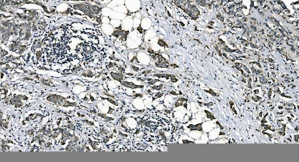
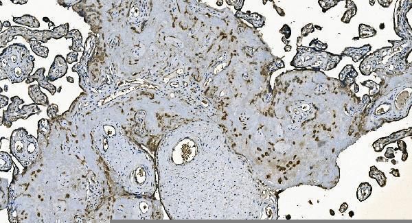
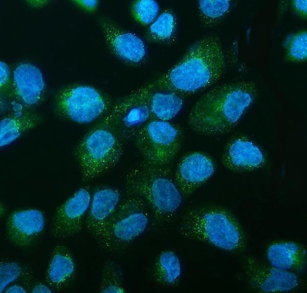
A07954-1 has real IHC data from human paraffin sections of breast cancer, placenta, prostate cancer and gastric cancer (catalog IHC captions), plus IF data from A431 cells (catalog IF caption).

Real IHC data IHC analysis of DNAJC10 using anti-DNAJC10 antibody (A07954-1). DNAJC10 was detected in paraffin-embedded section of human breast cancer tissue. Heat mediated antigen retrieval was performed in EDTA buffer (pH8.0, epitope retrieval solution). The tissue section was blocked with 10% goat serum. The tissue section was then incubated with 2μg/ml rabbit anti-DNAJC10 Antibody (A07954-1) overnight at 4°C. Biotinylated goat anti-rabbit IgG was used as secondary antibody and incubated for 30 minutes at 37°C. The tissue section was developed using Strepavidin-Biotin-Complex (SABC) (Catalog # SA1022) with DAB as the chromogen.
Anti-DNAJC10 Antibody ®
Cat # A07954-1

A07954-1 is listed for IHC and IF/ICC, with human, mouse and rat reactivity (catalog applications and reactivity). Its IHC images show human paraffin sections, while its IF image shows A431 cells (catalog IHC and IF captions).

Which to pick: Choose A07954-1 for human paraffin-section IHC: its own captions document staining at 2 μg/ml, within the listed 2–5 μg/ml IHC range (catalog IHC captions; catalog dilution). The same SKU is listed for IF/ICC at 5 μg/ml and has an IF image from A431 cells (catalog applications, dilution and IF caption). It lists mouse and rat reactivity, although the supplied images show human samples; the IHC captions do not report the fixative, and clonality is unreported (catalog reactivity, IHC and IF captions; catalog clone field).

Each figure is that product's own IHC / IF validation image from its datasheet.

References

  1. UniProt Consortium. UniProt entry Q8IXB1 (DJC10_HUMAN, Endoplasmic reticulum disulfide reductase DNAJC10).
  2. Human Protein Atlas. DNAJC10 tissue IHC expression (reliability: Approved).
  3. Human Protein Atlas. DNAJC10 subcellular location (ICC-IF): Highest expression in PODO/TERT256: 89.0 nTPM.
  4. Human Protein Atlas. DNAJC10 antibody validation summary (1 antibodies).
  5. DNAJC10 correlates with tumor immune characteristics and predicts the prognosis of glioma patients. Bioscience reports 2022 — PMC8766825.
  6. Endoplasmic reticulum-resident protein DNAJC10 inhibits glioblastoma metastasis by suppressing XBP-1s-driven EGFR transcription. Molecular biomedicine 2025 — PMC12589755.
  7. MCM8 promotes lung cancer progression through upregulating DNAJC10. Journal of cellular and molecular medicine 2024 — PMC11190951.
  8. Unveiling FLNC variants: iPSC-derived myogenic cells as a model to study disease mechanisms. Skeletal muscle 2026 — PMC13063712.
  9. PubMed PMID:12411443 — UniProt-cited evidence.
  10. PubMed PMID:14587667 — UniProt-cited evidence.
  11. PubMed PMID:12975309 — UniProt-cited evidence.