This website uses cookies to ensure you get the best experience on our website.


- Table of Contents
Plan DNPH1 chromogenic IHC in paraffin sections around the predominantly cytoplasmic tissue pattern (HPA tissue IHC). Use high-staining duodenal glandular cells and kidney proximal tubules as positive references, and score staining by cell type and compartment (HPA tissue IHC).

Expected localisation, validated protocols, controls and antibodies — the at-a-glance facts below, then the full design guide.
| Expected localisation | Cytoplasmic tissue staining; nuclear location annotated (HPA tissue IHC; UniProt) | |
| Staining pattern | Mostly cytoplasmic; high in duodenal glandular cells (HPA tissue IHC) | |
| Antigen retrieval | Tris-EDTA pH 9.0 HIER, heat-mediated (datasheet A30621) | |
| Positive control | Duodenum+4 more · see all | |
| Negative control | Adipose tissue+2 more · see all |
| Fixation | Keep fixation consistent across paraffin sections. (standard IHC practice; not target-specific) | |
| Caveat | Splenic red pulp is unstained despite reported high splenic expression (HPA tissue IHC; UniProt) | |
| Regulation | Overexpressed in some breast cancers (UniProt) | |
| Isoform / epitope | 2 isoforms; check antibody epitope coverage (UniProt) |
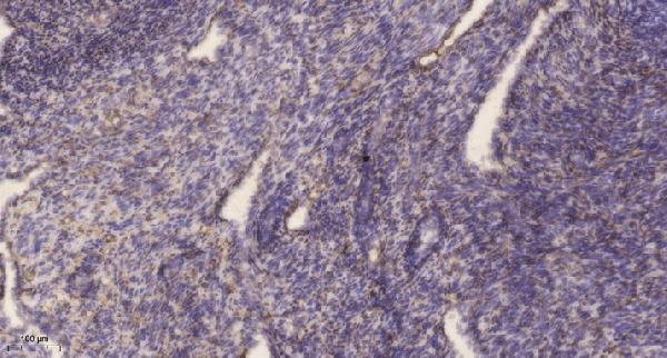
The catalog antibody has an IHC-P protocol (datasheet A30621). One published DNPH1 IHC protocol for human tissue microarrays is shown below (PMC12990376).
| Sample | Paraffin-embedded human uterus tissue; fixative not specified (datasheet A30621) |
| Fixation | Image fixative and duration unreported (datasheet A30621); verify before use. |
| Sectioning | 4–5 µm sections on charged slides (standard) |
| Deparaffinisation | Xylene, graded ethanol series to water (standard) |
| Antigen retrieval | Heat retrieval: Tris-EDTA pH 9.0 (datasheet A30621); 20 min, 95–100 °C (standard) |
| Peroxidase block | 3% H2O2, 10 min, room temperature (standard) |
| Blocking | 10% normal serum of the secondary host, 30 min, room temperature (standard) |
| Primary antibody | Rabbit anti-DNPH1, 1:50 recommended; image 1:200 (datasheet A30621) |
| Primary incubation | Overnight at 4 °C (standard) |
| Detection | HRP-polymer secondary, DAB chromogen 5–10 min (standard) |
| Counterstain | Hematoxylin, blue, dehydrate and mount (standard) |
| Expected result | DNPH1-positive staining in glandular cells of duodenum (HPA tissue IHC: High). HPA tissue profile: Cytoplasmic expression in most tissues. No signal in the no-primary control. |
In paraffin-section IHC, expect predominantly cytoplasmic DNPH1 staining in glandular cells of duodenum, gallbladder and small intestine, and in kidney proximal tubules (HPA tissue IHC: High; cytoplasmic expression in most tissues). UniProt also places DNPH1 in the nucleus and reports no transmembrane segment (UniProt O43598). HPA rates tissue IHC reliability Enhanced, while noting medium consistency between antibody staining and RNA expression (HPA tissue IHC).
| Clear cytoplasmic staining in kidney proximal-tubule cell bodies or glandular cells of duodenum, gallbladder or small intestine. | This fits the reported high-staining cell populations and the predominantly cytoplasmic tissue pattern (HPA tissue IHC: High; cytoplasmic expression in most tissues). Interpret intensity within the named cells, rather than assigning one score to the whole section (general IHC practice). |
| Predominantly nuclear staining in paraffin-section IHC, with little staining in the expected cytoplasmic compartment. | This differs from the reported tissue IHC pattern (HPA tissue IHC: cytoplasmic expression in most tissues). Nuclear DNPH1 remains plausible because UniProt lists the nucleus and HPA ICC-IF reports mainly nucleoplasmic localization (UniProt O43598; HPA subcellular). Check the IHC controls before calling it an artifact (general IHC practice). |
| Strong staining in adipocytes, chondrocytes or splenic red-pulp cells, especially when expected positive cells are weak. | Those cell populations were reported as not detected (HPA tissue IHC). Consider cross-reactivity or endogenous detection activity, then inspect the no-primary control and expected positive tissue (general IHC practice). A different specimen is not automatically a definitive DNPH1-negative control. |
| Diffuse chromogen across cells, extracellular space or the whole section, obscuring cell boundaries. | A widespread haze is difficult to reconcile with the reported cell-resolved cytoplasmic pattern (HPA tissue IHC). Review blocking, washes, detection reagents and chromogen development with controls (general IHC practice); the appearance alone cannot identify which step caused the background. |
| No convincing stain in kidney proximal tubules or high-staining glandular cells. | Absence in these reported positive populations makes the run inconclusive before it supports a biological negative call (HPA tissue IHC: High). Check tissue preservation, antibody dilution, retrieval and detection performance against the run controls (general IHC practice); no DNPH1-specific fixation sensitivity is reported here. |
| Assay-specific localization | Q: Should the IHC pattern define the IF/ICC result? A: No. Tissue IHC is predominantly cytoplasmic, whereas ICC-IF is mainly nucleoplasmic with additional cytosol signal (HPA tissue IHC; HPA subcellular). UniProt lists both cytoplasm and nucleus (UniProt O43598). Interpret the assays in their own specimens. |
| Strength of the IHC evidence | Two listed rabbit polyclonal antibodies have Enhanced IHC status (HPA antibodies: HPA029675, HPA029676). The tissue profile is also rated Enhanced, with medium consistency against RNA expression (HPA tissue IHC). Use that support for the reported pattern, while evaluating each new staining run with its controls (general IHC practice). |
| Cell-level tissue variation | High staining is reported for selected glandular cells and kidney proximal tubules; adrenal glandular and bronchial respiratory epithelial cells are Medium (HPA tissue IHC). Adipocytes and chondrocytes are Not detected (HPA tissue IHC). Compare the relevant cell population rather than treating every cell in an organ as equivalent. |
| Protein abundance versus tissue staining | UniProt describes high expression in spleen, yet HPA reports no detectable staining in splenic red-pulp cells (UniProt O43598 tissue specificity; HPA tissue IHC: Not detected). These statements concern different measurements and cell scopes; do not designate red pulp as an IHC positive solely from the UniProt tissue-level description. |
| Isoforms and antibody coverage | Two DNPH1 isoforms are listed, but the supplied records give no antibody epitope coordinates or isoform-specific staining results (UniProt O43598: isoforms 1 and 2; HPA antibodies). Do not infer that a stained or unstained cell identifies one isoform, or that both catalog antibodies detect each isoform equally. |
| Topology and processing | DNPH1 has no annotated transmembrane segment or signal peptide; its annotated chain is residues 2–174 (UniProt O43598 topology and processing). A sharply membrane-restricted pattern would therefore need scrutiny, but these annotations do not establish an epitope location or any DNPH1-specific effect of fixation or antigen retrieval. |
| Situation | Likely cause | Next action |
|---|---|---|
| Expected positive cells are blank in the test section. | The run may have lost detectable signal; the cause cannot be assigned from one negative slide (general IHC practice). | Check a section containing kidney proximal tubules or reported high-staining glandular cells (HPA tissue IHC: High). Review antibody dilution, retrieval, detection and section quality against run controls (general IHC practice). |
| Most cells show broad brown haze or deposit. | Excess background can obscure the reported cytoplasmic pattern (HPA tissue IHC; general IHC practice). | Compare with a no-primary control, then review blocking, washing and chromogen development one step at a time (general IHC practice). Reassess whether cell borders and the expected compartment can be resolved. |
| A reported negative cell population stains strongly. | Possible nonspecific binding or endogenous detection activity (general IHC practice); adipocytes, chondrocytes and splenic red-pulp cells were Not detected (HPA tissue IHC). | Examine the no-primary control and a reported positive population in the same run; verify that the signal tracks the expected cell type and compartment before scoring it (HPA tissue IHC; general IHC practice). |
| Nuclear signal dominates the IHC section. | The pattern differs from predominantly cytoplasmic tissue IHC, although nuclear localization is documented by UniProt and ICC-IF (HPA tissue IHC; UniProt O43598; HPA subcellular). | Compare positive and no-primary controls, inspect morphology and report nuclear staining separately from cytoplasmic staining (general IHC practice). Do not treat the ICC-IF pattern as an IHC acceptance criterion. |
| The spleen seems inconsistent with the expected positive tissue list. | UniProt reports high spleen expression, while HPA found no staining in splenic red-pulp cells (UniProt O43598 tissue specificity; HPA tissue IHC: Not detected). | Identify the splenic cell population being scored and use an HPA high-staining IHC population as the positive comparator (HPA tissue IHC). Avoid interpreting a red-pulp negative as assay failure by itself. |
| Two antibody runs give different compartment or cell-type patterns. | Both listed antibodies have Enhanced IHC status, but that status does not establish identical staining in a new run (HPA antibodies; general IHC practice). | Compare matched tissue areas and controls, then assess each result against the reported cytoplasmic cell pattern (HPA tissue IHC; general IHC practice). Record the disagreement rather than assigning it to an unreported isoform or fixation effect. |


Comprehensive Human Protein Atlas IHC scoring per tissue (reliability: Enhanced — Medium consistency between antibody staining and RNA expression data.). Rows are taken directly from the HPA tissue chart — click any row's HPA link to view the source.
| Tissue | Cell type | Level | Evidence | Source |
|---|---|---|---|---|
| Duodenum | Glandular cells | High | Protein (IHC) | HPA → |
| Gallbladder | Glandular cells | High | Protein (IHC) | HPA → |
| Kidney | Proximal tubules (cell body) | High | Protein (IHC) | HPA → |
| Small intestine | Glandular cells | High | Protein (IHC) | HPA → |
| Adrenal gland | Glandular cells | Medium | Protein (IHC) | HPA → |
Troubleshoot DNPH1 staining in paraffin sections by checking retrieval, cellular distribution, controls and scoring before interpreting differences between tissues.
Three DNPH1/RCL catalog antibodies list human IHC (catalog: applications/reactivity). Only A30621 has an IHC figure of paraffin-embedded human uterus (A30621 image caption); none lists IF/ICC (catalog: applications).
A30621 lists IHC for human, mouse and rat and has an IHC image of paraffin-embedded human uterus (A30621 catalog: applications/reactivity; image caption). M08676 and M08676-1 list human IHC, but neither has an IHC image caption (catalog: applications/reactivity; image fields).
Which to pick: For tissue IHC, choose A30621 when an image from paraffin-embedded human uterus is relevant; its caption reports 1:200 primary antibody overnight at 4 °C and Tris-EDTA retrieval at pH 9.0, but does not report the fixative (A30621 image caption). For cross-species work, A30621 lists human, mouse and rat reactivity, while the monoclonals M08676 (clone 27D38) and M08676-1 list human reactivity only (catalog: reactivity/clone). No listed SKU has IF/ICC validation, so there is no catalog-supported IF/ICC pick (catalog: applications; IF image fields).