This website uses cookies to ensure you get the best experience on our website.


- Table of Contents
Plan DOCK3 chromogenic IHC in paraffin sections using the IHC-validated antibody and cerebral cortex neuronal cells as a positive reference (HPA tissue IHC). The guide covers fixation consistency, staining controls and interpretation of the expected cytoplasmic pattern (HPA tissue IHC).

Expected localisation, validated protocols, controls and antibodies — the at-a-glance facts below, then the full design guide.
| Expected localisation | Cytoplasmic staining in CNS (HPA tissue IHC) | |
| Staining pattern | Cytoplasmic staining in CNS neuronal cells (HPA tissue IHC) | |
| Antigen retrieval | EDTA pH 8.0 HIER, heat-mediated (datasheet A08904) | |
| Positive control | Cerebral cortex+4 more · see all | |
| Negative control | Adrenal gland+4 more · see all |
| Fixation | Keep fixation consistent across sections (standard IHC practice; not target-specific). Selected-image fixative and duration unreported (datasheet A08904); verify before use. | |
| Caveat | Staining has medium concordance with RNA data (HPA tissue IHC) | |
| Regulation | Expression regulation unreported (UniProt) | |
| Isoform / epitope | No listed isoforms or processing; epitope site unspecified (UniProt) |
The catalog antibody has a paraffin-section IHC protocol (datasheet A08904). One published Dock3 IHC protocol uses frozen optic-nerve sections (PMC4454328).
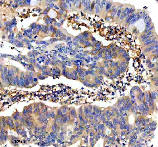
| Sample | Paraffin-embedded human colon cancer tissue; fixative not specified (datasheet A08904) |
| Fixation | Image fixative and duration unreported (datasheet A08904); verify before use. |
| Sectioning | 4–5 µm sections on charged slides (standard) |
| Deparaffinisation | Xylene, graded ethanol series to water (standard) |
| Antigen retrieval | Heat retrieval: EDTA pH 8.0 (datasheet A08904); 20 min, 95–100 °C (standard) |
| Peroxidase block | 3% H2O2, 10 min, room temperature (standard) |
| Blocking | 10% goat serum (datasheet A08904) |
| Primary antibody | Rabbit anti-DOCK3, 2-5 μg/ml (datasheet A08904) |
| Primary incubation | Overnight at 4 °C (datasheet A08904) |
| Detection | HRP-conjugated secondary, DAB chromogen (datasheet A08904) |
| Counterstain | Hematoxylin, blue, dehydrate and mount (standard) |
| Expected result | DOCK3-positive staining in neuronal cells of cerebral cortex (HPA tissue IHC: High). HPA tissue profile: Cytoplasmic expression in CNS. No signal in the no-primary control. |
DOCK3 is primarily cytoplasmic and has no transmembrane segment (UniProt Q8IZD9). In paraffin sections, expect the clearest staining in cerebral cortex neuronal cells, with weaker or variable staining elsewhere (HPA: High in cerebral cortex neuronal cells; tissue IHC reliability Enhanced). HPA describes cytoplasmic expression in the CNS, but its tissue profile has medium consistency with RNA data and awaits external verification (HPA: tissue IHC profile and reliability description).
| Neuronal cytoplasm stains strongly in cerebral cortex; nearby cells and structures vary. | This fits the strongest listed tissue result (HPA: High in cerebral cortex neuronal cells). Neuropil staining can also fit normal brain distribution (UniProt Q8IZD9: normal brain tissue specificity). Judge the cell type and compartment together; a brown signal alone does not identify DOCK3 (general IHC practice). |
| Predominantly nuclear or sharply outlined membrane staining replaces the expected cytoplasmic pattern. | A dominant nuclear pattern conflicts with the reported cytoplasmic location (UniProt Q8IZD9; HPA: cytoplasmic expression in CNS). DOCK3 can become membrane associated through PSEN1 interaction, so limited membrane proximity is plausible; a dominant membrane outline needs control-based review (UniProt Q8IZD9: PSEN1 interaction; general IHC practice). |
| Strong staining appears in a cell population listed as undetected by HPA. | For example, strong adrenal glandular-cell staining conflicts with that listed result (HPA: Not detected in adrenal glandular cells). Consider nonspecific antibody binding or endogenous chromogenic activity, then compare a no-primary control (general IHC practice). Do not treat every non-neuronal cell as suspect: several non-neuronal populations have Medium staining (HPA: skin, stomach, testis). |
| Pale brown color spreads across cells and tissue without a discernible cytoplasmic pattern. | Diffuse color is difficult to score as DOCK3 because the expected pattern is compartmental (HPA: cytoplasmic expression in CNS; UniProt Q8IZD9: cytoplasm). Check the no-primary control and detection reagents, then optimize blocking, washing and antibody concentration as general IHC practice; no DOCK3-specific cause follows from this appearance alone. |
| Cerebral cortex neuronal cells show no convincing signal. | This is unexpected relative to the strongest listed tissue result (HPA: High in cerebral cortex neuronal cells). First verify tissue identity and the presence of evaluable neurons; then check the IHC antibody, retrieval and detection workflow (general IHC practice). HPA's Enhanced rating still has medium RNA–staining consistency and pending external verification (HPA: reliability description). |
| Tissue and cell selection | Cerebral cortex neuronal cells offer the strongest listed IHC signal; caudate neurons are Medium and hippocampal neurons Low (HPA: tissue IHC). A weak hippocampal result alone is less informative than a failed cerebral cortex control. HPA also lists Medium staining in skin keratinocytes, stomach glandular cells and testis Leydig cells (HPA: tissue IHC). |
| Compartment and membrane association | DOCK3 is cytoplasmic and lacks a transmembrane segment (UniProt Q8IZD9). Its PSEN1 interaction can mediate membrane association, so some signal near a membrane can be biologically plausible (UniProt Q8IZD9: subunit and subcellular location). This does not establish that a dominant membrane outline is the expected paraffin-section pattern. |
| Antibody evidence | HPA037543 has Enhanced IHC validation, meaning its pattern was reproduced by independent antibodies or orthogonal data (HPA: antibody validation summary). HPA037544 has an Enhanced ICC rating but no supplied IHC rating (HPA: antibody validation summary). Interpret either rating alongside the tissue profile's medium RNA–staining consistency and pending external verification (HPA: reliability description). |
| Processing and epitope limits | UniProt reports one 1–2030 chain, no signal peptide, no propeptide and no listed isoforms (UniProt Q8IZD9). Those annotations do not identify the antibody epitope or establish an antigen-retrieval condition. No DOCK3-specific fixation sensitivity is supplied; select and document retrieval using the IHC antibody's validated procedure (general IHC practice). |
| IF/ICC question: what pattern is expected? | Predominantly cytosolic fluorescence is the HPA ICC-IF result, with an Enhanced cytosol location (HPA: subcellular summary). HPA lists ICC-IF images in A-431, U-251MG and U2OS, and Enhanced ICC ratings for HPA037543 and HPA037544 (HPA: subcellular and antibody records). Those ICC observations do not establish an IHC retrieval or chromogenic protocol. |
| Situation | Likely cause | Next action |
|---|---|---|
| Cerebral cortex has no neuronal staining. | The strongest listed positive population is missing (HPA: High in cerebral cortex neuronal cells); the specific technical cause cannot be inferred from the image alone. | Confirm evaluable neurons and tissue identity, then check primary-antibody use, retrieval and chromogenic detection against the IHC procedure; include a working positive control (general IHC practice). |
| Nuclei stain more strongly than cytoplasm. | Dominant nuclear staining conflicts with the reported cytoplasmic location (UniProt Q8IZD9; HPA: CNS cytoplasmic profile). | Compare with a no-primary control and inspect counterstain and chromogen separately; reassess antibody specificity before scoring nuclear signal as DOCK3 (general IHC practice). |
| Strong signal appears in an HPA-undetected population. | An unexpected cell pattern may reflect nonspecific binding or endogenous detection activity (general IHC practice); HPA lists adrenal glandular cells as Not detected (HPA: tissue IHC). | Run a no-primary control, check detection chemistry and blocking, and compare the suspect cells with cerebral cortex neurons on the same staining run (general IHC practice). |
| Brown haze obscures cell boundaries. | Diffuse background can arise from the staining workflow; its appearance alone does not identify a DOCK3-specific mechanism (general IHC practice). | Inspect the no-primary control, improve washing or blocking if indicated, and titrate the primary antibody within its validated IHC procedure (general IHC practice). |
| Hippocampus is weak while cerebral cortex stains clearly. | The difference matches the listed levels: Low in hippocampal neurons and High in cerebral cortex neuronal cells (HPA: tissue IHC). | Score each tissue against its own expected level and compartment; retain cerebral cortex as the stronger positive comparator (HPA: tissue IHC; general IHC practice). |
| A strong membrane rim is scored as positive. | PSEN1-mediated membrane association is possible, but DOCK3 is primarily cytoplasmic and has no transmembrane segment (UniProt Q8IZD9). | Review the cytoplasmic signal and control sections before accepting the rim; record membrane enrichment separately from the expected cytoplasmic pattern (UniProt Q8IZD9; general IHC practice). |


Comprehensive Human Protein Atlas IHC scoring per tissue (reliability: Enhanced — Medium consistency between antibody staining and RNA expression data. Pending external verification.). Rows are taken directly from the HPA tissue chart — click any row's HPA link to view the source.
| Tissue | Cell type | Level | Evidence | Source |
|---|---|---|---|---|
| Cerebral cortex | Neuronal cells | High | Protein (IHC) | HPA → |
| Caudate | Neuronal cells | Medium | Protein (IHC) | HPA → |
| Skin | Keratinocytes | Medium | Protein (IHC) | HPA → |
| Stomach | Glandular cells | Medium | Protein (IHC) | HPA → |
| Testis | Leydig cells | Medium | Protein (IHC) | HPA → |
| Tissue | Cell type | Level | Evidence | Source |
|---|---|---|---|---|
| Adrenal gland | Glandular cells | Not detected | Protein (IHC) | HPA → |
| Appendix | Endocrine cells | Not detected | Protein (IHC) | HPA → |
| Bone marrow | Hematopoietic cells | Not detected | Protein (IHC) | HPA → |
| Breast | Adipocytes | Not detected | Protein (IHC) | HPA → |
| Bronchus | Respiratory epithelial cells | Not detected | Protein (IHC) | HPA → |
Troubleshoot DOCK3 staining in paraffin sections using the catalog antibody’s tissue example, expected cytoplasmic pattern, and appropriate controls.
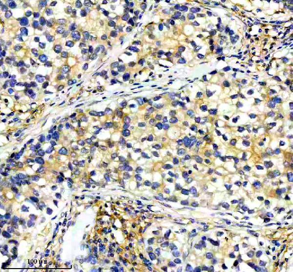
A08904 has chromogenic IHC images from paraffin-embedded human colon and lung cancer sections (catalog IHC captions). No IF/ICC images are supplied (catalog images).
A08904 is listed for IHC and shows staining in paraffin-embedded human colon and lung cancer sections (catalog applications; catalog IHC captions). Its listed reactivity is Human, Mouse and Rat, but the supplied IHC images show human tissue only (catalog reactivity; catalog IHC captions).
Which to pick: Choose A08904 for tissue IHC: it is a rabbit antibody with human paraffin-section IHC images and a recommended concentration of 2–5 μg/ml (catalog host; catalog IHC captions; datasheet: 2–5 μg/ml). The captions do not report a fixative (catalog IHC captions). There is no IF/ICC recommendation because A08904 has no listed IF/ICC application or image; Mouse and Rat are listed as reactive species, but their tissue IHC performance is not shown here (catalog applications; catalog images; catalog reactivity).