This website uses cookies to ensure you get the best experience on our website.


- Table of Contents
Plan DOK7 IHC in paraffin sections using heart muscle as a strong positive tissue (HPA tissue IHC). This guide covers staining patterns, tissue controls, fixation consistency and chromogenic detection with the IHC-validated antibody (datasheet A05165-1).

Expected localisation, validated protocols, controls and antibodies — the at-a-glance facts below, then the full design guide.
| Expected localisation | Cytoplasmic staining in most tissues (HPA tissue IHC) | |
| Staining pattern | Strong cytoplasmic staining in cardiomyocytes (HPA tissue IHC) | |
| Antigen retrieval | EDTA pH 8.0 HIER, heat-mediated (datasheet A05165-1) | |
| Positive control | Heart muscle+4 more · see all | |
| Negative control | Adipose tissue+4 more · see all |
| Fixation | Keep fixation consistent across sections (standard IHC practice; not target-specific) | |
| Caveat | Presumed off-target binding; restaining pending (HPA tissue IHC) | |
| Regulation | Expression enriched in heart and skeletal muscle (UniProt) | |
| Isoform / epitope | Four isoforms; check epitope coverage across variants (UniProt) |
The catalog antibody’s IHC-P protocol is supplemented by one published DOK7 staining protocol using mouse tumor sections (PMC10756110).
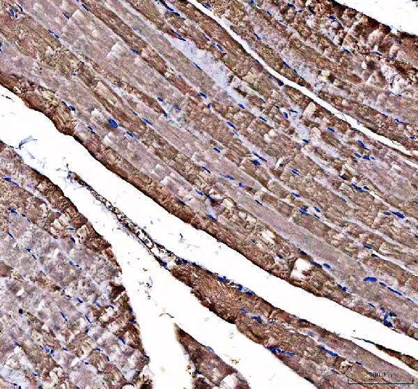
| Sample | Paraffin-embedded human skeletal muscle tissue; fixative not specified (datasheet A05165-1) |
| Fixation | Image fixative and duration unreported (datasheet A05165-1); verify before use. |
| Sectioning | 4–5 µm sections on charged slides (standard) |
| Deparaffinisation | Xylene, graded ethanol series to water (standard) |
| Antigen retrieval | Heat retrieval: EDTA pH 8.0 (datasheet A05165-1); 20 min, 95–100 °C (standard) |
| Peroxidase block | 3% H2O2, 10 min, room temperature (standard) |
| Blocking | 10% goat serum (datasheet A05165-1) |
| Primary antibody | Rabbit anti-DOK7, 2-5 μg/ml (datasheet A05165-1) |
| Primary incubation | Overnight at 4 °C (datasheet A05165-1) |
| Detection | HRP-conjugated secondary, DAB chromogen (datasheet A05165-1) |
| Counterstain | Hematoxylin, blue, dehydrate and mount (standard) |
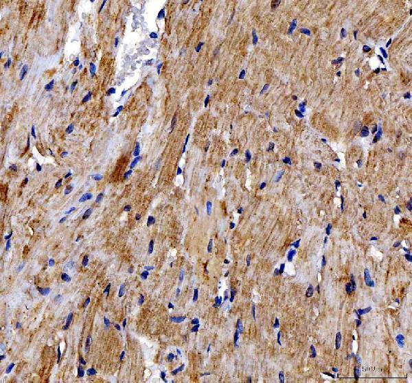
| Expected result | DOK7-positive staining in cardiomyocytes of heart muscle (HPA tissue IHC: High). HPA tissue profile: Cytoplasmic expression in most tissues. No signal in the no-primary control. |
DOK7 should give predominantly cytoplasmic staining in tissue sections, with the clearest supplied positive example in heart cardiomyocytes (HPA: cytoplasmic expression in most tissues; High in cardiomyocytes). Enrichment near neuromuscular junctions is biologically plausible, but is not an established routine IHC pattern here (UniProt Q18PE1: accumulates at neuromuscular junctions). DOK7 has no transmembrane segment (UniProt Q18PE1: topology). HPA rates its tissue IHC profile Approved while noting presumed off target binding and pending restaining (HPA: tissue IHC reliability).
| Cytoplasmic chromogen is prominent in heart cardiomyocytes, with interpretable cellular boundaries and little staining in the negative control. | This matches the strongest listed tissue result: High cardiomyocyte staining (HPA: heart muscle IHC). Interpret the compartment as tissue cytoplasm; neuromuscular junction accumulation is a separate UniProt annotation, not proof that ordinary heart sections must show a junctional pattern (UniProt Q18PE1: subcellular location). |
| Staining is confined to nuclei, or forms a uniform surface rim, in otherwise readable heart sections. | Neither is the reported dominant tissue IHC pattern (HPA: cytoplasmic expression in most tissues). Review morphology and controls before calling it DOK7. A nuclear signal alone is not conclusive evidence of artefact because the separate ICC-IF dataset reports enhanced nucleoplasmic localization (HPA: subcellular ICC-IF). |
| Alveolar cells or adipocytes stain strongly while heart cardiomyocytes show little or no signal. | This reverses the supplied tissue comparison (HPA: alveolar cells and adipocytes Not detected; cardiomyocytes High). Consider cross-reactivity, nonspecific detection, or section handling; the unexpected stain cannot be assigned to DOK7 from morphology alone, especially while the tissue profile awaits restaining (HPA: reliability description). |
| A diffuse brown haze covers several structures without a clear cellular compartment. | The result cannot be scored against the reported cytoplasmic pattern (HPA: tissue IHC profile). In chromogenic IHC, nonspecific antibody binding or endogenous detection activity can create background (standard IHC practice). Compare the matched detection-only control and inspect whether chromogen follows cells rather than section-wide deposits. |
| Heart cardiomyocytes remain unstained although tissue architecture and counterstain are intact. | This is a failed positive control for the listed IHC pattern, not evidence that DOK7 is absent from all heart tissue (HPA: cardiomyocytes High; UniProt Q18PE1: heart protein expression). Check the catalog antibody's IHC-P instructions and the staining run before interpreting weaker tissues. |
| Tissue and cell choice | Heart cardiomyocytes provide the strongest listed positive comparison; adipocytes and lung alveolar cells are listed as Not detected (HPA: tissue IHC). Heart and skeletal muscle are preferential expression sites in the protein record, but the supplied HPA list gives no skeletal-muscle IHC staining level (UniProt Q18PE1: tissue specificity). |
| Antibody validation and reliability | HPA059449 is Approved for IHC, whereas its Enhanced designation applies to ICC; HPA062780 has no listed IHC status (HPA: antibody validation). The tissue profile is Approved but notes presumed off target binding and pending restaining, so unexpected cells need independent control-based assessment (HPA: reliability description). |
| Compartment and topology | Tissue IHC is described as cytoplasmic (HPA: tissue IHC profile). UniProt places DOK7 at the cell membrane and synapse, with neuromuscular junction accumulation and no transmembrane segment (UniProt Q18PE1: location and topology). These annotations do not justify demanding a continuous membrane rim in a paraffin section. |
| Isoforms and epitope coverage | UniProt lists 4 isoforms (UniProt Q18PE1: isoforms). An antibody could recognize a region shared by some isoforms or restricted to one; no epitope map is supplied here. Therefore, isoform-specific staining or absence cannot be inferred from this record, and any such claim requires antibody documentation. |
| Retrieval and processing limits | No target-specific antigen-retrieval condition or fixation sensitivity is provided (supplied UniProt and HPA records). UniProt lists no signal peptide, propeptide, glycosylation sites, or modified residues for this record (UniProt Q18PE1: processing and modifications); these facts do not predict paraffin-section retrieval performance. |
| Situation | Likely cause | Next action |
|---|---|---|
| Heart positive control has no specific stain. | The result conflicts with the listed High cardiomyocyte signal (HPA: heart muscle IHC); an ineffective staining run is possible (standard IHC practice). | Confirm tissue identity and cardiomyocyte preservation, then follow the catalog antibody's IHC-P instructions for retrieval, dilution, and detection; include a run control. No DOK7-specific retrieval setting is supplied. |
| Diffuse chromogen obscures cytoplasmic detail. | Background can arise from nonspecific binding or endogenous enzyme activity in chromogenic IHC (standard IHC practice). HPA's cytoplasmic profile cannot be evaluated through a section-wide haze (HPA: tissue IHC profile). | Compare detection-only and other appropriate negative controls; review blocking, reagent dilution, washes, and endogenous activity blocking as relevant to the detection system (standard IHC practice). |
| Strong staining appears in adipocytes or alveolar cells. | Both are listed as Not detected, so strong signal there is discordant and may reflect nonspecific staining (HPA: adipose tissue and lung IHC). The HPA tissue profile also notes presumed off target binding (HPA: reliability description). | Check cell identity and negative controls, then compare with a heart positive section in the same run. Report the discordance rather than assigning the unexpected cells as DOK7-positive. |
| Only nuclei or a sharp cell-surface rim stain in heart. | The dominant reported tissue pattern is cytoplasmic (HPA: tissue IHC profile). A nuclear ICC-IF location is reported separately and does not validate nuclear-only paraffin IHC (HPA: subcellular ICC-IF). | Recheck cellular morphology, counterstain, and control sections; assess whether the signal is reproducible with an independently validated approach before treating the compartment as target-specific. |
| Heart stains, but a second tissue gives weak or mixed signal. | The supplied tissue levels differ by cell type: heart cardiomyocytes are High, several glandular or neural cell types Medium, and liver hepatocytes Low (HPA: tissue IHC). A whole-section average can hide that distribution. | Score the named cell population and compartment within each tissue, using the same run controls. Describe weak or mixed staining at cell level rather than extrapolating the heart intensity to every tissue. |
| IF/ICC Q&A: why does a nuclear signal differ from the IHC expectation? | HPA reports enhanced nucleoplasmic and supported mitochondrial localization in ICC-IF, while tissue IHC is described as cytoplasmic (HPA: subcellular ICC-IF; tissue IHC profile). The datasets use different applications. | Interpret each result within its application and validation evidence. Do not use the ICC-IF location alone to reclassify a nuclear-only paraffin IHC result; consult the separate IF/ICC guide for that application. |


Comprehensive Human Protein Atlas IHC scoring per tissue (reliability: Approved — Presumed off target binding observed and disregarded. Pending restaining.). Rows are taken directly from the HPA tissue chart — click any row's HPA link to view the source.
| Tissue | Cell type | Level | Evidence | Source |
|---|---|---|---|---|
| Heart muscle | Cardiomyocytes | High | Protein (IHC) | HPA → |
| Adrenal gland | Glandular cells | Medium | Protein (IHC) | HPA → |
| Appendix | Glandular cells | Medium | Protein (IHC) | HPA → |
| Breast | Glandular cells | Medium | Protein (IHC) | HPA → |
| Bronchus | Respiratory epithelial cells | Medium | Protein (IHC) | HPA → |
| Tissue | Cell type | Level | Evidence | Source |
|---|---|---|---|---|
| Adipose tissue | Adipocytes | Not detected | Protein (IHC) | HPA → |
| Bone marrow | Hematopoietic cells | Not detected | Protein (IHC) | HPA → |
| Epididymis | Glandular cells | Not detected | Protein (IHC) | HPA → |
| Lung | Alveolar cells | Not detected | Protein (IHC) | HPA → |
| Lymph node | Germinal center cells | Not detected | Protein (IHC) | HPA → |
Use the catalog antibody’s paraffin-section IHC conditions as the starting point, then interpret DOK7 staining against muscle controls and compartment-specific evidence.
A05165-1 has real paraffin-section IHC images in human skeletal muscle and rat heart (catalog IHC captions). No IF image is supplied (catalog IF image alts).
A05165-1 has IHC images of paraffin-embedded human skeletal muscle and rat heart (catalog IHC captions). It lists IHC and human, mouse and rat reactivity (catalog applications; reactivity); IF/ICC validation is not listed (catalog applications; IF image alts).
Which to pick: For tissue IHC, choose A05165-1, a rabbit polyclonal listed for IHC (catalog host; dilution_raw; applications); its paraffin-section images used EDTA pH 8 retrieval and 2 μg/ml primary antibody, and the fixative is unreported (catalog IHC captions). For IF/ICC, A05165-1 has no listed IF/ICC application or IF image (catalog applications; IF image alts), so it requires separate validation. For cross-species IHC, A05165-1 lists human, mouse and rat reactivity (catalog reactivity), with paraffin-section IHC images for human and rat (catalog IHC captions).