Click image to see more details
-

-
-
-
-
+1
Product Info Summary
| SKU: | A05165-1 |
|---|---|
| Size: | 100 μg/vial |
| Reactive Species: | Human, Mouse, Rat |
| Host: | Rabbit |
| Application: | Flow Cytometry, IHC, WB |
Customers Who Bought This Also Bought

Product info
Product Name
Anti-DOK7 Antibody Picoband®
SKU/Catalog Number
A05165-1
Size
100 μg/vial
Form
Lyophilized
Description
Boster Bio Anti-DOK7 Antibody Picoband® catalog # A05165-1. Tested in Flow Cytometry, IHC, WB applications. This antibody reacts with Human, Mouse, Rat. The brand Picoband indicates this is a premium antibody that guarantees superior quality, high affinity, and strong signals with minimal background in Western blot applications. Only our best-performing antibodies are designated as Picoband, ensuring unmatched performance.
Storage & Handling
Store at -20˚C for one year from date of receipt. After reconstitution, at 4˚C for one month. It can also be aliquotted and stored frozen at -20˚C for six months. Avoid repeated freeze-thaw cycles.
Cite This Product
Anti-DOK7 Antibody Picoband® (Boster Biological Technology, Pleasanton CA, USA, Catalog # A05165-1)
Host
Rabbit
Contents
Each vial contains 4 mg Trehalose, 0.9 mg NaCl and 0.2 mg Na2HPO4.
Clonality
Polyclonal
Isotype
Rabbit IgG
Immunogen
A synthetic peptide corresponding to a sequence in the middle region of human DOK7, which shares 86.2% amino acid (aa) sequence identity with mouse DOK7.
Cross-reactivity
No cross-reactivity with other proteins.
Reactive Species
A05165-1 is reactive to DOK7 in Human, Mouse, Rat
Observed Molecular Weight
53 kDa
Calculated molecular weight
53.1 kDa
Background of DOK7
Dok-7 is a non-catalytic cytoplasmic adaptor protein that is expressed specifically in muscle and is essential for the formation of neuromuscular synapses. Further, Dok-7 contains pleckstrin homology (PH) and phosphotyrosine-binding (PTB) domains that are critical for Dok-7 function. It is mapped to 4p16.3. The protein encoded by this gene is essential for neuromuscular synaptogenesis. The protein functions in aneural activation of muscle-specific receptor kinase, which is required for postsynaptic differentiation, and in the subsequent clustering of the acetylcholine receptor in myotubes. This protein can also induce autophosphorylation of muscle-specific receptor kinase. Mutations in this gene are a cause of familial limb-girdle myasthenia autosomal recessive, which is also known as congenital myasthenic syndrome type 1B. Alternative splicing results in multiple transcript variants.
Antibody Validation
Boster validates all antibodies on WB, IHC, ICC, Immunofluorescence, and ELISA with known positive control and negative samples to ensure specificity and high affinity, including thorough antibody incubations.
Application & Images
Applications
A05165-1 is guaranteed for Flow Cytometry, IHC, WB Boster Guarantee
Assay Dilutions Recommendation
The recommendations below provide a starting point for assay optimization. The actual working concentration varies and should be decided by the user.
Western blot, 0.1-0.5μg/ml
Immunohistochemistry (Paraffin-embedded Section), 2-5μg/ml
Flow Cytometry(Fixed), 1-3 μg/1x106 cells
Positive Control
WB: human MCF-7 whole cell, human RT4 whole cell, rat skeletal muscle tissue lysates, rat brain tissue lysates, rat heart tissue lysates, rat L6 whole cell lysates, mouse skeletal muscle tissue lysates, mouse brain tissue lysates, mouse heart tissue lysates, mouse C2C12 whole cell lysates
IHC: human skeletal muscle, rat heart
FCM: RT4 cell
Validation Images & Assay Conditions
Click image to see more details
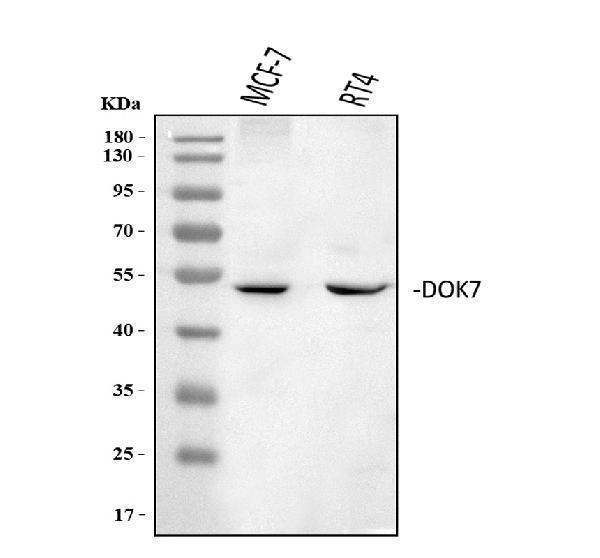
Western blot analysis of DOK7 using anti-DOK7 antibody (A05165-1).
Electrophoresis was performed on a 5-20% SDS-PAGE gel at 70V (Stacking gel) / 90V (Resolving gel) for 2-3 hours. The sample well of each lane was loaded with 30 ug of sample under reducing conditions.
Lane 1: human MCF-7 whole cell lysates,
Lane 2: human RT4 whole cell lysates.
After electrophoresis, proteins were transferred to a nitrocellulose membrane at 150 mA for 50-90 minutes. Blocked the membrane with 5% non-fat milk/TBS for 1.5 hour at RT. The membrane was incubated with rabbit anti-DOK7 antigen affinity purified polyclonal antibody (Catalog # A05165-1) at 0.5 μg/mL overnight at 4°C, then washed with TBS-0.1%Tween 3 times with 5 minutes each and probed with a goat anti-rabbit IgG-HRP secondary antibody at a dilution of 1:5000 for 1.5 hour at RT. The signal is developed using an Enhanced Chemiluminescent detection (ECL) kit (Catalog # EK1002) with Tanon 5200 system. A specific band was detected for DOK7 at approximately 53 kDa. The expected band size for DOK7 is at 53 kDa.

Click image to see more details
Western blot analysis of DOK7 using anti-DOK7 antibody (A05165-1).
Electrophoresis was performed on a 5-20% SDS-PAGE gel at 70V (Stacking gel) / 90V (Resolving gel) for 2-3 hours. The sample well of each lane was loaded with 30 ug of sample under reducing conditions.
Lane 1: rat skeletal muscle tissue lysates,
Lane 2: rat brain tissue lysates,
Lane 3: rat heart tissue lysates,
Lane 4: rat L6 whole cell lysates,
Lane 5: mouse skeletal muscle tissue lysates,
Lane 6: mouse brain tissue lysates,
Lane 7: mouse heart tissue lysates,
Lane 8: mouse C2C12 whole cell lysates.
After electrophoresis, proteins were transferred to a nitrocellulose membrane at 150 mA for 50-90 minutes. Blocked the membrane with 5% non-fat milk/TBS for 1.5 hour at RT. The membrane was incubated with rabbit anti-DOK7 antigen affinity purified polyclonal antibody (Catalog # A05165-1) at 0.5 μg/mL overnight at 4°C, then washed with TBS-0.1%Tween 3 times with 5 minutes each and probed with a goat anti-rabbit IgG-HRP secondary antibody at a dilution of 1:5000 for 1.5 hour at RT. The signal is developed using an Enhanced Chemiluminescent detection (ECL) kit (Catalog # EK1002) with Tanon 5200 system. A specific band was detected for DOK7 at approximately 53 kDa. The expected band size for DOK7 is at 53 kDa.

Click image to see more details
IHC analysis of DOK7 using anti-DOK7 antibody (A05165-1).
DOK7 was detected in a paraffin-embedded section of human skeletal muscle tissue. Heat mediated antigen retrieval was performed in EDTA buffer (pH 8.0, epitope retrieval solution). The tissue section was blocked with 10% goat serum. The tissue section was then incubated with 2 μg/ml rabbit anti-DOK7 Antibody (A05165-1) overnight at 4°C. Peroxidase Conjugated Goat Anti-rabbit IgG was used as secondary antibody and incubated for 30 minutes at 37°C. The tissue section was developed using HRP Conjugated Rabbit IgG Super Vision Assay Kit (Catalog # SV0002) with DAB as the chromogen.

Click image to see more details
IHC analysis of DOK7 using anti-DOK7 antibody (A05165-1).
DOK7 was detected in a paraffin-embedded section of rat heart tissue. Heat mediated antigen retrieval was performed in EDTA buffer (pH 8.0, epitope retrieval solution). The tissue section was blocked with 10% goat serum. The tissue section was then incubated with 2 μg/ml rabbit anti-DOK7 Antibody (A05165-1) overnight at 4°C. Peroxidase Conjugated Goat Anti-rabbit IgG was used as secondary antibody and incubated for 30 minutes at 37°C. The tissue section was developed using HRP Conjugated Rabbit IgG Super Vision Assay Kit (Catalog # SV0002) with DAB as the chromogen.

Click image to see more details
Flow Cytometry analysis of RT4 cells using anti-DOK7 antibody (A05165-1).
Overlay histogram showing RT4 cells stained with A05165-1 (Blue line). The cells were fixed with 4% paraformaldehyde and blocked with 10% normal goat serum. And then incubated with rabbit anti-DOK7 Antibody (A05165-1, 1 μg/1x106 cells) for 30 min at 20°C. DyLight®488 conjugated goat anti-rabbit IgG (BA1127, 5-10 μg/1x106 cells) was used as secondary antibody for 30 minutes at 20°C. Isotype control antibody (Green line) was rabbit IgG (1 μg/1x106) used under the same conditions. Unlabelled sample without incubation with primary antibody and secondary antibody (Red line) was used as a blank control.
Specific Publications For Anti-DOK7 Antibody Picoband® (A05165-1)
Loading publications
Recommended Resources
Here are featured tools and databases that you might find useful.
- Boster's Pathways Library
- Protein Databases
- Bioscience Research Protocol Resources
- Data Processing & Analysis Software
- Photo Editing Software
- Scientific Literature Resources
- Research Paper Management Tools
- Molecular Biology Software
- Primer Design Tools
- Bioinformatics Tools
- Phylogenetic Tree Analysis
Customer Reviews
Have you used Anti-DOK7 Antibody Picoband®?
Share your experimental results or join a short interview to earn up to $1,000 in product credits or other rewards.
0 Reviews For Anti-DOK7 Antibody Picoband®
Customer Q&As
Have a question?
Find answers in Q&As, reviews.
Can't find your answer?
Submit your question
3 Customer Q&As for Anti-DOK7 Antibody Picoband®
Question
We are currently using anti-DOK7 antibody A05165-1 for mouse tissue, and we are happy with the ICC results. The species of reactivity given in the datasheet says human, mouse, rat. Is it likely that the antibody can work on pig tissues as well?
Verified Customer
Verified customer
Asked: 2019-08-09
Answer
The anti-DOK7 antibody (A05165-1) has not been validated for cross reactivity specifically with pig tissues, but there is a good chance of cross reactivity. We have an innovator award program that if you test this antibody and show it works in pig you can get your next antibody for free. Please contact me if I can help you with anything.
Boster Scientific Support
Answered: 2019-08-09
Question
Is this A05165-1 anti-DOK7 antibody reactive to the isotypes of DOK7?
B. Miller
Verified customer
Asked: 2018-05-25
Answer
The immunogen of A05165-1 anti-DOK7 antibody is A synthetic peptide corresponding to a sequence of human DOK7(STVEERVAQEALETLQLEKRLSLLSHAGR). Could you tell me which isotype you are interested in so I can help see if the immunogen is part of this isotype?
Boster Scientific Support
Answered: 2018-05-25
Question
Does A05165-1 anti-DOK7 antibody work on parafin embedded sections? If so, which fixation method do you recommend we use (PFA, paraformaldehyde, other)?
J. Anderson
Verified customer
Asked: 2013-06-14
Answer
As indicated on the product datasheet, A05165-1 anti-DOK7 antibody as been validated on IHC-P. It is best to use PFA for fixation because it has better tissue penetration ability. PFA needs to be prepared fresh before use. Long term stored PFA turns into formalin, as the PFA molecules congregate and become formalin.
Boster Scientific Support
Answered: 2013-06-14
