This website uses cookies to ensure you get the best experience on our website.
- Table of Contents
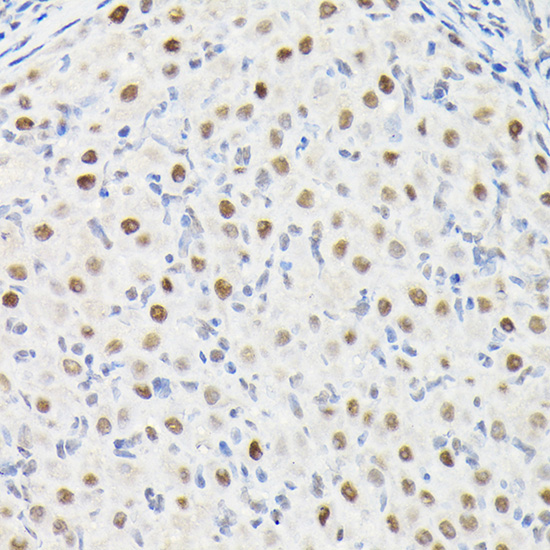
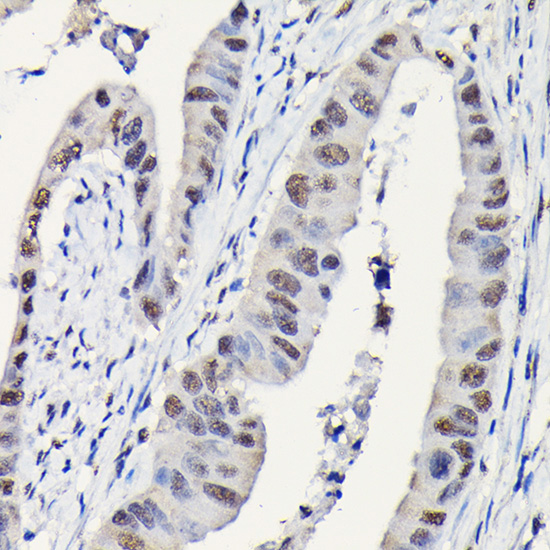
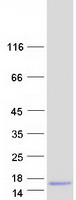
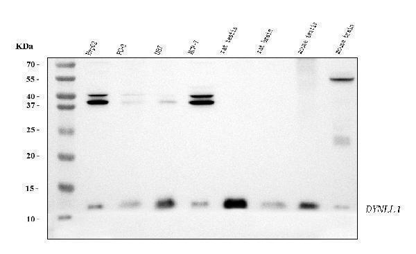
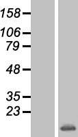
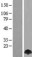
Facts about Dynein light chain 1, cytoplasmic.
May play a role in changing or maintaining the spatial distribution of cytoskeletal structures. .
Human | |
---|---|
Gene Name: | DYNLL1 |
Uniprot: | P63167 |
Entrez: | 8655 |
Belongs to: |
---|
dynein light chain family |
cytoplasmic dynein light polypeptide; DLC1; DLC1DNCLC1; DLC8; DLC8MGC126137; DNCL1; DNCL1MGC126138; DNCLC1; dynein light chain 1, cytoplasmic; Dynein light chain LC8-type 1; dynein, cytoplasmic, light polypeptide 1; dynein, light chain, LC8-type 1,8 kDa dynein light chain; DYNLL1; hdlc1; LC8; LC8a; PIN; PINLC8a; Protein inhibitor of neuronal nitric oxide synthase
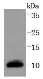
Mass (kDA):
10.366 kDA
Human | |
---|---|
Location: | 12q24.31 |
Sequence: | 12; NC_000012.12 (120469842..120498493) |
Ubiquitous.
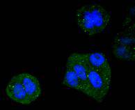
Cytoplasm, cytoskeleton, microtubule organizing center, centrosome. Cytoplasm, cytoskeleton. Nucleus. Mitochondrion. Upon induction of apoptosis translocates together with BCL2L11 to mitochondria.
The DYNLL1 marker is a versatile probe for gene expression that detects the presence endothelial cell. The marker's sensitivity and specificity make it an excellent choice for diagnostic applications in cancer and other human diseases. Endothelial cells express a high amount of the DYNLL1 genes. This gene can be used for identification of many pathogenic microorganisms.
The DYNLL1 genetic variant is a common occurrence and is associated with schizophrenia, bipolar disorder, certain physiological parameters, and other disorders. It is located in human DNA's 12q22-24 region. It has an estimated genetic heritability of as high as 81%. The DYNLL1 protein is a multifunctional protein and acts as an inhibitor to NOS-I. It interacts well with KIBRA and NUDEL/DISC1.
DYNLL1 regulates 53BP1-dependent NHEJ. It also controls cell expression of the DYNLL1 protein. It is found all over the body, including in prostate cancer. It is used most often in cancer research. Here are some of its advantages:
The production of the protein 53BP1 requires the DYNLL1 genes. This gene promotes the recruitment of 53BP1 to DSB sites, thereby promoting the growth of cancer cells. 53BP1 is an essential component of adaptive immunity. However, in BRCA1 mutant cancer cells, it is considered oncogenic. It is involved the intrachromosomal dual-strand break joining event. It also promotes genomic instability and toxic NHEJ in BRCA1 mutant cells. The DYNLL1 proteins complex is organized around a dimerization Hub that combines DYNLL1 &53BP1. The proteins are bound together to promote their interaction with DSB-associated chromatin.
In addition to its role in DNA repair, DYNLL1 also helps in the recruitment of 53BP1 to DSB sites. 53BP1/ MCF-7 cell line lines expressed a fragment with mC2-tag that possesses either the OD motif or the LC8 motif. Mutations in the OD and LC8 motif decreased binding ability of DYNLL1 protein.
The DYNLL1 gene can be found in the glomeruli and has a variety of physiological functions. Dynll1 increases expression in the GBM of sclerosing glomeruli, while its expression decreases in the ki/ki mice. Clinical applications of the DYNLL1 marker have been developed to diagnose renal failure. Dynll1 has been detected in the nephrin pullingdown and biotinylated renal nephrin.
Subjects may have direct access to the data using an electronic communication system if they are DYNLL1. The results may allow the subject to choose further counseling and intervention. The data could also be used in research to optimize the markers as indicators. It is a very useful indicator for a number of health conditions. This tool will prove to be helpful in assessing the severity of renal failure.
In vivo, we analyzed the RNA expression and protein data of endothelial-cells. We examined the heterogeneity of RNA gene expression and the corresponding protein expression profiles of endothelial cells in homeostatic laminar flow. Our results showed an increase in RNA heterogeneity compared to controls. Additionally, the protein profile of the subset of genes and the level of heterogeneity in the gene expression levels revealed decreased heterogeneity. This was due to the coordinated changes among ECs involved in the flow.
The expression data of ECs within different tissues is highly heterogeneous, and may reflect tissue-specific differentiation. To better understand this, we need to compare gene expression levels of human and mouse ECs. Although it is difficult to compare EC profiles in human and mice tissues, it can be used to identify tissue-specific ECs. These gene expression levels may be due to age, sex, or species differences. The vessel organization and morphology of mice and humans differ, and their hemodynamics will likely be reflected in their transcriptomes. Hence, an atlas of human ECs will be a valuable tool for answering these questions.
We further studied the EC distribution in the human aorta. Two studies used single cells mRNA sequencing in order to identify ECs. Pecam1, Cdh5, as well as Tie1 were high in the aorta. This confirmed their aorta specificity. These data were validated through single molecular in situ hybridization. We identified 170 ECs and their expression patterns in the aorta by using the UMAP graph.
Our gene analysis revealed that many major developmental pathways are involved with endothelial function. These pathways can be found in many organs. However, most genes associated to these pathways are unique to organ-specific ECs. We found that Wnt signaling was highly regulated in the liver, brain, and heart ECs. Our data also showed the presence of various genes involved in the Wnt signaling pathway, such as Apc and Ep300.
Interestingly, LARS2 protein expression and the Lars2 gene are not found in all ECs. They are most abundant in male ECs in all organs, but we didn't notice any differences in expression in other parts of the body. Furthermore, our data suggested LARS2 was expressed in heart chamber cells and brain, ECs, as also in aortas.
Doctors can detect disorders and diseases by testing for the presence DYNLL1 in a patient’s blood. This marker can be used to diagnose diseases and disorders. It requires synthetic antibodies to be created based on an in-depth knowledge of the various environments where viruses are produced. Some of these tests are quantitative and some are qualitative. They are used to determine if a patient is infected.
They are no more hyperlocal as they are increasingly used in precision medical. These tests, which were developed in large commercial companies, are now able to diagnose and monitor a wide range conditions. They are subjected at different levels to regulation, which can sometimes lead to inaccuracies in diagnosis and delays in treatment. Inaccurate results could lead to unnecessary treatments and delays in timely diagnosis. It is important that regulators ensure that all diagnostic tests and analyses are clinically and analytically valid, and have the authority of requesting full evidence to support their validity.
Another study found that DYNLL1 promotes oligomerization 53BP1. Purified DYNLL1 was titrated in binding reactions with a 53BP1 fragment containing two LC8-binding motifs and a mutated OD domain. The amount of higher-molecular-weight protein complexes in the presence of DYNLL1 was determined by native PAGE.
Despite its low diagnostic value the DYNLL1 genome has shown great promise in research. WHO has made it a priority of developing and using point-of care immunodiagnostic tests based upon the DYNLL1 genetic. The testing of COVID-19 antibody levels may eventually provide a definitive diagnosis of the disease and thereby save precious time and money. These tests, when done correctly, can help clinicians determine if a person is a candidate to receive vaccines.
PMID: 8628263 by Dick T., et al. Cytoplasmic dynein (ddlc1) mutations cause morphogenetic defects and apoptotic cell death in Drosophila melanogaster.
PMID: 10198631 by Puthalakath H., et al. The proapoptotic activity of the Bcl-2 family member Bim is regulated by interaction with the dynein motor complex.