DYSF / Dysferlin · IHC design guide

Design Immunohistochemistry for DYSF

Plan paraffin DYSF IHC using skeletal muscle myocytes as a positive reference (HPA tissue IHC). The catalog antibody is listed at 2–5 μg/mL (datasheet A01234-3); assess cytoplasmic tissue staining (HPA tissue IHC), with sarcolemmal localization as a molecular expectation (UniProt).

Evidence assembled Oct 2026 · For research use; verify linked source records and product datasheet before use
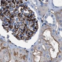
Immunohistochemistry protocol sheet for DYSF (IHC for DYSF): expected localisation Cytoplasmic staining in tissue cells (HPA tissue IHC), antibody A01234-3, validated IHC image, and IHC protocol steps
Printable DYSF IHC protocol sheet — expected localisation Cytoplasmic staining in tissue cells (HPA tissue IHC), antibody A01234-3, controls and protocol steps. Open the full DYSF IHC guide →

DYSF Immunohistochemistry Experimental Design Guide

Expected localisation, validated protocols, controls and antibodies — the at-a-glance facts below, then the full design guide.

Must know before staining
Expected localisation Cytoplasmic staining in tissue cells (HPA tissue IHC)
Staining pattern Cytoplasmic myocytes, trophoblasts and endothelial cells (HPA tissue IHC)
Antigen retrieval EDTA pH 8.0 HIER, heat-mediated (datasheet A01234-3)
Positive control ⓘ Cerebellum+4 more · see all
Negative control ⓘ Adipose tissue+4 more · see all
Important caveats
Reasons your staining may differ from the expected pattern.
Fixation Matched tissue-IHC evidence does not establish the fixation claim. Validate the specimen-specific method before use. (selected-SKU IHC image A01234-3)
Caveat Endothelial and hematopoietic staining can confound scoring (HPA tissue IHC)
Regulation Myotube injury focally enriches DYSF (UniProt)
Isoform / epitope 15 isoforms; epitope side: cytoplasmic/extracellular (UniProt)
Section 1

Recommended DYSF IHC & IF Protocols

The catalog antibody’s paraffin IHC protocol (datasheet A01234-3) is accompanied by published dysferlin muscle staining protocols (PMC7674836; PMC10216094).

Recommended immunohistochemistry (IHC-P) protocol parameters
SampleParaffin-embedded human skeletal muscle tissue; fixative not specified (datasheet A01234-3)
FixationImage fixative and duration unreported (datasheet A01234-3); verify before use.
Sectioning4–5 µm sections on charged slides (standard)
DeparaffinisationXylene, graded ethanol series to water (standard)
Antigen retrievalHeat retrieval: EDTA pH 8.0 (datasheet A01234-3); 20 min, 95–100 °C (standard)
Peroxidase block3% H2O2, 10 min, room temperature (standard)
Blocking10% goat serum (datasheet A01234-3)
Primary antibodyRabbit anti-DYSF, 2-5μg/ml (datasheet A01234-3)
Primary incubationOvernight at 4 °C (datasheet A01234-3)
DetectionStreptavidin-biotin complex (SABC), DAB chromogen (datasheet A01234-3)
CounterstainHematoxylin, blue, dehydrate and mount (standard)
Expected resultDYSF-positive staining in gLUC cells - cytoplasm/membrane of cerebellum (HPA tissue IHC: High). HPA tissue profile: Cytoplasmic expression in cardiac and skeletal muscle, hematopoietic cells, trophoblasts and endothelial cells. No signal in the no-primary control.
💡Decision noteFor paraffin sections, start with heat-mediated EDTA pH 8.0 retrieval (datasheet A01234-3). The frozen-section protocol does not specify retrieval (PMC7674836).
Section 2

What Is the Expected DYSF Staining Pattern?

DYSF is a single-pass membrane protein with a large cytoplasmic region and a short extracellular tail (UniProt O75923 topology). In paraffin sections, expect cytoplasmic or membrane-associated staining in skeletal myocytes, cardiomyocytes, trophoblasts, endothelial cells and hematopoietic cells (HPA tissue IHC; reliability: Enhanced). Sarcolemmal staining is consistent with its reported muscle localisation (UniProt O75923 subcellular location).

What am I looking at on my slide?
Skeletal myocytes show strong cytoplasmic or sarcolemmal staining.This fits the high myocyte signal in skeletal muscle (HPA tissue IHC) and sarcolemmal localisation (UniProt O75923). Assess cell boundaries as well as overall intensity; a membrane rim and cytoplasmic signal can both fit the supplied localisation evidence (HPA tissue IHC; UniProt O75923).
Endometrial or lung endothelial cells, placental cytotrophoblasts, or glomerular cells stain strongly.These are reported high-staining cell populations (HPA tissue IHC). Score the named cells within each tissue; an isolated stained cell elsewhere does not establish the same pattern (HPA tissue IHC; general IHC interpretation).
Staining is predominantly nuclear in an otherwise positive section.A dominant nuclear pattern conflicts with the reported cytoplasmic, sarcolemmal and membrane locations (HPA tissue IHC; UniProt O75923). Treat it as questionable until the compartment pattern is checked with controls or another IHC-validated antibody (general IHC practice).
Adipocytes or bronchial respiratory epithelial cells stain strongly.Those cell populations were reported as not detected (HPA tissue IHC). Consider nonspecific binding or endogenous detection activity, especially if staining also appears in a negative-reagent control (HPA tissue IHC; general IHC practice).
Known-positive skeletal myocytes show no signal.Skeletal myocytes were high in the HPA tissue profile (HPA tissue IHC). Check section quality, reagent performance and detection controls before interpreting the absence as a biological loss (general IHC practice); the supplied sources do not establish DYSF-specific fixation sensitivity.
💡Expected DYSF appearanceCall a convincing positive when skeletal myocytes show high cytoplasmic or sarcolemmal signal (HPA tissue IHC; UniProt O75923); dominant nuclear signal or similarly strong staining in HPA-negative cell populations is questionable (HPA tissue IHC; UniProt O75923).
How each factor affects the staining
Membrane topologyResidues 1–2046 are cytoplasmic, followed by a transmembrane segment and a short extracellular tail (UniProt O75923 topology). The antibody epitope is unspecified here, so topology alone cannot predict its access in a paraffin section.
Tissue and cell selectionSkeletal myocytes are high, cardiomyocytes medium, and adipocytes not detected in the reported tissue images (HPA tissue IHC). Compare the specified cell populations, since whole-section intensity can obscure where the signal occurs (general IHC interpretation).
Antibody validationHPA017071, HPA021945 and CAB002510 each have Enhanced IHC status (HPA antibodies). That supports comparison with HPA tissue patterns; it does not establish that every antibody or staining condition will reproduce them (HPA antibodies; general IHC interpretation).
Isoforms and epitope coverageUniProt lists 15 DYSF isoforms (UniProt O75923 isoforms). Their detection by a particular IHC antibody cannot be inferred without its epitope information; interpret a discordant result cautiously rather than assigning it to an isoform.
Antigen retrievalUse the validated IHC-P retrieval condition for the chosen antibody and tissue when available (general IHC practice). HPA staining levels and UniProt topology do not identify an optimal retrieval method or establish DYSF-specific fixation effects (HPA tissue IHC; UniProt O75923 topology).
Endogenous chromogenic activityTissue activity can create apparent chromogenic signal if the detection system is insufficiently controlled (general IHC practice). A negative-reagent control helps distinguish that signal from antibody-dependent staining (general IHC practice).
IF/ICC Q: Where should a corroborating signal localise?A plasma-membrane signal is supported; centriolar satellite and mid-piece locations are uncertain (HPA subcellular ICC-IF). ICC-IF localisation does not by itself define the appearance or detection conditions of paraffin IHC (HPA subcellular ICC-IF; general IHC interpretation).
Why is my staining missing, weak or wrong?
SituationLikely causeNext action
No staining in skeletal muscle myocytes.A failed stain or detection step is possible; myocytes are reported high (HPA tissue IHC; general IHC practice).Review the section and reagent controls, then repeat using the chosen antibody's validated IHC-P conditions (general IHC practice). Do not infer a DYSF-specific fixation effect from this result.
Strong nuclear staining dominates.The compartment conflicts with reported DYSF cytoplasmic and membrane localisation (HPA tissue IHC; UniProt O75923).Check a negative-reagent control and compare the same cell type with an independently IHC-validated antibody if available (general IHC practice; HPA antibodies).
Adipocytes or bronchial respiratory epithelial cells stain.These populations are reported as not detected; nonspecific or endogenous signal is possible (HPA tissue IHC; general IHC practice).Inspect negative-reagent controls and score the exact cell population before calling the signal DYSF-positive (general IHC practice; HPA tissue IHC).
Brown signal spreads across tissue without clear cell boundaries.Diffuse background can mask the expected cell-associated pattern (HPA tissue IHC; general IHC interpretation).Check blocking, washes and detection controls, then reassess localisation in a reported positive cell population (general IHC practice; HPA tissue IHC).
Cardiomyocytes look weaker than skeletal myocytes.The reported levels are medium in cardiomyocytes and high in skeletal myocytes (HPA tissue IHC).Compare each cell type with its own HPA expectation and the same run's controls before treating the difference as assay failure (HPA tissue IHC; general IHC practice).
A paraffin IHC result differs from an ICC-IF image.The assays examine different preparations; HPA supports plasma-membrane ICC-IF localisation, while tissue IHC reports cell-specific staining (HPA subcellular ICC-IF; HPA tissue IHC).Judge the paraffin section against the tissue IHC cell and compartment pattern, and use the ICC-IF result only as a localisation cross-check (HPA tissue IHC; HPA subcellular ICC-IF).

Sample controls for DYSF IHC & IF

🧪Run lung first and score endothelial cells, which HPA rates High for DYSF (HPA: High in lung endothelial cells). Use bronchus respiratory epithelial cells as the tissue negative (HPA: Not detected in bronchus respiratory epithelial cells); on the lung slide, compare nonendothelial cells with the no-primary background without assuming they are DYSF-negative, since HPA specifies only the endothelial compartment in lung (HPA: lung row).
Positive control tissue: Cerebellum (GLUC cells - cytoplasm/membrane, HPA High)
Negative control tissue: Adipose tissue (HPA Not detected)
ICC-IF cell lines (HPA subcellular resource): HPA ICC-IF images show DYSF in Rh30, U2OS, Sperm, with annotated localisation: Plasma membrane (supported), Centriolar satellite (uncertain), Mid piece (uncertain) (HPA subcellular).
Technical controls: Include no-primary and secondary-only slides, a rabbit IgG isotype control matched to the primary antibody’s clonality and concentration, and a DYSF-knockout sample or a validated immunizing-peptide block as a biological specificity control (caption: rabbit primary; standard IHC practice). For lung DAB staining, quench endogenous peroxidase and check or block endogenous biotin when using the caption’s streptavidin–biotin detection system (caption: SABC/DAB; standard IHC practice).
⚠️Feasibility: No target-specific fixation window or fixation effect is reported, and the selected A01234-3 paraffin-section caption does not state a fixative (selected tissue-IHC caption). That caption uses heat retrieval in EDTA, pH 8.0, but does not establish that retrieval is required; the supplied evidence does not establish whether frozen sections or IF are easier (selected tissue-IHC caption). In lung, endogenous peroxidase or biotin can mimic signal with the caption’s DAB/SABC detection, so interpret endothelial staining against the control slides (caption: SABC/DAB; standard IHC practice).

HPA tissue IHC evidence for DYSF

Comprehensive Human Protein Atlas IHC scoring per tissue (reliability: Enhanced — High consistency between antibody staining and RNA expression data.). Rows are taken directly from the HPA tissue chart — click any row's HPA link to view the source.

Positive expression · recommended positive controls

TissueCell typeLevelEvidenceSource
Cerebellum GLUC cells - cytoplasm/membrane High Protein (IHC) HPA →
Endometrium Endothelial cells High Protein (IHC) HPA →
Kidney Cells in glomeruli High Protein (IHC) HPA →
Lung Endothelial cells High Protein (IHC) HPA →
Placenta Cytotrophoblasts High Protein (IHC) HPA →

Undetected expression · recommended negative controls

TissueCell typeLevelEvidenceSource
Adipose tissue Adipocytes Not detected Protein (IHC) HPA →
Adrenal gland Glandular cells Not detected Protein (IHC) HPA →
Appendix Glandular cells Not detected Protein (IHC) HPA →
Breast Adipocytes Not detected Protein (IHC) HPA →
Bronchus Respiratory epithelial cells Not detected Protein (IHC) HPA →
Section 3

Advanced DYSF IHC Tips

Troubleshoot DYSF staining in paraffin sections by checking retrieval, cellular compartment, controls, and scoring before interpreting signal as dysferlin expression.

What should I change when DYSF staining is weak after antigen retrieval?
Start with heat-mediated retrieval in EDTA buffer at pH 8.0 for paraffin sections (datasheet A01234-3). The documented skeletal-muscle staining then used 2 μg/ml primary antibody overnight at 4°C, so match those conditions before changing retrieval (datasheet A01234-3). If signal remains weak, vary heating duration on adjacent sections while keeping antibody concentration, detection, and imaging conditions fixed (standard IHC practice). Use known positive skeletal muscle to distinguish inadequate retrieval from low expression in the specimen (HPA: High in skeletal-muscle myocytes). Compare tissue preservation and background after each change, since excessive heating can damage morphology and raise nonspecific staining (standard IHC practice).
Could fixation explain inconsistent DYSF staining between paraffin blocks?
The selected paraffin-section caption does not state a fixative, so target-specific fixation sensitivity is unknown (datasheet A01234-3). Record each block’s fixative and fixation duration, then compare sections processed and stained together; this tests whether processing differences track the inconsistent signal (standard IHC practice). Keep EDTA retrieval at pH 8.0 and the documented 2 μg/ml overnight primary incubation constant during that comparison (datasheet A01234-3). Examine tissue preservation alongside staining, because damaged sections can make intensity comparisons unreliable (standard IHC practice). Do not assign a DYSF-specific fixation effect from expression patterns or protein topology alone (HPA: tissue IHC profile; UniProt O75923 topology).
Should DYSF stain the sarcolemma or the cytoplasm in skeletal muscle?
Assess the sarcolemmal outline and intracellular staining separately: dysferlin is reported at the sarcolemma and on cytoplasmic vesicle membranes (UniProt O75923 subcellular location). Cytoplasmic staining in skeletal-muscle myocytes is also reported in tissue IHC, so cytoplasmic signal alone is not automatically discordant (HPA: skeletal-muscle profile). Its single transmembrane segment spans residues 2047–2067, with most of the protein on the cytoplasmic side (UniProt O75923 topology). Compare adjacent fibers at the same magnification and exposure, and check whether staining follows intact cell boundaries rather than folds or torn edges (standard IHC practice). Record membrane and cytoplasmic patterns separately when reporting results (standard IHC practice).
Why might two DYSF antibodies give different staining patterns?
DYSF has 15 listed isoforms, so antibody results can differ if their epitopes are not shared across the expressed forms (UniProt O75923 isoforms). The supplied caption does not identify the catalog antibody’s epitope; verify its mapped sequence before assigning an isoform-specific explanation (datasheet A01234-3). Most of dysferlin is cytoplasmic, while only residues 2068–2080 follow the membrane-spanning segment on the extracellular side (UniProt O75923 topology). Phosphorylation is listed at residues 166 and 197, but its effect on staining is not established here (UniProt O75923 modified residues). Compare antibodies on adjacent sections under matched retrieval and detection conditions before interpreting discordance (standard IHC practice).
How can IF help assess ambiguous DYSF localisation seen by chromogenic IHC?
Use a separate IF experiment to compare DYSF with a validated myocyte marker when skeletal-muscle IHC cannot resolve which cells carry the signal (HPA: High in skeletal-muscle myocytes; standard IF practice). Choose well-separated fluorophores and favor a far-red channel if tissue autofluorescence obscures shorter-wavelength signal; include an unstained section to assess autofluorescence (standard IF practice). For an antibody against a cytoplasmic epitope, test permeabilisation, whereas an antibody against the short extracellular tail may be assessed without it (UniProt O75923 topology; standard IF practice). Confirm the antibody’s actual epitope first, because it is not specified in the supplied IHC caption (datasheet A01234-3). Keep IF fixation choices separate from the paraffin IHC caption, which reports no fixative (datasheet A01234-3).
How do I troubleshoot diffuse brown background in DYSF IHC?
Run a no-primary control through the same secondary, streptavidin–biotin complex, and DAB steps to locate detection-related background (datasheet A01234-3; standard IHC practice). The documented section used 10% goat serum blocking and a biotinylated secondary for 30 minutes at 37°C; match those conditions while assessing background (datasheet A01234-3). Check endogenous peroxidase blocking and, with this biotin-based system, assess endogenous biotin as possible sources of brown signal (standard IHC practice). Compare intact myocytes with surrounding structures instead of treating uniform section-wide color as DYSF expression (HPA: High in skeletal-muscle myocytes; standard IHC practice). Improve washes or titrate primary concentration only after identifying which control reproduces the background (standard IHC practice).
How should I quantify DYSF staining across skeletal-muscle sections? ⚠ ANSWER MARKED FOR VERIFICATION
Score intact myocytes and report the percentage positive plus staining intensity, or calculate an H-score from intensity categories for a 0–300 range (HPA: High in skeletal-muscle myocytes; standard IHC practice). Define membrane and cytoplasmic positivity separately because both compartments have supporting localisation evidence (UniProt O75923 subcellular location; HPA: tissue IHC profile). Normalize positive-cell counts to the total number of evaluable myocytes, or stained area to the measured tissue area, and state which denominator was used (standard IHC practice). Apply the same threshold, retrieval at pH 8.0, detection run, and image settings to comparison sections (datasheet A01234-3; standard IHC practice). Exclude folds, tears, and necrotic areas using a rule set before scoring (standard IHC practice).
When should a positive DYSF IHC result be considered artefactual?
Treat staining as more credible when it occurs in intact skeletal-muscle myocytes with a reproducible sarcolemmal or cytoplasmic pattern (UniProt O75923 subcellular location; HPA: High in skeletal-muscle myocytes). DYSF is also reported in hematopoietic cells and endothelial cells, so identify the stained cell type before assigning signal to myocytes (HPA: tissue IHC profile). Question signal confined to section edges, folds, or necrotic tissue, and compare it with a no-primary control for DAB or endogenous-enzyme background (standard IHC practice). A solely nuclear pattern lacks support from the supplied DYSF localisation record and warrants further validation (UniProt O75923 subcellular location). Confirm an unexpected pattern on another section under matched retrieval and detection conditions (standard IHC practice).
Boster reagents

Best DYSF / Dysferlin IHC Antibodies

A01234-3 has pictured paraffin-section IHC in human and mouse skeletal muscle and IF/ICC in SiHa cells (catalog image captions); rat reactivity is listed (catalog reactivity).

Real IHC data IHC analysis of Dysferlin/DYSF using anti-Dysferlin/DYSF antibody (A01234-3). Dysferlin/DYSF was detected in a paraffin-embedded section of human skeletal muscle tissue. Heat mediated antigen retrieval was performed in EDTA buffer (pH 8.0, epitope retrieval solution). The tissue section was blocked with 10% goat serum. The tissue section was then incubated with 2 μg/ml rabbit anti-Dysferlin/DYSF Antibody (A01234-3) overnight at 4°C. Biotinylated goat anti-rabbit IgG was used as secondary antibody and incubated for 30 minutes at 37°C. The tissue section was developed using Strepavidin-Biotin-Complex (SABC) (Catalog # SA1022) with DAB as the chromogen.
Anti-Dysferlin/DYSF Antibody ®
Cat # A01234-3

A01234-3 will render with an IHC figure from a human skeletal-muscle paraffin section; its captions also document mouse skeletal-muscle IHC and SiHa-cell IF/ICC (A01234-3 image captions). M01234 will render with IHC and IF/ICC listed for human and mouse, but no image is supplied (M01234 applications, reactivity and image fields).

Which to pick: For tissue IHC, choose A01234-3: its captions document human and mouse skeletal-muscle paraffin sections; the fixative is unreported (A01234-3 IHC captions). For IF/ICC, A01234-3 has a SiHa-cell image, while M01234 is a rabbit monoclonal with IF/ICC listed but no supplied image (A01234-3 IF caption; M01234 clone, applications and image fields). For cross-species work, A01234-3 lists human, mouse and rat reactivity, versus human and mouse for M01234; the pictured A01234-3 IHC covers human and mouse only (catalog reactivity; A01234-3 IHC captions).

Each figure is that product's own IHC / IF validation image from its datasheet.

References

  1. UniProt Consortium. UniProt entry O75923 (DYSF_HUMAN, Dysferlin).
  2. Human Protein Atlas. DYSF tissue IHC expression (reliability: Enhanced).
  3. Human Protein Atlas. DYSF subcellular location (ICC-IF): Localized to the plasma membrane, centriolar satellites and mid piece..
  4. Human Protein Atlas. DYSF antibody validation summary (3 antibodies).
  5. Clinical and Genomic Evaluation of 207 Genetic Myopathies in the Indian Subcontinent. Frontiers in neurology 2020 — PMC7674836.
  6. A Dysferlin Exon 32 Nonsense Mutant Mouse Model Shows Pathological Signs of Dysferlinopathy. Biomedicines 2023 — PMC10216094.
  7. Multiple sclerosis severity variant in DYSF-ZNF638 locus associates with neuronal loss and inflammation. iScience 2025 — PMC12063138.
  8. Whole Exome Sequencing Reveals DYSF, FKTN, and ISPD Mutations in Congenital Muscular Dystrophy Without Brain or Eye Involvement. Journal of neuromuscular diseases 2015 — PMC4373448.
  9. PubMed PMID:9731526 — UniProt-cited evidence.
  10. PubMed PMID:16896923 — UniProt-cited evidence.
  11. PubMed PMID:19221801 — UniProt-cited evidence.