EBI3 / Interleukin-27 subunit beta · IHC design guide

Design Immunohistochemistry for EBI3

Plan paraffin-section EBI3 IHC around cytoplasmic staining in placental cytotrophoblasts (HPA tissue IHC). Start the catalog antibody at 2.5 μg/mL (datasheet A07190) and compare staining with a negative tissue control.

Evidence assembled Oct 2026 · For research use; verify linked source records and product datasheet before use
Immunohistochemistry protocol sheet for EBI3 (IHC for EBI3): expected localisation Cytoplasm in placental trophoblasts (HPA tissue IHC); secreted (UniProt), antibody A07190, validated IHC image, and IHC protocol steps
Printable EBI3 IHC protocol sheet — expected localisation Cytoplasm in placental trophoblasts (HPA tissue IHC); secreted (UniProt), antibody A07190, controls and protocol steps. Open the full EBI3 IHC guide →

EBI3 Immunohistochemistry Experimental Design Guide

Expected localisation, validated protocols, controls and antibodies — the at-a-glance facts below, then the full design guide.

Must know before staining
Expected localisation Cytoplasm in placental trophoblasts (HPA tissue IHC); secreted (UniProt)
Staining pattern High cytoplasmic staining in placental cytotrophoblasts (HPA tissue IHC)
Antigen retrieval Citrate pH 6 HIER, heat-mediated (datasheet A07190)
Positive control ⓘ Placenta
Negative control ⓘ Adipose tissue+4 more · see all
Important caveats
Reasons your staining may differ from the expected pattern.
Fixation Formaldehyde-fixed tissue (selected-SKU IHC image A07190); keep fixation consistent (standard IHC practice; not target-specific)
Caveat Secreted EBI3: RNA and protein locations may differ (HPA tissue IHC)
Regulation Expression regulation not specified (UniProt)
Isoform / epitope No isoforms listed; the mature chain starts at residue 21 (UniProt)
Section 1

Recommended EBI3 IHC & IF Protocols

The catalog antibody’s IHC-P protocol (datasheet A07190) is accompanied by two published EBI3 IHC protocols (PMC3169615; PMC4444442).

Recommended immunohistochemistry (IHC-P) protocol parameters
SampleFormaldehyde-fixed, paraffin-embedded Human Spleen tissue (datasheet A07190)
FixationImage formalin-fixed; duration unreported (datasheet A07190); verify before use.
Sectioning4–5 µm sections on charged slides (standard)
DeparaffinisationXylene, graded ethanol series to water (standard)
Antigen retrievalHeat retrieval: Citrate pH 6 (datasheet A07190); 20 min, 95–100 °C (standard)
Peroxidase block3% H2O2, 10 min, room temperature (standard)
Blocking10% serum (datasheet A07190)
Primary antibodyRabbit anti-EBI3, 2.5 μg/mL (datasheet A07190)
Primary incubationOvernight at 4 °C (standard)
DetectionHRP-polymer secondary, DAB chromogen 5–10 min (standard)
CounterstainHematoxylin, blue, dehydrate and mount (standard)
Expected resultEBI3-positive staining in cytotrophoblasts of placenta (HPA tissue IHC: High). HPA tissue profile: Cytoplasmic expression in placental trophoblasts. No signal in the no-primary control.
💡Decision noteStart with citrate pH 6 heat retrieval (datasheet A07190); both published protocols also used citrate retrieval (PMC3169615; PMC4444442).
Section 2

What Is the Expected EBI3 Staining Pattern?

EBI3 is a secreted protein with no transmembrane segment (UniProt Q14213 topology). In paraffin section IHC, expect cytoplasmic staining in placental trophoblasts, with high staining reported specifically in cytotrophoblasts (HPA: placental profile; High in cytotrophoblasts). HPA rates the tissue IHC evidence Enhanced, while noting medium consistency with RNA data and that secreted protein may appear away from its site of RNA expression (HPA: reliability description).

What am I looking at on my slide?
Strong cytoplasmic signal in placental cytotrophoblasts, with recognizable cell boundaries.This matches the reported positive cell type, compartment and high staining level (HPA: cytoplasmic placental profile; High in cytotrophoblasts). Compare cells within the same section before assigning a positive call.
Predominantly nuclear staining, or staining confined to cell membranes.Treat this as a suspect compartment pattern: HPA reports cytoplasmic trophoblast staining, and UniProt reports no transmembrane segment (HPA: placental profile; UniProt Q14213 topology). Review controls and morphology before interpreting it as EBI3.
Strong signal in cells outside the expected trophoblast population.Check for cross-reactivity or endogenous detection activity. HPA reports presumed off-target binding that was disregarded in its tissue assessment (HPA: reliability description). A secreted protein’s location can differ from its RNA source, so cell identity alone does not prove an artefact (HPA: reliability description).
Diffuse stain across many cells or tissue spaces without a clear cytotrophoblast pattern.This does not reproduce the reported cell-associated cytoplasmic pattern (HPA: placental profile). Use a detection-only control and inspect blocking, washing and chromogen development for nonspecific signal (general IHC practice).
No cytoplasmic signal in an adequately preserved placenta section.A negative result conflicts with the reported high cytotrophoblast staining (HPA: High in cytotrophoblasts). First confirm that cytotrophoblasts are present and identifiable; then check antibody and detection performance with appropriate controls (general IHC practice).
💡Expected EBI3 appearanceCall the section positive when placental cytotrophoblasts show clear, high cytoplasmic staining (HPA: High in cytotrophoblasts; cytoplasmic profile); isolated nuclear, membrane-only or indiscriminate background signal is suspect against that reference pattern (HPA: placental profile).
How each factor affects the staining
Cell type and reference tissuePlacenta provides the reported positive reference: cytotrophoblasts stain at a high level (HPA: High in cytotrophoblasts). HPA’s negative calls apply to named cells, such as adipocytes in adipose tissue, rather than every cell in those tissues (HPA: Not detected in adipocytes).
Secretory topology and processingEBI3 has a signal sequence at residues 1–20, a mature chain at 21–229 and no transmembrane segment (UniProt Q14213 processing and topology). Interpret its observed cytoplasmic IHC pattern in light of secretion; extracellular staining alone is not the HPA reference pattern (HPA: placental profile).
Protein modificationsUniProt lists glycosylation sites at residues 55 and 105 (UniProt Q14213 glycosylation). The supplied sources do not identify this antibody’s epitope or show whether these sites change IHC staining; do not infer an antigen retrieval requirement from them.
Strength and limits of validationThe listed antibody HPA046635 has Enhanced IHC validation (HPA: antibody validation). HPA also reports medium consistency between staining and RNA data and disregarded presumed off-target binding (HPA: reliability description); use the reported cell and compartment pattern to judge a slide.
Antigen retrieval and fixationNo EBI3-specific retrieval condition or fixation sensitivity is supplied (HPA: tissue IHC; UniProt Q14213). If signal is weak, compare documented IHC retrieval conditions with controls as general IHC practice; do not attribute a change specifically to EBI3 epitope masking.
Why is my staining missing, weak or wrong?
SituationLikely causeNext action
Placental cytotrophoblasts show no detectable stain.The expected positive cells may be absent from the sampled area, or the staining run may have failed (HPA: High in cytotrophoblasts; general IHC practice).Verify cell identity on the counterstained section, then assess the antibody, detection reagents and retrieval conditions against a positive control (general IHC practice).
Signal is weak or patchy in recognizable cytotrophoblasts.A weak run may obscure the reported high cytoplasmic pattern; the sources provide no EBI3-specific fixation or retrieval effect (HPA: High in cytotrophoblasts; UniProt Q14213).Compare controlled retrieval and antibody dilution conditions using the catalog antibody’s IHC-P instructions, and retain the condition that gives specific cytoplasmic contrast (general IHC practice).
The entire section has brown haze or granular background.Excess detection signal, incomplete blocking or residual endogenous peroxidase activity can create background in chromogenic IHC (general IHC practice).Inspect detection-only and peroxidase-block controls; adjust blocking, washing or development consistently across comparison sections (general IHC practice).
A prominent nuclear or membrane-only pattern dominates.That compartment differs from HPA’s cytoplasmic trophoblast profile; EBI3 has no transmembrane segment (HPA: placental profile; UniProt Q14213 topology).Check cell boundaries and counterstain, examine a negative detection control, and withhold an EBI3-positive call unless the expected cytoplasmic pattern is demonstrable.
Unexpected cells stain strongly while cytotrophoblasts do not.Off-target binding is plausible because HPA reports presumed off-target binding in its assessment (HPA: reliability description). Endogenous detection activity is another general IHC possibility.Compare a placenta positive reference with cell-specific HPA negatives, such as adipocytes in adipose tissue, and check detection-only controls (HPA: High in cytotrophoblasts; Not detected in adipocytes).
Can IF/ICC establish the same subcellular pattern?HPA labels EBI3 secreted but supplies no ICC-IF images or main subcellular location, and lists no ICC validation for HPA046635 (HPA: subcellular summary; antibody validation).Use the separate IF/ICC guide for that application. Treat cytoplasmic placental staining as an IHC observation, not an IF/ICC validation claim (HPA: placental profile; subcellular summary).

Sample controls for EBI3 IHC & IF

🧪Run placenta first: cytotrophoblasts should stain strongly (HPA: High in placental cytotrophoblasts). Use adipose tissue adipocytes as a negative tissue (HPA: Not detected in adipocytes); on the placenta slide, assess cells outside the cytotrophoblast population against background without assuming they are EBI3-negative (HPA: only cytotrophoblasts listed as High).
Positive control tissue: Placenta (Cytotrophoblasts, HPA High)
Negative control tissue: Adipose tissue (HPA Not detected)
ICC-IF cell lines (HPA subcellular resource): HPA carries no ICC-IF cell line for EBI3; derive a cell-line control from the positive tissue's cell type (Cytotrophoblasts) and confirm it by RNA or western blot first.
Technical controls: Include no-primary (secondary-only), a host-species- and clonality-matched isotype control, and an EBI3 knockout sample or validated peptide-block control (standard IHC practice). In placenta, check for endogenous peroxidase background before interpreting weak chromogen staining (standard IHC practice).
⚠️Feasibility: A target-specific fixation window or fixation effect is unreported, and the selected A07190 record lists its fixative as unreported (selected A07190 caption metadata). The caption uses heat retrieval in citrate buffer at pH 6, but does not establish that retrieval is required (selected A07190 caption). The supplied evidence does not show that frozen sections or IF are easier; placenta background from endogenous peroxidase needs checking (HPA: no ICC-IF cell-line images; standard IHC practice).

HPA tissue IHC evidence for EBI3

Comprehensive Human Protein Atlas IHC scoring per tissue (reliability: Enhanced — Medium consistency between antibody staining and RNA expression data. Secreted protein, tissue location of RNA and protein is expected to differ. Presumed off target binding observed and disregarded.). Rows are taken directly from the HPA tissue chart — click any row's HPA link to view the source.

Positive expression · recommended positive controls

TissueCell typeLevelEvidenceSource
Placenta Cytotrophoblasts High Protein (IHC) HPA →

Undetected expression · recommended negative controls

TissueCell typeLevelEvidenceSource
Adipose tissue Adipocytes Not detected Protein (IHC) HPA →
Adrenal gland Glandular cells Not detected Protein (IHC) HPA →
Appendix Glandular cells Not detected Protein (IHC) HPA →
Bone marrow Hematopoietic cells Not detected Protein (IHC) HPA →
Breast Adipocytes Not detected Protein (IHC) HPA →
Section 3

Advanced EBI3 IHC Tips

Troubleshoot EBI3 staining in paraffin-section chromogenic IHC, using the selected antibody’s tissue image and independent localisation evidence as reference points.

Which retrieval conditions should I start with for EBI3 paraffin IHC?
Start with heat-mediated citrate antigen retrieval at pH 6 for paraffin sections (datasheet A07190). Use matched sections and a consistent heating and cooling schedule so handling differences do not obscure the comparison (standard IHC practice). The selected spleen image used this retrieval, 2.5 μg/ml primary antibody overnight at 4 °C, and hematoxylin counterstain (A07190 tissue-IHC caption). If staining remains weak, titrate retrieval duration before trying another buffer on parallel sections, while holding antibody and detection conditions fixed (standard IHC practice). Assess whether changes improve cytoplasmic staining in placental cytotrophoblasts without increasing background (HPA: High in placental cytotrophoblasts; HPA: cytoplasmic expression).
How should I investigate weak EBI3 staining when fixation histories differ?
Target-specific sensitivity of EBI3 staining to fixative choice or fixation duration is unknown from the supplied evidence (evidence payload: no fixation-sensitivity comparison). For paraffin IHC, document the fixative, time before fixation, processing history, and section thickness for each specimen (standard IHC practice). Compare similarly processed sections first, keeping retrieval, antibody concentration, and detection conditions constant (standard IHC practice). If archived blocks permit a processing comparison, change one variable at a time and include a separate process control (standard IHC practice). Do not attribute a negative stain to overfixation without checking section morphology and control staining (standard IHC practice).
Where should credible EBI3 staining appear in tissue sections?
Expect interpretable cellular staining chiefly in the cytoplasm of placental trophoblasts, with high signal reported in cytotrophoblasts (HPA: cytoplasmic expression in placental trophoblasts; HPA: High in placental cytotrophoblasts). EBI3 is secreted and has no transmembrane segment, so a sharp membrane-only pattern needs independent confirmation (UniProt Q14213: secreted; UniProt Q14213 topology). Examine intact cells and nearby extracellular material separately rather than combining them into one score (standard IHC practice). Do not require protein staining to match the exact cells expressing RNA, because tissue protein and RNA locations may differ for this secreted protein (HPA: reliability description). Compare suspicious patterns with the placental control and matched negative controls (HPA: High in placental cytotrophoblasts; standard IHC practice).
Could epitope accessibility explain inconsistent EBI3 IHC?
No alternative EBI3 isoforms are listed in the supplied record, so an isoform-specific staining explanation lacks support here (UniProt Q14213: isoforms 0). The precursor has a 1–20 signal peptide and a 21–229 mature chain (UniProt Q14213: processing). It also has reported glycosylation sites at residues 55 and 105 and two fibronectin type-III domains (UniProt Q14213: glycosylation and domains). The catalog antibody’s binding epitope is unspecified in the supplied evidence, so these features cannot establish which treatment exposes it (evidence payload: no epitope map). Compare retrieval conditions on adjacent sections and judge both specific staining and background (standard IHC practice).
How can I adapt the EBI3 question to multiplex IF?
Pair EBI3 with a separately validated cytotrophoblast marker when examining placenta, and confirm that each channel remains interpretable alone (HPA: High in placental cytotrophoblasts; standard IF practice). Choose spectrally separated fluorophores, placing a weak EBI3 signal in a channel with low measured tissue autofluorescence (standard IF practice). Because EBI3 is secreted and lacks a transmembrane segment, the relevant epitope’s accessibility must be established for the chosen staining goal (UniProt Q14213: secreted; UniProt Q14213 topology). Test mild permeabilisation for intracellular staining; omit it initially when testing accessible extracellular antigen (standard IF practice). Treat this as an IF optimisation, since the supplied HPA subcellular record includes no ICC/IF images (HPA subcellular: no cell lines with ICC/IF images).
How do I reduce diffuse or misleading EBI3 chromogenic staining?
Begin by checking a no-primary section and whether chromogen appears in tissue compartments unrelated to the expected cytoplasmic pattern (standard IHC practice; HPA: cytoplasmic expression in placental trophoblasts). Include a peroxidase block before HRP detection and assess endogenous pigment separately from chromogen (standard IHC practice). The selected spleen image used 10% serum for 1 hour at room temperature and an HRP secondary at 1:250 (A07190 tissue-IHC caption). If background persists, titrate primary antibody below the caption’s 2.5 μg/ml while keeping detection exposure consistent (A07190 tissue-IHC caption; standard IHC practice). Disregard edge-heavy, necrotic, or no-primary-positive deposits when judging EBI3 signal (standard IHC practice).
How should I score EBI3 IHC across specimens? ⚠ ANSWER MARKED FOR VERIFICATION
Define the cell population and cytoplasmic scoring boundary before reviewing outcomes; placental cytotrophoblasts provide an evidence-backed positive reference (HPA: High in placental cytotrophoblasts; HPA: cytoplasmic expression). For cellular staining, record the percentage of positive target cells and intensity, or calculate an H-score from those observations (standard IHC practice). Where dispersed positive cells are the endpoint, report their density per mm² of viable tissue (standard IHC practice). Normalise each measurement to the eligible cells or viable tissue area, excluding folds, edges, and necrosis by the same rule throughout (standard IHC practice). Report extracellular deposits separately because EBI3 is secreted (UniProt Q14213: secreted).
What separates true EBI3 positivity from tissue artefact?
Give greatest weight to reproducible cytoplasmic staining in intact placental cytotrophoblasts, the reported high-expression population (HPA: High in placental cytotrophoblasts; HPA: cytoplasmic expression). A membrane-only pattern is less consistent with EBI3’s secreted, non-transmembrane topology and warrants confirmation (UniProt Q14213: secreted; UniProt Q14213 topology). Reject deposits concentrated at section edges, folds, or necrotic areas, and investigate signal retained in a no-primary control as possible endogenous enzyme or detection background (standard IHC practice). Interpret extracellular staining cautiously because secreted protein need not remain at its cell of origin (UniProt Q14213: secreted; HPA: reliability description). EBI3 positivity alone does not identify IL-27 versus IL-35, since EBI3 can pair with IL27A or IL12A (UniProt Q14213: subunit).
Boster reagents

Best EBI3 / Interleukin-27 subunit beta IHC Antibodies

Two anti-EBI3 antibodies have IHC and IF images from human spleen tissue (catalog image captions); A07190-1 also lists mouse and rat reactivity (catalog reactivity).

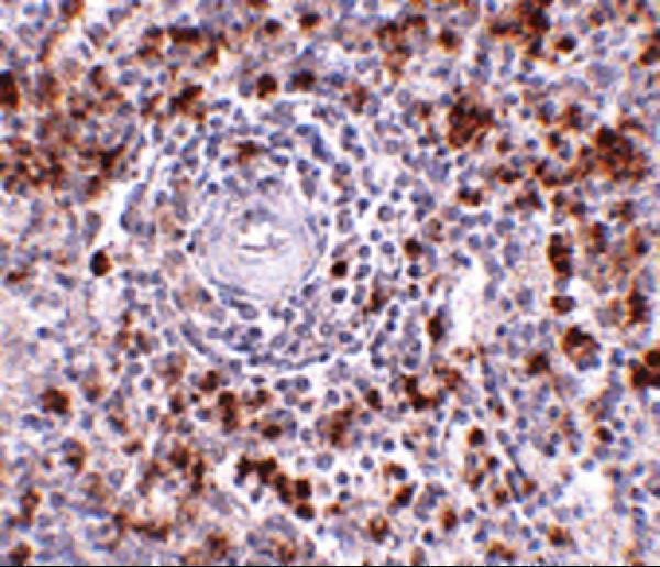
Real IHC data Immunohistochemistry Validation of EBI-3 in Human Spleen Tissue Immunohistochemical analysis of paraffin-embedded Human Spleen tissue using anti-EBI-3 antibody (A07190) at 2.5 μg/ml. Tissue was fixed with formaldehyde and blocked with 10% serum for 1 h at RT; antigen retrieval was by heat mediation with a citrate buffer (pH6). Samples were incubated with primary antibody overnight at 4˚C. A goat anti-rabbit IgG H&L (HRP) at 1/250 was used as secondary. Counter stained with Hematoxylin.
Anti-Interleukin-27 subunit beta EBI3 Antibody
Cat # A07190
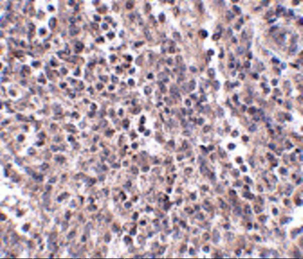
Real IHC data Immunohistochemistry Validation of EBI-3 in Human Spleen Tissue Immunohistochemical analysis of paraffin-embedded Human Spleen tissue using anti-EBI-3 antibody (A07190-1) at 2.5 μg/ml. Tissue was fixed with formaldehyde and blocked with 10% serum for 1 h at RT; antigen retrieval was by heat mediation with a citrate buffer (pH6). Samples were incubated with primary antibody overnight at 4˚C. A goat anti-rabbit IgG H&L (HRP) at 1/250 was used as secondary. Counter stained with Hematoxylin.
Anti-Interleukin-27 subunit beta EBI3 Antibody
Cat # A07190-1

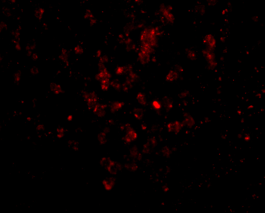
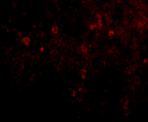
A07190 has IHC data from formaldehyde-fixed, paraffin-embedded human spleen and IF data from paraformaldehyde-fixed human spleen (A07190 image captions). A07190-1 has corresponding human spleen IHC and IF data (A07190-1 image captions); mouse and rat are listed as reactive species (catalog reactivity).

Which to pick: For tissue IHC, either SKU has its own formaldehyde-fixed, paraffin-embedded human spleen image at 2.5 μg/mL (A07190 and A07190-1 IHC captions). For IF, either has a human spleen image at 20 μg/mL (A07190 and A07190-1 IF captions); the payload provides no ICC image (catalog image captions). For mouse or rat work, consider A07190-1 because those species are listed as reactive, while its IHC and IF images document human tissue only (A07190-1 catalog reactivity and image captions).

Each figure is that product's own IHC / IF validation image from its datasheet.

References

  1. UniProt Consortium. UniProt entry Q14213 (IL27B_HUMAN, Interleukin-27 subunit beta).
  2. Human Protein Atlas. EBI3 tissue IHC expression (reliability: Enhanced).
  3. Human Protein Atlas. EBI3 subcellular location (ICC-IF): Secreted.
  4. Human Protein Atlas. EBI3 antibody validation summary (1 antibodies).
  5. Epstein-Barr virus-induced gene 3 (EBI3): a novel diagnosis marker in Burkitt lymphoma and diffuse large B-cell lymphoma. PloS one 2011 — PMC3169615.
  6. Interleukin 35 is an independent prognostic factor and a therapeutic target for nasopharyngeal carcinoma. Contemporary oncology (Poznan, Poland) 2015 — PMC4444442.
  7. Suppression of NK cell-mediated immunosurveillance by IL-35 drives tumor progression in EGFR-mutant non-small cell lung cancer. Frontiers in oncology 2025 — PMC12676456.
  8. Contribution of IL-12/IL-35 common subunit p35 to maintaining the testicular immune privilege. PloS one 2014 — PMC3997559.
  9. PubMed PMID:8551575 — UniProt-cited evidence.
  10. PubMed PMID:15057824 — UniProt-cited evidence.
  11. PubMed PMID:15489334 — UniProt-cited evidence.