ECHS1 / Enoyl-CoA hydratase, mitochondrial · IHC design guide

Design Immunohistochemistry for ECHS1

Plan chromogenic IHC for ECHS1 in paraffin sections using the observed granular cytoplasmic pattern (HPA tissue IHC). The guide supports selection of controls and scoring by cell type and staining intensity.

Evidence assembled Oct 2026 · For research use; verify linked source records and product datasheet before use
Immunohistochemistry protocol sheet for ECHS1 (IHC for ECHS1): expected localisation Granular cytoplasmic staining (HPA tissue IHC), antibody A04508-1, validated IHC image, and IHC protocol steps
Printable ECHS1 IHC protocol sheet — expected localisation Granular cytoplasmic staining (HPA tissue IHC), antibody A04508-1, controls and protocol steps. Open the full ECHS1 IHC guide →

ECHS1 Immunohistochemistry Experimental Design Guide

Expected localisation, validated protocols, controls and antibodies — the at-a-glance facts below, then the full design guide.

Must know before staining
Expected localisation Granular cytoplasmic staining (HPA tissue IHC)
Staining pattern Widespread granular cytoplasmic staining (HPA tissue IHC)
Antigen retrieval EDTA pH 8.0 HIER, heat-mediated (datasheet A04508-1)
Positive control ⓘ Adipose tissue+4 more · see all
Negative control ⓘ None in HPA (detected in all 45 tissues); use no-primary + isotype controls
Important caveats
Reasons your staining may differ from the expected pattern.
Fixation Keep fixation consistent across sections (standard IHC practice; not target-specific). Selected-image fixative and duration unreported (datasheet A04508-1); verify before use.
Caveat Widespread staining limits tissue specificity (HPA tissue IHC)
Regulation Liver-enhanced RNA expression (HPA tissue RNA)
Isoform / epitope No annotated isoforms; mature chain spans 28–290 (UniProt)
Section 1

Recommended ECHS1 IHC & IF Protocols

The catalog antibody’s IHC-P protocol (datasheet A04508-1) is accompanied by three published ECHS1 IHC protocols (PMC8494735; PMC12117712; PMC12352869).

Recommended immunohistochemistry (IHC-P) protocol parameters
SampleParaffin-embedded human breast cancer tissue; fixative not specified (datasheet A04508-1)
FixationImage fixative and duration unreported (datasheet A04508-1); verify before use.
Sectioning4–5 µm sections on charged slides (standard)
DeparaffinisationXylene, graded ethanol series to water (standard)
Antigen retrievalHeat retrieval: EDTA pH 8.0 (datasheet A04508-1); 20 min, 95–100 °C (standard)
Peroxidase block3% H2O2, 10 min, room temperature (standard)
Blocking10% goat serum (datasheet A04508-1)
Primary antibodyRabbit anti-ECHS1, 2-5 μg/ml (datasheet A04508-1)
Primary incubationOvernight at 4 °C (datasheet A04508-1)
DetectionHRP-conjugated secondary, DAB chromogen (datasheet A04508-1)
CounterstainHematoxylin, blue, dehydrate and mount (standard)
Expected resultECHS1-positive staining in adipocytes of adipose tissue (HPA tissue IHC: High). HPA tissue profile: Ubiquitous cytoplasmic expression with a granular pattern. No signal in the no-primary control.
💡Decision noteStart with heat retrieval in EDTA pH 8.0 for the catalog antibody (datasheet A04508-1); use each article’s stated retrieval when reproducing its protocol (PMC12117712; PMC12352869).
Section 2

What Is the Expected ECHS1 Staining Pattern?

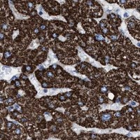
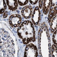
ECHS1 is a mitochondrial matrix protein with no transmembrane segment (UniProt P30084 topology). In paraffin-section IHC, expect granular cytoplasmic staining across many cell types (HPA: ubiquitous cytoplasmic expression with a granular pattern). Reported high-staining examples include adipocytes, adrenal glandular cells, and bronchial respiratory epithelial cells (HPA: High in each). The tissue IHC profile has Enhanced reliability, reflecting consistency between antibody staining and RNA expression (HPA: Enhanced reliability).

What am I looking at on my slide?
Granular cytoplasmic staining in adipocytes, adrenal glandular cells, or bronchial respiratory epithelial cells (HPA: High in each).This fits the reported IHC pattern and mitochondrial location (HPA: granular cytoplasm; UniProt P30084: matrix). Score the specified cells and their staining intensity; the examples do not define an intensity threshold for every tissue (HPA: selected High examples).
Strong nuclear, membrane-rim, or extracellular staining dominates while granular cytoplasm is absent.Those compartments conflict with the reported mitochondrial pattern (UniProt P30084: matrix; HPA: granular cytoplasm). Treat the result as suspect and compare it with a known-positive section and detection controls before interpreting it (standard IHC practice).
An unexpected cell population stains strongly while the expected cells in the same section remain unstained.Possible causes include cross-reactivity or endogenous detection activity (standard IHC practice). ECHS1 staining is broadly distributed, so staining in an additional cell type alone does not establish an artefact (HPA: ubiquitous cytoplasmic expression).
A uniform haze covers cells and surrounding tissue, obscuring intracellular granules.Diffuse background prevents a reliable localisation call (standard IHC practice). Check blocking, washes, antibody concentration, and the detection-only control; the supplied sources do not identify an ECHS1-specific cause of this appearance.
A known-positive control lacks granular cytoplasmic staining (HPA: High in adipocytes, for example).The absent control signal makes a negative result in a test section hard to interpret (standard IHC practice). Check retrieval, primary antibody application, and detection performance before concluding that the test tissue lacks ECHS1.
💡Expected ECHS1 appearanceCall a result positive when identifiable cells show granular cytoplasmic staining, especially in a reported High-staining cell population (HPA: granular cytoplasm; HPA: High examples); isolated dominant nuclear or extracellular colour is suspect (UniProt P30084: mitochondrial matrix).
How each factor affects the staining
Compartment and morphologyECHS1 is in the mitochondrial matrix (UniProt P30084), and tissue IHC is described as granular cytoplasmic (HPA). Use both features when judging whether a signal matches the expected pattern.
Choice of tissue comparatorHPA reports High staining in adipocytes, adrenal glandular cells, and bronchial respiratory epithelial cells (HPA: High in each). Liver has tissue-enhanced RNA expression (HPA: tissue enhanced, Liver), which is a different measurement from an IHC intensity score.
Antibody evidenceHPA021995, HPA022476, and CAB003783 each have Enhanced IHC validation (HPA: antibody validation). This supports the reported pattern; assess the staining and controls for the antibody used in the experiment (standard IHC practice).
Protein processing and epitopeUniProt annotates the mature ECHS1 chain as residues 28–290 and reports no transmembrane segment (UniProt P30084). The supplied record does not locate the antibody epitope, so it cannot predict an epitope-specific retrieval requirement.
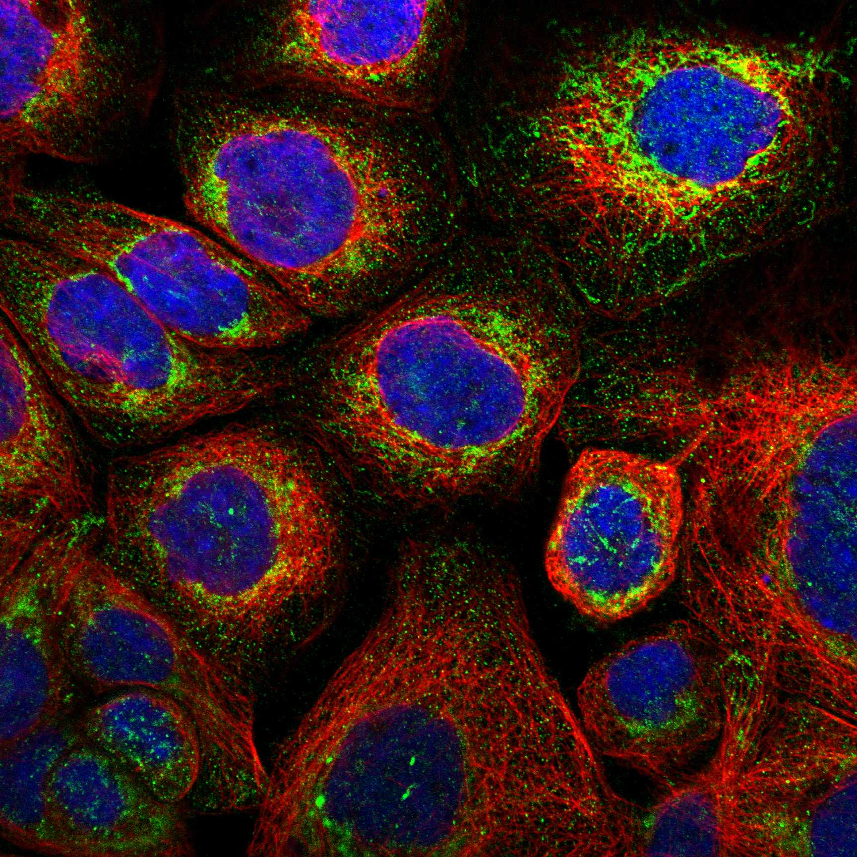
IF/ICC Q&A: What localisation should IF show?Mitochondrial staining is the expected IF/ICC localisation (HPA: mitochondria, Enhanced; UniProt P30084: matrix). HPA lists A-431, U-251MG, U2OS, and NIH 3T3 images; this is a localisation comparison, not an IF protocol recommendation.
Why is my staining missing, weak or wrong?
SituationLikely causeNext action
The positive control and test section show no signal.A shared staining-step failure is possible (standard IHC practice); the supplied ECHS1 sources do not identify a target-specific fixation sensitivity.Verify primary antibody application, detection reagents, and the run control; review retrieval conditions as a general IHC workflow check (standard IHC practice).
Granules are weak in an expected High-staining cell population (HPA: High examples).Weak detection or an unsuitable working dilution may reduce visible contrast (standard IHC practice); the payload provides no dilution to prescribe.Compare with a concurrently stained known-positive section and optimise the antibody dilution and detection conditions using the antibody's IHC instructions (standard IHC practice).
Nuclear or membrane staining overwhelms cytoplasmic granules.That distribution conflicts with the mitochondrial matrix assignment (UniProt P30084) and HPA's granular cytoplasmic IHC profile.Review staining specificity with a known-positive section and an appropriate control lacking primary antibody; record the discordant compartment rather than scoring it as expected ECHS1 (standard IHC practice).
Colour appears broadly in tissue even when primary antibody is omitted.The signal may reflect endogenous detection activity or nonspecific detection chemistry (standard IHC practice).Review the detection-only control and the detection system's blocking steps; interpret ECHS1 only where specific granular cytoplasmic staining remains (standard IHC practice; HPA: granular cytoplasm).
Background haze makes cellular granules difficult to resolve.Excess reagent, inadequate blocking, or insufficient washing can produce diffuse IHC background (standard IHC practice).Review blocking and wash steps and optimise primary antibody concentration; judge localisation only after background permits a clear cellular pattern (standard IHC practice).
A test tissue stains differently from a named High-staining HPA example.The supplied HPA list gives selected cell-specific High observations, not a universal intensity standard (HPA: selected positive examples).Identify the stained cell population and compare its compartment and morphology with the HPA profile; use a separate known-positive control to assess run performance (HPA: granular cytoplasm; standard IHC practice).

Sample controls for ECHS1 IHC & IF

🧪Run breast tissue first and assess staining in glandular cells (HPA: High in breast glandular cells). HPA lists no negative tissue because ECHS1 is detected in all 45 scored tissues (HPA: no negative rows); use no-primary and isotype controls for the negative reference, and treat cells without discernible granular cytoplasmic staining on the positive slide as low-staining comparators, not proven ECHS1-negative cells (UniProt P30084: mitochondrial matrix localisation).
Positive control tissue: Adipose tissue (Adipocytes, HPA High)
Negative control tissue: None in HPA: ECHS1 is detected in all 45 scored tissues. Use a no-primary (secondary-only) and an isotype control instead.
ICC-IF cell lines (HPA subcellular resource): HPA ICC-IF images show ECHS1 in A-431, U-251MG, U2OS, NIH 3T3, with annotated localisation: Mitochondria (enhanced) (HPA subcellular).
Technical controls: Include a secondary-only slide, a concentration-matched nonimmune rabbit IgG control with matched clonality if known, and ECHS1-knockout material where available (caption: rabbit anti-ECHS1; clonality unreported). Block endogenous peroxidase and check blood-containing areas for background before interpreting DAB staining (caption: HRP/DAB detection; standard IHC practice).
⚠️Feasibility: The selected A04508-1 paraffin-section caption reports heat retrieval in EDTA at pH 8.0 and antibody incubation at 2 μg/ml overnight at 4°C; it does not establish whether retrieval is required for all specimens (caption: A04508-1 tissue IHC). Its fixative is unreported, and no target-specific fixation window or fixation effect is supplied (caption: fixative not stated). Relative ease of frozen-section IHC or IF is unreported; ICC-IF images support mitochondrial localisation, while blood-associated endogenous peroxidase can complicate interpretation of breast-section DAB staining (HPA: mitochondrial ICC-IF localisation; caption: HRP/DAB detection; standard IHC practice).

HPA tissue IHC evidence for ECHS1

Comprehensive Human Protein Atlas IHC scoring per tissue (reliability: Enhanced — High consistency between antibody staining and RNA expression data.). Rows are taken directly from the HPA tissue chart — click any row's HPA link to view the source.

Positive expression · recommended positive controls

TissueCell typeLevelEvidenceSource
Adipose tissue Adipocytes High Protein (IHC) HPA →
Adrenal gland Glandular cells High Protein (IHC) HPA →
Appendix Glandular cells High Protein (IHC) HPA →
Bone marrow Hematopoietic cells High Protein (IHC) HPA →
Breast Glandular cells High Protein (IHC) HPA →

Undetected expression · recommended negative controls

TissueCell typeLevelEvidenceSource
None in HPA: ECHS1 is detected in all 45 scored tissues. Use a no-primary (secondary-only) and an isotype control instead.
Section 3

Advanced ECHS1 IHC Tips

Troubleshoot ECHS1 staining in paraffin sections by checking retrieval, mitochondrial pattern, controls, and scoring before interpreting chromogenic signal.

Which retrieval condition should I try first for weak ECHS1 staining?
Start with heat-mediated retrieval in EDTA at pH 8.0 (datasheet A04508-1). The selected paraffin-section example then used 10% goat serum, 2 μg/ml primary antibody overnight at 4°C, and DAB detection (datasheet A04508-1). If staining remains weak, vary heating duration on matched sections while holding antibody concentration and detection conditions constant (standard IHC practice). Evaluate recovery by looking for granular cytoplasmic staining in intact cells, alongside a no-primary control; widespread nuclear or diffuse extracellular color does not match the reported ECHS1 pattern (HPA tissue IHC; standard IHC practice).
How should I assess whether fixation caused weak or patchy ECHS1 staining?
The selected image documents a paraffin-embedded section but does not report its fixative, so target-specific ECHS1 fixation sensitivity is unknown (datasheet A04508-1). Record the fixative, fixation duration, tissue thickness, and processing history for each specimen before comparing stain intensity (standard IHC practice). Compare similarly processed sections using the same EDTA pH 8.0 retrieval and 2 μg/ml primary concentration reported for the catalog antibody (datasheet A04508-1). If staining tracks with processing batches or section edges, repeat with matched handling and appropriate controls before attributing the difference to ECHS1 abundance (standard IHC practice).
What staining pattern supports ECHS1 localisation in a paraffin section?
Expect a granular cytoplasmic pattern in chromogenic sections because ECHS1 is assigned to the mitochondrial matrix and HPA reports granular cytoplasmic tissue staining (UniProt P30084 subcellular location; HPA tissue IHC). Mitochondrial staining may appear as fine cytoplasmic granules rather than individually resolved organelles under routine brightfield microscopy (standard IHC practice). Compare cells within the same well-preserved section, including glandular cells where HPA reports high staining, to judge whether the pattern is coherent (HPA tissue IHC). Predominantly nuclear, cell-surface, or extracellular DAB warrants review of counterstain, background controls, and tissue integrity before calling it ECHS1 (UniProt P30084 subcellular location; standard IHC practice).
How could processing or epitope location affect my ECHS1 result?
ECHS1 is annotated with a mature chain spanning residues 28–290, no annotated isoforms, and no transmembrane segment (UniProt P30084 processing; UniProt P30084 isoforms; UniProt P30084 topology). The supplied evidence does not locate the catalog antibody’s epitope, so a negative section cannot establish whether its epitope was inaccessible after processing (datasheet A04508-1; standard IHC practice). Document antibody identity and retrieval conditions when comparing sections, especially across staining runs (standard IHC practice). Annotated modifications include residues 46, 101, 114, and 115, but their effect on this antibody’s tissue staining is unreported (UniProt P30084 modified residues; datasheet A04508-1).
How can IF help assess an ambiguous chromogenic ECHS1 pattern?
Use IF as a complementary localisation check: HPA reports mitochondrial ECHS1 in ICC/IF images, whereas the selected catalog example documents chromogenic staining in a paraffin section (HPA subcellular; datasheet A04508-1). Multiplex ECHS1 with a mitochondrial marker and, for glandular regions, an epithelial cell marker to identify the stained compartment and cell population (UniProt P30084 subcellular location; HPA tissue IHC; standard IF practice). Choose spectrally separated fluorophores, favoring a far-red channel when tissue autofluorescence obscures shorter wavelengths, and include single-label controls (standard IF practice). For an epitope in the mitochondrial matrix, validate permeabilisation that provides access across cellular and mitochondrial membranes without losing morphology; the antibody’s epitope location is unspecified (UniProt P30084 subcellular location; datasheet A04508-1; standard IF practice).
What should I check when DAB obscures granular ECHS1 staining?
Include a no-primary section to identify staining from the detection system and endogenous tissue activity (standard IHC practice). Peroxidase blocking and careful DAB development are general chromogenic IHC steps; the selected example reports peroxidase-conjugated detection with DAB but does not establish an ECHS1-specific blocking requirement (datasheet A04508-1; standard IHC practice). If color spreads across stroma or section edges, inspect washing, tissue drying, and chromogen development before adjusting the primary antibody (standard IHC practice). Compare the result with the reported granular cytoplasmic pattern and the catalog example’s 2 μg/ml primary concentration, while keeping exposure and controls matched (HPA tissue IHC; datasheet A04508-1; standard IHC practice).
How should I score ECHS1 staining across paraffin sections? ⚠ ANSWER MARKED FOR VERIFICATION
Define the cell population and viable tissue area before scoring, then use the same thresholds and imaging settings across sections (standard IHC practice). For cellular intensity, report an H-score on a 0–300 scale or the percentage of positive cells; for discrete populations, report positive cells per mm² (standard IHC practice). Normalise counts to the annotated viable area, and compare intensity within the same cell type and processing batch (standard IHC practice). Record granular cytoplasmic signal separately from diffuse background, since HPA describes ubiquitous granular cytoplasmic expression and high staining in several distinct cell populations (HPA tissue IHC).
How do I distinguish convincing ECHS1 signal from artefact?
A convincing result has granular cytoplasmic staining in intact cells, consistent with mitochondrial matrix localisation and the HPA tissue pattern (UniProt P30084 subcellular location; HPA tissue IHC). Check whether the stained population fits the section: HPA reports high staining in breast glandular cells and adipocytes, among other cell types, but intensity can differ across populations (HPA tissue IHC). Treat edge-concentrated color, necrotic debris, and signal persisting on a no-primary section as reasons to investigate processing or detection background (standard IHC practice). Review peroxidase blocking and DAB development when diffuse color masks granules; enzyme-related background cannot establish ECHS1 expression (standard IHC practice).
Boster reagents

Best ECHS1 / Enoyl-CoA hydratase, mitochondrial IHC Antibodies

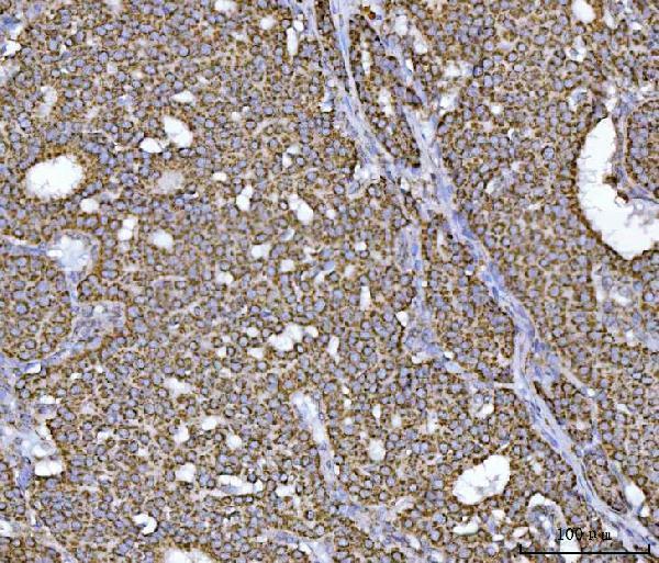
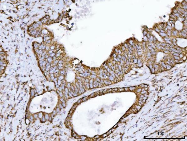
A04508-1 has IHC images from human paraffin tissue and IF images from CACO-2 cells and human paraffin tissue (catalog image captions). Listed reactivity includes human, mouse and rat (catalog reactivity).

Real IHC data IHC analysis of ECHS1 using anti-ECHS1 antibody (A04508-1). ECHS1 was detected in a paraffin-embedded section of human breast cancer tissue. Heat mediated antigen retrieval was performed in EDTA buffer (pH 8.0, epitope retrieval solution). The tissue section was blocked with 10% goat serum. The tissue section was then incubated with 2 μg/ml rabbit anti-ECHS1 Antibody (A04508-1) overnight at 4°C. Peroxidase Conjugated Goat Anti-rabbit IgG was used as secondary antibody and incubated for 30 minutes at 37°C. The tissue section was developed using HRP Conjugated Rabbit IgG Super Vision Assay Kit (Catalog # SV0002) with DAB as the chromogen.
Anti-ECHS1 Antibody ®
Cat # A04508-1

A04508-1 is listed for IHC, IF and ICC (catalog applications). Its IHC images show human breast, colorectal, esophageal and liver cancer paraffin sections; its IF images show CACO-2 cells and human intestinal and lung cancer paraffin sections (catalog image captions).

Which to pick: For tissue IHC, choose A04508-1: its human paraffin section captions document EDTA pH 8 retrieval and 2 μg/ml primary antibody; the fixative is unreported (catalog IHC image captions). For IF/ICC, A04508-1 has cell and tissue IF images using 5 μg/ml primary antibody (catalog IF image captions). For cross-species work, A04508-1 lists human, mouse and rat reactivity, while the supplied IHC/IF images are human only; its host is rabbit and clonality is unreported (catalog reactivity, host and image captions).

Each figure is that product's own IHC / IF validation image from its datasheet.

References

  1. UniProt Consortium. UniProt entry P30084 (ECHM_HUMAN, Enoyl-CoA hydratase, mitochondrial).
  2. Human Protein Atlas. ECHS1 tissue IHC expression (reliability: Enhanced).
  3. Human Protein Atlas. ECHS1 subcellular location (ICC-IF): Localized to the mitochondria..
  4. Human Protein Atlas. ECHS1 antibody validation summary (3 antibodies).
  5. ECHS1, an interacting protein of LASP1, induces sphingolipid-metabolism imbalance to promote colorectal cancer progression by regulating ceramide glycosylation. Cell death & disease 2021 — PMC8494735.
  6. Acetylation-induced degradation of ECHS1 enhances BCAA accumulation and proliferation in KRAS-mutant colorectal cancer. Journal of experimental & clinical cancer research : CR 2025 — PMC12117712.
  7. Nicotinamide Mononucleotide Alleviates Cardiomyopathy Phenotypes Caused by Short-Chain Enoyl-Coa Hydratase 1 Deficiency. JACC. Basic to translational science 2022 — PMC9079797.
  8. Interplay of YEATS2 and GCDH regulates histone crotonylation and drives EMT in head and neck cancer. eLife 2025 — PMC12352869.
  9. PubMed PMID:8012501 — UniProt-cited evidence.
  10. PubMed PMID:9073515 — UniProt-cited evidence.
  11. PubMed PMID:15164054 — UniProt-cited evidence.