This website uses cookies to ensure you get the best experience on our website.


- Table of Contents
Plan EEF1A2 paraffin-section IHC using neuronal cells with high staining as a positive control (HPA tissue IHC). Assess cytoplasmic staining in neurons and skeletal myocytes, while accounting for the antibody’s potential to target proteins from more than one gene (HPA tissue IHC).

Expected localisation, validated protocols, controls and antibodies — the at-a-glance facts below, then the full design guide.
| Expected localisation | Cytoplasmic in neurons and skeletal myocytes (HPA tissue IHC) | |
| Staining pattern | Cytoplasmic neuronal and skeletal myocyte staining (HPA tissue IHC) | |
| Antigen retrieval | Citrate pH 6.0 HIER, heat-mediated (datasheet A03304) | |
| Positive control | Caudate+4 more · see all | |
| Negative control | Adipose tissue+4 more · see all |
| Fixation | Keep fixation consistent across sections (standard IHC practice; not target-specific) | |
| Caveat | Antibody may target proteins from more than one gene (HPA tissue IHC) | |
| Regulation | Brain, heart, skeletal muscle expression (UniProt) | |
| Isoform / epitope | No isoforms listed; annotated chain spans residues 2–463 (UniProt) |
The catalog antibody’s IHC-P protocol (datasheet A03304) is followed by four published EEF1A2 IHC protocols (PMC11551808; PMC6586289; PMC1236916; PMC8007567).
| Sample | Paraffin-embedded rat brain tissue; fixative not specified (datasheet A03304) |
| Fixation | Image fixative and duration unreported (datasheet A03304); verify before use. |
| Sectioning | 4–5 µm sections on charged slides (standard) |
| Deparaffinisation | Xylene, graded ethanol series to water (standard) |
| Antigen retrieval | Heat retrieval: Citrate pH 6.0 (datasheet A03304); 20 min, 95–100 °C (standard) |
| Peroxidase block | 3% H2O2, 10 min, room temperature (standard) |
| Blocking | 10% normal serum of the secondary host, 30 min, room temperature (standard) |
| Primary antibody | Rabbit anti-EEF1A2, 1:50-1:200 (datasheet A03304) |
| Primary incubation | Overnight at 4 °C (standard) |
| Detection | HRP-polymer secondary, DAB chromogen 5–10 min (standard) |
| Counterstain | Hematoxylin, blue, dehydrate and mount (standard) |
| Expected result | EEF1A2-positive staining in neuronal cells of caudate (HPA tissue IHC: High). HPA tissue profile: Cytoplasmic expression in neuronal cells and skeletal myocytes. No signal in the no-primary control. |
In paraffin-section IHC, expect predominantly cytoplasmic staining in neuronal cells and skeletal myocytes (HPA: tissue IHC profile). HPA reports cytosolic localisation by ICC-IF (HPA: subcellular localisation), while UniProt lists an endoplasmic reticulum membrane association without a transmembrane segment (UniProt Q05639: subcellular location and topology). Interpret the pattern cautiously: HPA rates tissue IHC Approved but reports low staining-to-RNA consistency and possible detection of protein from more than one gene (HPA: reliability description).
| Neuronal cytoplasm stains strongly in caudate, cerebral cortex or hippocampus (HPA: High in neuronal cells at these sites). | This matches the reported tissue pattern (HPA: tissue IHC). Judge the neuronal compartment and surrounding cells separately; a uniformly coloured section cannot establish cell-specific localisation (standard IHC practice). |
| Signal appears confined to nuclei or forms a sharp cell-surface outline, with little cytoplasmic staining. | That differs from the reported cytoplasmic tissue pattern and mainly cytosolic ICC-IF pattern (HPA: tissue IHC and subcellular localisation). Check morphology, counterstain and detection controls before interpreting it as EEF1A2 (standard IHC practice). |
| Adipocytes in adipose tissue stain strongly, or broad staining replaces a neuronal pattern (HPA: Not detected in adipocytes). | Treat the result as unexpected relative to the sampled HPA pattern (HPA: tissue IHC). Cross-reactivity or endogenous detection activity are possibilities to investigate, not diagnoses from appearance alone (standard IHC practice). |
| Colour spreads across stroma or empty regions and obscures cell boundaries. | Diffuse background prevents a reliable cell-specific call (standard IHC practice). Review blocking, washes and detection controls; HPA tissue levels do not identify the cause of background in a new assay (HPA: tissue IHC). |
| No signal appears in morphologically intact neuronal cells from a selected positive section. | This conflicts with High neuronal staining reported for several brain regions (HPA: tissue IHC). First assess the positive control and assay conditions; one negative run does not establish absence of EEF1A2 (standard IHC practice). |
| Which cells offer reference patterns? | Caudate, cerebral cortex and hippocampus show High neuronal staining; skeletal myocytes and cardiomyocytes show Medium staining (HPA: tissue IHC). Match the scored cell type to the reference, since a tissue name alone does not define every cell's expected result (standard IHC practice). |
| How should a negative reference be used? | HPA reports no detection in adipocytes from adipose tissue and glandular cells from appendix (HPA: tissue IHC). These are observations for specified cells; they do not prove that every cell in those organs is negative (HPA: tissue IHC). |
| What does localisation evidence support? | Tissue staining is described as cytoplasmic and ICC-IF localisation as mainly cytosolic (HPA: tissue IHC and subcellular localisation). UniProt also lists endoplasmic reticulum membrane localisation but no transmembrane segment (UniProt Q05639: location and topology); these records do not specify a distinctive membrane outline in paraffin IHC. |
| How firm is antibody-based interpretation? | Three listed antibodies have Approved IHC status: HPA051759, HPA056990 and CAB034019 (HPA: antibody validation). The tissue profile still carries low staining-to-RNA consistency and a multi-gene detection caution (HPA: reliability description); resolve unexpected patterns with controls or independent evidence (standard IHC practice). |
| Is target-specific fixation sensitivity known? | No target-specific fixation effect is supplied by the UniProt or HPA records in this guide (UniProt Q05639; HPA: tissue IHC). Retrieval conditions may be checked during general paraffin IHC optimization, without assuming that EEF1A2 has a documented fixation-sensitive epitope (standard IHC practice). |
| Can IF/ICC inform this IHC reading? | Yes: HPA reports an Approved, mainly cytosolic ICC-IF location and lists ICC approval for HPA051759, HPA053862, HPA056990 and CAB034019 (HPA: subcellular localisation and antibody validation). Use that localisation as supporting context; IF/ICC workflow belongs to its own guide. |
| Situation | Likely cause | Next action |
|---|---|---|
| The known-positive brain section has no neuronal colour (HPA: High in neuronal cells). | An IHC workflow or detection failure is possible; the slide alone cannot identify which step failed (standard IHC practice). | Check control tissue integrity, antibody application, retrieval settings and chromogen development against the assay record (standard IHC practice). |
| Neuronal nuclei dominate while cytoplasm is faint. | The distribution conflicts with cytoplasmic tissue staining and mainly cytosolic ICC-IF localisation (HPA: tissue IHC and subcellular localisation). | Recheck cell boundaries and counterstain; compare a positive control and detection-only control before scoring nuclei (standard IHC practice). |
| Adipocytes stain in a proposed negative reference (HPA: Not detected in adipocytes). | Cross-reactivity or endogenous detection activity may contribute; HPA warns that staining may target protein from more than one gene (HPA: reliability description). | Run appropriate detection controls and compare the IHC-validated antibody's pattern with independent evidence where available (standard IHC practice). |
| Brown signal covers stroma and masks cellular detail. | Non-specific reagent binding, insufficient washing or detection background may obscure localisation (standard IHC practice). | Inspect detection-only controls, blocking, wash steps and chromogen development; score only interpretable cellular staining (standard IHC practice). |
| Skeletal myocytes stain less intensely than neurons. | That difference can fit the reference: myocytes are Medium and neurons in several brain regions are High (HPA: tissue IHC). | Score each cell type against its own reported pattern and the run's controls before calling the muscle result a failure (HPA: tissue IHC; standard IHC practice). |
| A convincing cytoplasmic pattern appears in an unexpected cell type. | HPA's cell-specific observations and multi-gene detection caution limit target attribution from colour alone (HPA: tissue IHC and reliability description). | Record the cell type and compartment, examine controls, and seek independent target evidence before assigning the signal to EEF1A2 (standard IHC practice). |
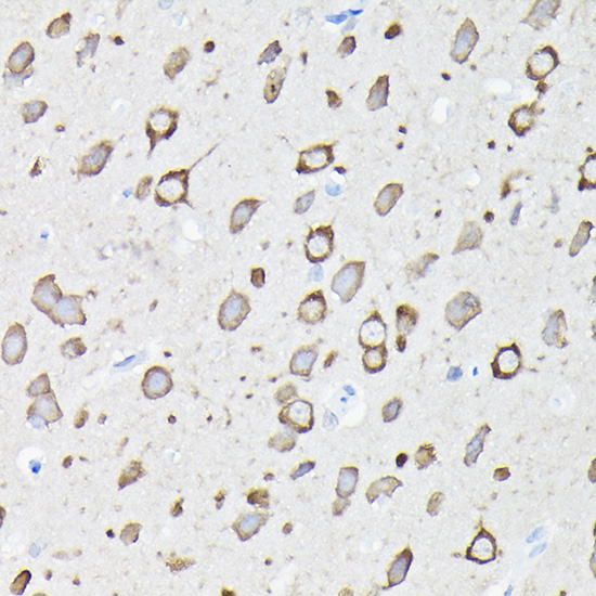
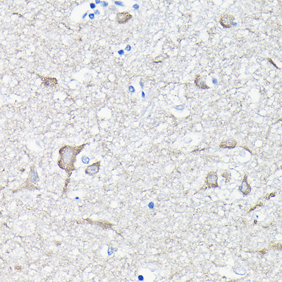


Comprehensive Human Protein Atlas IHC scoring per tissue (reliability: Approved — Low consistency between antibody staining and RNA expression data. Caution, targets protein from more than one gene.). Rows are taken directly from the HPA tissue chart — click any row's HPA link to view the source.
| Tissue | Cell type | Level | Evidence | Source |
|---|---|---|---|---|
| Caudate | Neuronal cells | High | Protein (IHC) | HPA → |
| Cerebellum | Cells in molecular layer | High | Protein (IHC) | HPA → |
| Cerebral cortex | Neuronal cells | High | Protein (IHC) | HPA → |
| Hippocampus | Neuronal cells | High | Protein (IHC) | HPA → |
| Adrenal gland | Glandular cells | Medium | Protein (IHC) | HPA → |
| Tissue | Cell type | Level | Evidence | Source |
|---|---|---|---|---|
| Adipose tissue | Adipocytes | Not detected | Protein (IHC) | HPA → |
| Appendix | Glandular cells | Not detected | Protein (IHC) | HPA → |
| Bone marrow | Hematopoietic cells | Not detected | Protein (IHC) | HPA → |
| Breast | Adipocytes | Not detected | Protein (IHC) | HPA → |
| Bronchus | Respiratory epithelial cells | Not detected | Protein (IHC) | HPA → |
Troubleshoot EEF1A2 staining in paraffin sections by checking retrieval, cell type, cytoplasmic signal and antibody specificity before comparing scores.
A03304 has IHC images of paraffin-embedded human, mouse, and rat brain (A03304 image captions). No IF data are supplied (catalog applications; IF image alts).
A03304 is listed for IHC and reactivity with human, mouse, and rat (catalog applications; reactivity). Its IHC captions show paraffin-embedded brain from all three species stained at 1:100 after high-pressure retrieval in 10 mM citrate buffer, pH 6.0 (A03304 image captions).
Which to pick: Choose A03304 for paraffin-section tissue IHC across human, mouse, and rat samples; its captions identify a rabbit polyclonal antibody and show IHC staining in each species (A03304 image captions). The catalog IHC range is 1:50–1:200, and the pictured sections used 1:100 with citrate retrieval; the fixative is unreported (catalog IHC dilution; A03304 image captions). For IF/ICC, this payload provides no listed IF application or IF figure for A03304 (catalog applications; IF image alts).