This website uses cookies to ensure you get the best experience on our website.


- Table of Contents
Plan EHD3 chromogenic IHC on paraffin sections using the catalog antibody's IHC-P conditions (datasheet A04576-1). Assess the cytoplasmic tissue pattern (HPA tissue IHC) while accounting for the warning that the antibody may detect proteins from more than one gene (HPA tissue IHC).

Expected localisation, validated protocols, controls and antibodies — the at-a-glance facts below, then the full design guide.
| Expected localisation | Cytoplasmic staining in selected tissue cells (HPA tissue IHC) | |
| Staining pattern | Cytoplasmic in endothelial, trophoblastic and some germinal center cells (HPA tissue IHC) | |
| Antigen retrieval | EDTA pH 8.0 HIER, heat-mediated (datasheet A04576-1) | |
| Positive control | Kidney+4 more · see all | |
| Negative control | Adrenal gland+4 more · see all |
| Fixation | Matched tissue-IHC evidence does not establish the fixation claim. Validate the specimen-specific method before use. (selected-SKU IHC image A04576-1) | |
| Caveat | Antibody may detect proteins from more than one gene (HPA tissue IHC) | |
| Regulation | Staining regulation unreported (UniProt) | |
| Isoform / epitope | 2 isoforms; no transmembrane segment; epitope coverage unknown (UniProt) |
The catalog antibody protocol is accompanied by two published chromogenic IHC protocols for EHD3 in rat liver and pituitary adenoma sections (PMC10418749; PMC12715928).
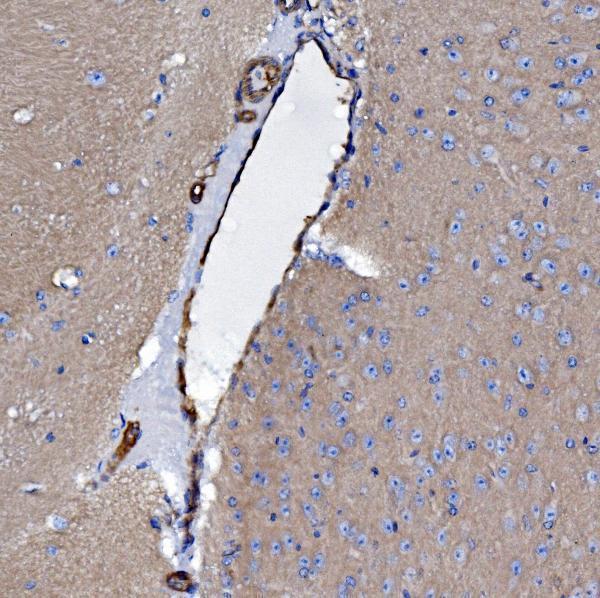
| Sample | Paraffin-embedded mouse brain tissue; fixative not specified (datasheet A04576-1) |
| Fixation | Image fixative and duration unreported (datasheet A04576-1); verify before use. |
| Sectioning | 4–5 µm sections on charged slides (standard) |
| Deparaffinisation | Xylene, graded ethanol series to water (standard) |
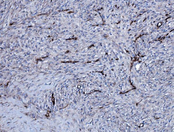
| Antigen retrieval | Heat retrieval: EDTA pH 8.0 (datasheet A04576-1); 20 min, 95–100 °C (standard) |
| Peroxidase block | 3% H2O2, 10 min, room temperature (standard) |
| Blocking | 10% goat serum (datasheet A04576-1) |
| Primary antibody | Rabbit anti-EHD3, 2μg/ml (datasheet A04576-1) |
| Primary incubation | Overnight at 4 °C (datasheet A04576-1) |
| Detection | Streptavidin-biotin complex (SABC), DAB chromogen (datasheet A04576-1) |
| Counterstain | Hematoxylin, blue, dehydrate and mount (standard) |
| Expected result | EHD3-positive staining in cells in glomeruli of kidney (HPA tissue IHC: High). HPA tissue profile: Cytoplasmic expression in endothelial, trophoblastic and a subset of germinal center cells. No signal in the no-primary control. |
EHD3 associates with recycling endosome, cell and cilium membranes despite having no transmembrane segment (UniProt Q9NZN3). In paraffin IHC, expect cytoplasmic staining in endothelial and trophoblastic cells and a subset of germinal center cells (HPA tissue IHC). Kidney glomerular cells show high staining (HPA tissue IHC). Treat this as a guide to interpretation: HPA rates the tissue pattern Approved, with medium RNA concordance and a warning that the antibodies target proteins from more than one gene (HPA tissue IHC).
| Cytoplasmic stain in endothelial or trophoblastic cells, or a subset of germinal center cells (HPA tissue IHC). | This fits the reported tissue pattern (HPA tissue IHC). Compare the stained cells with the section's anatomy; the pattern supports interpretation but does not prove that every signal is EHD3-specific, because HPA warns of antibodies targeting more than one gene (HPA tissue IHC). |
| Prominent nuclear stain or uniform staining across compartments, with little of the reported cytoplasmic pattern (HPA tissue IHC). | A dominant nuclear pattern lacks support from the reported membrane and recycling endosome locations (UniProt Q9NZN3; HPA subcellular ICC-IF). Check morphology, counterstain and controls before calling it positive; compartment mismatch alone does not identify the source of the signal (general IHC practice). |
| Strong chromogen in cell types listed as not detected, such as adrenal glandular or bronchial respiratory epithelial cells (HPA tissue IHC). | This conflicts with those HPA observations, although a listed negative is not an absolute absence claim (HPA tissue IHC). Consider antibody cross-reactivity, endogenous detection activity or misplaced cell identification; controls and tissue context distinguish these possibilities (general IHC practice). |
| Haze, edge staining or widespread deposit that obscures cell boundaries (general IHC practice). | Score this as background until a cell-associated pattern can be resolved (general IHC practice). Examine a no-primary control and the distribution of deposit; diffuse colour cannot establish the cytoplasmic, cell-selective HPA pattern (HPA tissue IHC). |
| No visible stain in kidney glomerular cells, even though they are an HPA high-staining reference (HPA tissue IHC). | A missing reference signal makes a negative result elsewhere hard to interpret (general IHC practice). First check that glomeruli are present and that detection controls worked; then review the IHC assay settings. HPA's high label describes its observation, not a guarantee for every antibody or section (HPA tissue IHC). |
| Tissue and cell context (HPA tissue IHC). | Kidney glomerular cells are high; placental trophoblastic and lymph node germinal center cells are medium, while heart cardiomyocytes are low in HPA IHC (HPA tissue IHC). UniProt reports high heart and brain tissue expression (UniProt Q9NZN3); that tissue-level statement does not assign a high IHC score to cardiomyocytes or every brain cell. |
| Compartment and resolution (UniProt Q9NZN3; HPA subcellular ICC-IF). | Recycling endosome, cell and cilium membranes are reported for EHD3 (UniProt Q9NZN3). HPA ICC-IF chiefly resolves plasma membrane, with primary cilium and transition-zone signals also supported (HPA subcellular ICC-IF). These fine structures may not be separately resolved in chromogenic tissue IHC (general IHC practice). |
| Antibody evidence and specificity limit (HPA tissue IHC; HPA antibodies). | Two listed antibodies have Approved IHC status, while the tissue summary reports medium consistency with RNA and cautions that antibodies target proteins from more than one gene (HPA antibodies; HPA tissue IHC). An anatomically plausible signal therefore remains an observation requiring appropriate controls, not a unique molecular identification. |
| Isoforms and epitope (UniProt Q9NZN3). | UniProt lists two EHD3 isoforms and an EH domain at residues 444–532 (UniProt Q9NZN3). The supplied antibody record gives no epitope positions or isoform coverage (HPA antibodies). Do not infer which isoform produced a stain, or claim that a particular retrieval condition exposes its epitope. |
| Processing and topology (UniProt Q9NZN3). | The record lists a 1–535 chain, no signal peptide or propeptide, and no transmembrane segment (UniProt Q9NZN3). Interpret membrane-associated staining alongside its reported membrane binding and trafficking role; these annotations provide no basis to expect a shed extracellular staining pattern (UniProt Q9NZN3). |
| Situation | Likely cause | Next action |
|---|---|---|
| Kidney glomeruli appear unstained despite their high HPA IHC level (HPA tissue IHC). | The section may lack evaluable glomeruli, or the assay may have failed; HPA's level is an observed reference rather than a guaranteed result (HPA tissue IHC; general IHC practice). | Confirm the cell population on the counterstained section and verify detection controls. Review the antibody's IHC instructions and the run's retrieval and detection records without assuming EHD3-specific fixation sensitivity (general IHC practice). |
| Colour is diffuse or also appears in a no-primary control (general IHC practice). | Background from the detection workflow can obscure cell-associated stain (general IHC practice). | Compare test and no-primary sections, inspect wash and blocking steps, and adjust the detection workflow according to its controls. Score EHD3 only where the reported cell-selective cytoplasmic pattern remains visible (general IHC practice; HPA tissue IHC). |
| Nuclear stain dominates while the reported cytoplasmic cell pattern is absent (HPA tissue IHC). | The dominant compartment does not match the reported EHD3 locations (UniProt Q9NZN3; HPA subcellular ICC-IF); its cause is unresolved. | Check the counterstain and negative control, then compare staining across the reported positive cell populations. Avoid assigning a nuclear EHD3 location from this slide alone (general IHC practice; HPA tissue IHC). |
| Strong stain appears in HPA not-detected adrenal glandular or bronchial epithelial cells (HPA tissue IHC). | Possible explanations include cross-reactivity, endogenous detection activity or cell identification error; the HPA antibody warning makes specificity a concern (HPA tissue IHC; general IHC practice). | Recheck anatomy and run an appropriate no-primary and detection control. Report the discrepancy rather than treating the unexpected cell population as confirmed EHD3 expression (general IHC practice; HPA tissue IHC). |
| Heart cardiomyocytes stain weakly although UniProt reports high expression in heart (UniProt Q9NZN3). | HPA specifically lists cardiomyocytes as low by tissue IHC; whole-tissue expression and cell-level stain intensity are different observations (HPA tissue IHC; UniProt Q9NZN3). | Use the HPA cardiomyocyte observation when judging this IHC pattern. Confirm assay performance with a reported high-staining population before interpreting absent cardiac signal (HPA tissue IHC; general IHC practice). |
| IF/ICC Q: Should a plasma-membrane or cilium signal be judged by the paraffin IHC pattern? | HPA's membrane, cilium and transition-zone localisation comes from ICC-IF; its tissue IHC summary describes cytoplasmic cell patterns (HPA subcellular ICC-IF; HPA tissue IHC). | A: Interpret IF/ICC against its own localisation evidence and controls; do not require the same visual resolution in chromogenic paraffin IHC. HPA flags multi-gene targeting for the subcellular result, so localisation alone does not settle specificity (HPA subcellular ICC-IF; general IHC practice). |


![HPA ICC-IF BJ [Human fibroblast] — EHD3 (HPA066751)](https://images.proteinatlas.org/66751/1667_E12_5_blue_red_green.jpg)
![HPA ICC-IF BJ [Human fibroblast] — EHD3 (HPA066751)](https://images.proteinatlas.org/66751/1667_E12_6_blue_red_green.jpg)
Comprehensive Human Protein Atlas IHC scoring per tissue (reliability: Approved — Medium consistency between antibody staining and RNA expression data. Caution, targets protein from more than one gene. Pending external verification.). Rows are taken directly from the HPA tissue chart — click any row's HPA link to view the source.
| Tissue | Cell type | Level | Evidence | Source |
|---|---|---|---|---|
| Kidney | Cells in glomeruli | High | Protein (IHC) | HPA → |
| Adipose tissue | Adipocytes | Medium | Protein (IHC) | HPA → |
| Appendix | Lymphoid tissue | Medium | Protein (IHC) | HPA → |
| Lung | Alveolar cells | Medium | Protein (IHC) | HPA → |
| Lymph node | Germinal center cells | Medium | Protein (IHC) | HPA → |
| Tissue | Cell type | Level | Evidence | Source |
|---|---|---|---|---|
| Adrenal gland | Glandular cells | Not detected | Protein (IHC) | HPA → |
| Bone marrow | Hematopoietic cells | Not detected | Protein (IHC) | HPA → |
| Bronchus | Respiratory epithelial cells | Not detected | Protein (IHC) | HPA → |
| Caudate | Glial cells | Not detected | Protein (IHC) | HPA → |
| Cerebellum | Cells in granular layer | Not detected | Protein (IHC) | HPA → |
Troubleshoot EHD3 staining in paraffin sections by checking retrieval, cellular context and controls before interpreting chromogenic signal.
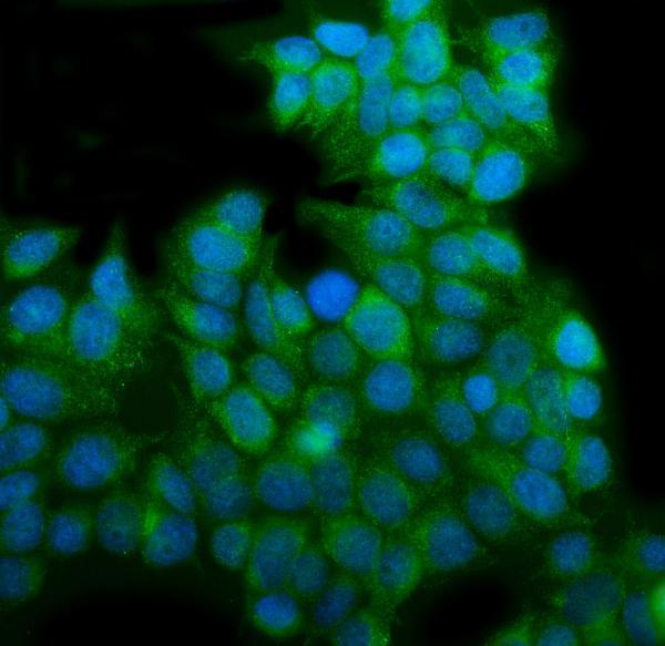
The IHC-validated anti-EHD3 antibody has paraffin-section images from mouse brain and human ovarian and renal tumors, plus an IF/ICC image from MCF-7 cells (catalog image captions).
A04576-1 has IHC images from paraffin-embedded mouse brain, human ovarian serous adenocarcinoma, and human renal clear cell carcinoma (A04576-1 IHC captions). A04576-1 is also listed for IF/ICC and has an IF image from MCF-7 cells; its listed reactivity is human, monkey, mouse, and rat (catalog applications, IF caption, and reactivity).
Which to pick: Choose A04576-1 for tissue IHC when its paraffin-section examples match your workflow: the captions describe EDTA retrieval at pH 8.0 and 2 µg/mL primary antibody, but do not report the fixative (A04576-1 IHC captions). The same SKU is listed for IF/ICC and has an MCF-7 IF image; its listed reactivity spans human, monkey, mouse, and rat, while the supplied IHC images show only human and mouse tissue (catalog applications, reactivity, and image captions).