This website uses cookies to ensure you get the best experience on our website.


- Table of Contents
Plan EI24 chromogenic IHC-P using the cytoplasmic pattern in most tissues and membranous staining in parathyroid glandular cells (HPA tissue IHC). The catalog antibody starts at 5 μg/mL in rat IHC; optimize it for human sections (datasheet: rat IHC).

Expected localisation, validated protocols, controls and antibodies — the at-a-glance facts below, then the full design guide.
| Expected localisation | Mostly cytoplasmic; parathyroid membranous (HPA tissue IHC) | |
| Staining pattern | Cytoplasmic in most tissues; membranous in parathyroid glandular cells (HPA tissue IHC) | |
| Antigen retrieval | Tris-EDTA pH 9.0 HIER, 95–98 °C, 20 min (rule: nuclear antigen) | |
| Positive control | Parathyroid gland+4 more · see all | |
| Negative control | Adipose tissue+3 more · see all |
| Fixation | Keep fixation consistent across sections. (standard IHC practice; not target-specific) | |
| Caveat | Antibody staining has low consistency with RNA expression (HPA tissue IHC) | |
| Regulation | p53-mediated apoptosis role; staining induction unknown (UniProt) | |
| Isoform / epitope | 2 isoforms; epitope differences and membrane side unknown (UniProt) |
The catalog antibody's IHC-P protocol is accompanied by published EI24 IHC methods for breast, esophageal and pancreatic cancer specimens (PMC13489907; PMC7471874; PMC6189671; PMC5386697).
| Sample | Tissue sections; selected-image fixative not specified (standard IHC workflow) |
| Fixation | Image fixative and duration unreported (datasheet A06183); verify before use. |
| Sectioning | 4–5 µm sections on charged slides (standard) |
| Deparaffinisation | Xylene, graded ethanol series to water (standard) |
| Antigen retrieval | Heat-induced epitope retrieval in Tris-EDTA buffer, pH 9.0, 20 min at 95–98 °C (standard rule: nuclear antigen) |
| Peroxidase block | 3% H2O2, 10 min, room temperature (standard) |
| Blocking | 10% normal serum of the secondary host, 30 min, room temperature (standard) |
| Primary antibody | Rabbit anti-EI24, 5 μg/mL (datasheet A06183) |
| Primary incubation | Overnight at 4 °C (standard) |
| Detection | HRP-polymer secondary, DAB chromogen 5–10 min (standard) |
| Counterstain | Hematoxylin, blue, dehydrate and mount (standard) |
| Expected result | EI24-positive staining in glandular cells of parathyroid gland (HPA tissue IHC: High). HPA tissue profile: Cytoplasmic expression in most tissues. Membranous expression in parathyroid gland. No signal in the no-primary control. |
EI24 staining is predominantly cytoplasmic across most tissues, with membranous staining reported in parathyroid glandular cells (HPA tissue IHC). Expect strong staining in parathyroid glandular cells and elongated or late spermatids (HPA: High). EI24 has five transmembrane segments and is annotated at the endoplasmic reticulum and nuclear membranes and in the cytoplasm (UniProt O14681 topology and subcellular location). HPA rates its tissue IHC evidence Approved, while reporting low consistency with RNA expression (HPA tissue IHC).
| Strong glandular staining in parathyroid, with a membranous pattern (HPA: High; membranous expression). | This matches a documented positive tissue and compartment. Compare staining with the glandular cells on the same section; a strong signal alone is less informative if its cellular pattern is unclear (HPA tissue IHC). |
| Cytoplasmic staining in other tissues, or strong staining in elongated or late spermatids (HPA tissue IHC). | Cytoplasmic staining fits HPA's pattern across most tissues; the spermatids provide another reported strong positive population (HPA: High). Score identifiable cells and their compartment rather than treating all staining on the slide as equivalent. |
| Predominantly nuclear staining where the expected cytoplasmic or parathyroid membranous pattern is absent (HPA tissue IHC). | Treat this as a pattern mismatch requiring investigation, not confirmed EI24 localization. UniProt annotates the nuclear membrane, but that annotation does not establish diffuse nuclear IHC staining (UniProt O14681 subcellular location). |
| Strong signal in adipocytes, marrow hematopoietic cells, chondrocytes or splenic red-pulp cells (HPA: Not detected). | These are reported negative cell populations. Check morphology and controls for cross-reactivity or endogenous detection activity before calling the signal EI24; HPA's negative observations are reference patterns, not proof that every specimen must be blank. |
| Diffuse stain across cells and tissue spaces, or no signal in parathyroid glandular cells (HPA: High). | Diffuse stain obscures compartment and cell-type assessment and calls for a background check. An absent signal in a reported strong positive calls for review of the IHC workflow and control section before interpreting other negative areas. |
| Subcellular reference pattern | HPA reports mainly cytoplasmic tissue staining and membranous staining in parathyroid gland (HPA tissue IHC). UniProt lists cytoplasm and endoplasmic reticulum and nuclear membranes, with five transmembrane segments (UniProt O14681); these annotations do not predict a uniquely visible membrane outline in every section. |
| Tissue and cell selection | Parathyroid glandular cells and elongated or late spermatids are reported High; several other listed cell populations are Medium (HPA tissue IHC). Select and score the named cells within each tissue. A tissue name alone does not establish that every cell should stain. |
| Strength of HPA tissue evidence | The tissue IHC profile is Approved, with low consistency between antibody staining and RNA expression (HPA tissue IHC). Treat the documented protein staining as a useful visual reference, while checking unexpected results with appropriate controls rather than claiming RNA agreement. |
| Antibody-specific validation | HPA047165 and HPA051029 each have Approved IHC status; only HPA051029 has Supported ICC status (HPA antibodies). Those classifications apply to the listed antibodies. They do not establish that an unlisted antibody will reproduce either staining pattern. |
| Assay and preparation | HPA's ICC-IF summary places EI24 mainly in cytosol, with additional endoplasmic reticulum and Golgi localization (HPA subcellular ICC-IF). That cellular image reference can inform a localization question, but tissue IHC remains the reference for scoring a paraffin section; no ICC-IF protocol is specified here. |
| Protein forms and fixation evidence | UniProt lists two isoforms, no signal peptide or propeptide, and no annotated glycosylation sites (UniProt O14681). These facts do not identify the antibody epitope or establish differential staining. Target-specific fixation sensitivity is unreported in the supplied sources. |
| Situation | Likely cause | Next action |
|---|---|---|
| Parathyroid glandular cells lack the expected strong signal (HPA: High). | The reference positive has failed, or the sampled section does not show comparable glandular cells; the supplied sources do not identify an EI24-specific fixation cause. | Confirm cell morphology and the positive control, then review the antibody's IHC-P instructions and routine retrieval, dilution and detection steps. Record any optimization as assay-specific rather than an established EI24 fixation effect. |
| Signal is diffuse, with no readable cellular boundary or cytoplasmic distribution. | General IHC background can arise from nonspecific binding or the detection system; diffuse staining alone does not establish EI24 localization. | Compare with a no-primary control and inspect blocking, washing and chromogen development. Reassess the specimen only after cell outlines and the expected compartment can be distinguished. |
| Strong stain appears in a reported negative cell population (HPA: Not detected). | Cross-reactivity or endogenous detection activity is possible; a single discordant cell population cannot identify which mechanism is responsible. | Check a no-primary control and cell morphology. For an enzyme-based chromogen, review the relevant endogenous-enzyme blocking step; if signal persists, assess antibody specificity with an independent suitable control. |
| Staining is predominantly diffuse nuclear rather than cytoplasmic or parathyroid membranous (HPA tissue IHC). | The compartment differs from the tissue reference. UniProt's nuclear-membrane annotation does not by itself explain staining throughout the nucleus (UniProt O14681). | Inspect counterstain and optical focus to verify the compartment, then compare a known-positive section and detection controls before assigning the nuclear signal to EI24. |
| A parathyroid section shows staining, but glandular cells lack the expected membranous emphasis (HPA tissue IHC). | The observed pattern differs from HPA's parathyroid description; intensity alone cannot settle localization, and the sources do not establish a target-specific preparation cause. | Score glandular cells separately from adjacent tissue, check section quality and control staining, and document the actual compartment instead of forcing a positive-pattern call. |
| ICC-IF shows cytosol plus organelle signal, while the paraffin IHC section looks mainly cytoplasmic. | These are distinct assay readouts: HPA reports cytosol as the main ICC-IF location, with additional endoplasmic reticulum and Golgi signal (HPA subcellular ICC-IF). | Use the tissue IHC pattern to interpret the paraffin section and the ICC-IF record for its own localization question. Do not treat an ICC-IF organelle pattern as an IHC-P scoring requirement. |


Comprehensive Human Protein Atlas IHC scoring per tissue (reliability: Approved — Low consistency between antibody staining and RNA expression data.). Rows are taken directly from the HPA tissue chart — click any row's HPA link to view the source.
| Tissue | Cell type | Level | Evidence | Source |
|---|---|---|---|---|
| Parathyroid gland | Glandular cells | High | Protein (IHC) | HPA → |
| Testis | Elongated or late spermatids | High | Protein (IHC) | HPA → |
| Adrenal gland | Glandular cells | Medium | Protein (IHC) | HPA → |
| Appendix | Glandular cells | Medium | Protein (IHC) | HPA → |
| Breast | Glandular cells | Medium | Protein (IHC) | HPA → |
| Tissue | Cell type | Level | Evidence | Source |
|---|---|---|---|---|
| Adipose tissue | Adipocytes | Not detected | Protein (IHC) | HPA → |
| Bone marrow | Hematopoietic cells | Not detected | Protein (IHC) | HPA → |
| Soft tissue | Chondrocytes | Not detected | Protein (IHC) | HPA → |
| Spleen | Cells in red pulp | Not detected | Protein (IHC) | HPA → |
Troubleshoot EI24 staining in paraffin sections by checking retrieval, tissue controls, compartment patterns and scoring before interpreting chromogenic signal.
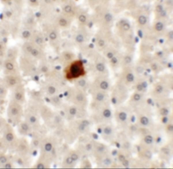
One anti-EI24 antibody has real IHC data from rat liver tissue (catalog image caption); human and mouse reactivity is listed, but IHC validation is reported only in rat samples (catalog datasheet).
A06183 will render with an IHC image of rat liver tissue stained at 5 μg/mL (catalog image caption). Its listed applications include IHC-P, and its listed reactivity covers human, mouse and rat (catalog application and reactivity lists).
Which to pick: Choose A06183 for paraffin-section IHC: IHC-P is listed, and its own image shows rat liver staining at 5 μg/mL (catalog application list; catalog image caption). The image caption does not report the fixative or tissue processing, and clonality is unreported (catalog image caption; catalog clone field). For IF/ICC or human or mouse IHC, no validation is shown; A06183 lists human and mouse reactivity but provides neither an IF application nor IF data (catalog application and reactivity lists; catalog image fields).