This website uses cookies to ensure you get the best experience on our website.


- Table of Contents
Plan EIF1AY paraffin-section IHC using the catalog antibody's 1:100–1:300 dilution range (datasheet A30678). Interpret cytoplasmic and nuclear staining cautiously because HPA rates the tissue-IHC evidence uncertain and warns that the antibody targets protein from more than one gene (HPA tissue IHC).

Expected localisation, validated protocols, controls and antibodies — the at-a-glance facts below, then the full design guide.
| Expected localisation | Tissue: cytoplasm and nuclei (HPA tissue IHC); molecular: cytoplasm (UniProt) | |
| Staining pattern | Glandular, lymphoid and hematopoietic cells: cytoplasm and nuclei (HPA tissue IHC) | |
| Antigen retrieval | Tris-EDTA pH 8.0 HIER, heat-mediated (datasheet A30678) | |
| Positive control | Adrenal gland+4 more · see all | |
| Negative control | Skeletal muscle+1 more · see all |
| Fixation | Keep fixation consistent across sections. (standard IHC practice; not target-specific) | |
| Caveat | Cross-gene staining is possible; reliability is uncertain (HPA tissue IHC) | |
| Regulation | RNA enhanced in heart muscle and tongue (HPA tissue RNA) | |
| Isoform / epitope | No isoforms reported; one 1–144 chain (UniProt) |
The catalog antibody protocol (datasheet A30678) and one published EIF1AY IHC protocol (PMC13018312) provide starting conditions for paraffin sections.
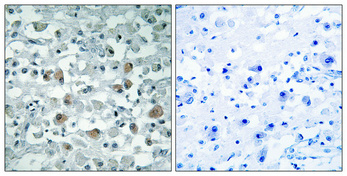
| Sample | Paraffin-embedded Human brain tissue; fixative not specified (datasheet A30678) |
| Fixation | Image fixative and duration unreported (datasheet A30678); verify before use. |
| Sectioning | 4–5 µm sections on charged slides (standard) |
| Deparaffinisation | Xylene, graded ethanol series to water (standard) |
| Antigen retrieval | Heat retrieval: Tris-EDTA pH 8.0 (datasheet A30678); 20 min, 95–100 °C (standard) |
| Peroxidase block | 3% H2O2, 10 min, room temperature (standard) |
| Blocking | 10% normal serum of the secondary host, 30 min, room temperature (standard) |
| Primary antibody | Rabbit anti-EIF1AY, 1:100 - 1:300 (datasheet A30678) |
| Primary incubation | Overnight at 4 °C (standard) |
| Detection | HRP-polymer secondary, DAB chromogen 5–10 min (standard) |
| Counterstain | Hematoxylin, blue, dehydrate and mount (standard) |
| Expected result | EIF1AY-positive staining in glandular cells of adrenal gland (HPA tissue IHC: High). HPA tissue profile: General cytoplasmic and nuclear expression. No signal in the no-primary control. |
EIF1AY is a soluble cytoplasmic translation initiation factor with no transmembrane segment (UniProt O14602: cytoplasm; topology). In paraffin-section IHC, look for cytoplasmic staining in cell populations reported as high, including adrenal glandular cells, esophageal squamous epithelial cells, and bone-marrow hematopoietic cells (HPA: High). Interpret that pattern cautiously: HPA rates its tissue IHC Uncertain because the antibody targets proteins from more than one gene (HPA: reliability).
| Cytoplasmic staining in adrenal glandular cells, esophageal squamous epithelium, or bone-marrow hematopoietic cells (HPA: High). | This agrees with the reported tissue distribution and UniProt cytoplasmic location (HPA: High; UniProt O14602: cytoplasm). It supports a plausible staining pattern, but does not identify EIF1AY specifically: HPA rates the tissue IHC Uncertain because the antibody targets proteins from more than one gene (HPA: reliability). |
| Predominantly nuclear staining, with little or no cytoplasmic signal in a reported high-staining cell population (HPA: High). | Check for an artefact or a detection problem before calling this the expected pattern: UniProt places EIF1AY in the cytoplasm, and HPA ICC-IF places the detected signal in the cytosol (UniProt O14602; HPA: ICC-IF cytosol). Nuclear signal alone is not proof of artefact, however; HPA also describes general nuclear expression in tissue IHC (HPA: tissue profile, Uncertain). |
| Strong staining in skeletal myocytes or smooth-muscle cells (HPA: Not detected). | That conflicts with the supplied HPA tissue IHC observations and warrants a specificity check (HPA: Not detected; HPA: IHC Uncertain). Consider cross-reactivity or endogenous chromogen-producing activity as possible causes (standard IHC practice); the slide alone cannot distinguish them, and the HPA profile does not prove an EIF1AY-specific negative result. |
| Diffuse colour across tissue, empty areas, or many unrelated structures, with little cell-level distinction (standard IHC practice). | Treat this as background until controls show otherwise. Nonspecific antibody binding, incomplete blocking, or residual endogenous detection activity can obscure a cytoplasmic pattern (standard IHC practice). The supplied HPA data describe cellular patterns and validation status; they do not attribute diffuse background to a particular EIF1AY property (HPA: tissue profile). |
| No staining in an adrenal glandular-cell section that is otherwise interpretable (HPA: High). | Check whether the positive control and detection system worked before interpreting the test section (standard IHC practice). HPA reports high staining in adrenal glandular cells, but its IHC reliability is Uncertain; one negative slide cannot establish biological absence or confirm antibody specificity (HPA: High; HPA: reliability). |
| Antibody validation (HPA: HPA002561 IHC Uncertain) | HPA flags targeting of proteins from more than one gene; a matching tissue pattern cannot by itself assign the chromogen to EIF1AY (HPA: reliability; HPA002561 IHC Uncertain). |
| Compartment evidence (UniProt O14602: cytoplasm; HPA: ICC-IF cytosol) | Cytoplasmic signal fits UniProt and the approved HPA ICC-IF location. HPA tissue IHC also reports general nuclear expression, so nuclear signal needs cautious interpretation (HPA: tissue profile, Uncertain). |
| Tissue distribution (HPA: High; Low; Not detected) | HPA reports high glandular, lymphoid, hematopoietic, and esophageal epithelial staining; low glomerular-cell and cardiomyocyte staining; and no detected muscle-cell staining. These are observations from uncertain IHC, not validated EIF1AY-only controls (HPA: tissue profile; reliability). |
| Protein architecture (UniProt O14602: topology; processing) | UniProt lists a 144-residue chain, no transmembrane segment, and no signal peptide or propeptide. These support interpretation as a cellular, soluble target; they do not predict retrieval conditions or fixation sensitivity (UniProt O14602: topology; processing). |
| IF/ICC evidence (HPA: cytosol approved) | HPA's cytosol call is approved for ICC-IF, but it carries a multiple-gene-targeting caution. It helps compare compartments; it does not validate the paraffin-section IHC signal as EIF1AY-specific (HPA: subcellular summary; HPA002561 IHC Uncertain). |
| Situation | Likely cause | Next action |
|---|---|---|
| No chromogen in a reported high-staining tissue, such as adrenal glandular cells (HPA: High). | The assay may have failed, or this section may differ from the HPA observation; HPA IHC is Uncertain (standard IHC practice; HPA: reliability). | Review the positive-control section, tissue preservation, primary-antibody step, and detection reagents before scoring absence (standard IHC practice). Do not infer an EIF1AY-specific negative from one slide (HPA: reliability). |
| Nuclear signal dominates while cytoplasm is nearly blank (UniProt O14602: cytoplasm). | Detection artefact is possible, although HPA's uncertain tissue IHC also records general nuclear expression (standard IHC practice; HPA: tissue profile). | Compare the cytoplasm with a reported high-staining control and inspect negative controls (standard IHC practice; HPA: High). Record nuclear staining separately rather than automatically counting it as EIF1AY-positive (HPA: IHC Uncertain). |
| Smooth-muscle cells or skeletal myocytes stain strongly (HPA: Not detected). | Cross-reactivity or endogenous detection activity may produce discordant staining (standard IHC practice). HPA's multiple-gene warning makes target assignment especially uncertain (HPA: reliability). | Check a no-primary control for detection background and compare the tissue pattern with HPA's Not detected entries (standard IHC practice; HPA: tissue profile). Treat persistent staining as unresolved until independently validated. |
| Diffuse chromogen prevents cell-level scoring (standard IHC practice). | Nonspecific binding or endogenous activity can raise background in chromogenic IHC (standard IHC practice). The supplied sources identify no EIF1AY-specific cause (UniProt O14602; HPA: tissue profile). | Inspect no-primary controls and review blocking, washing, and detection steps (standard IHC practice). Score only distinguishable cellular staining; do not convert a uniform haze into a positive tissue call. |
| Cardiomyocytes look weaker than adrenal glandular cells (HPA: Low in cardiomyocytes; High in adrenal glandular cells). | The difference agrees with HPA's observed levels, although those levels come from uncertain tissue IHC (HPA: tissue profile; reliability). | Score intensity by cell type with comparable slide processing and a positive control (standard IHC practice). Do not require every tissue to match the strongest HPA category or infer EIF1AY abundance from chromogen alone (HPA: reliability). |
| Does approved HPA ICC-IF cytosol staining establish an IHC-P protocol or confirm EIF1AY specificity? (HPA: ICC-IF approved). | No: the ICC-IF location has a multiple-gene-targeting caution, and HPA002561 remains IHC Uncertain (HPA: subcellular summary; HPA: antibody validation). | Use the ICC-IF result only as compartment context. Evaluate paraffin-section IHC with its own controls and validation; this section supplies no IF/ICC protocol option (HPA: ICC-IF approved; HPA: IHC Uncertain). |


Comprehensive Human Protein Atlas IHC scoring per tissue (reliability: Uncertain — Caution, targets protein from more than one gene. Pending external verification.). Rows are taken directly from the HPA tissue chart — click any row's HPA link to view the source.
| Tissue | Cell type | Level | Evidence | Source |
|---|---|---|---|---|
| Adrenal gland | Glandular cells | High | Protein (IHC) | HPA → |
| Appendix | Lymphoid tissue | High | Protein (IHC) | HPA → |
| Bone marrow | Hematopoietic cells | High | Protein (IHC) | HPA → |
| Cervix | Glandular cells | High | Protein (IHC) | HPA → |
| Endometrium | Glandular cells | High | Protein (IHC) | HPA → |
Use the paraffin-section evidence as a starting point, then evaluate compartment, controls and cross-reactivity before attributing staining to EIF1AY (caption A30678; HPA tissue IHC).
The rendered anti-EIF1AY card shows paraffin-embedded human brain IHC (A30678 IHC image caption). IF/ICC applications and human, mouse, and rat reactivity are catalog-listed (catalog: A30678 applications/reactivity).
A30678 is the rendered SKU, with an IHC image of paraffin-embedded human brain at 1:100 after Tris-EDTA pH 8.0 retrieval (A30678 IHC image caption). IHC, IF, and ICC are listed applications, but no IF image is supplied (catalog: A30678 applications/if_image_alts).
Which to pick: For tissue IHC, choose A30678: its own image caption documents paraffin-embedded human brain and a peptide pre-absorption control; the fixative is unreported (A30678 IHC image caption). For IF/ICC, A30678 is the polyclonal, application-listed option with an IF dilution of 1:50, although no IF image is supplied (catalog: A30678 dilution_raw/applications/if_dilution/if_image_alts). Both A30678 and A14440 list human, mouse, and rat reactivity; A14440 lists IHC but has no IHC image in the payload (catalog: A30678 and A14440 reactivity/applications/ihc_image_alts).