This website uses cookies to ensure you get the best experience on our website.


- Table of Contents
Plan EIF2S1 paraffin IHC around the widespread cytoplasmic tissue pattern (HPA tissue IHC). Use total EIF2S1 staining for protein distribution and phospho-S51 staining when assessing its stress response (UniProt; catalog P04387).

Expected localisation, validated protocols, controls and antibodies — the at-a-glance facts below, then the full design guide.
| Expected localisation | Cytoplasmic staining in tissue (HPA tissue IHC) | |
| Staining pattern | Widespread cytoplasmic staining across cell types (HPA tissue IHC) | |
| Antigen retrieval | Citrate pH 6.0 HIER, 95–98 °C, 20 min (rule: cytoplasmic / membrane antigen) | |
| Positive control | Adipose tissue+4 more · see all | |
| Negative control | None in HPA (detected in all 45 tissues); use no-primary + isotype controls |
| Fixation | Matched tissue-IHC evidence does not establish the fixation claim. Validate the specimen-specific method before use. (selected-SKU IHC image M04387-6) | |
| Caveat | Phospho-S51 staining varies with stress state (UniProt; catalog P04387) | |
| Regulation | Stress induces EIF2S1 phosphorylation (UniProt) | |
| Isoform / epitope | 0 annotated isoforms; one 1–315 chain (UniProt) |
The catalog antibody protocol is followed by published EIF2S1 IHC methods for intestinal-type adenocarcinoma tissue arrays and paraffin-embedded liver sections (PMC8616251; PMC10079382).
| Sample | Tissue sections; selected-image fixative not specified (standard IHC workflow) |
| Fixation | Image fixative and duration unreported (datasheet M04387-6); verify before use. |
| Sectioning | 4–5 µm sections on charged slides (standard) |
| Deparaffinisation | Xylene, graded ethanol series to water (standard) |
| Antigen retrieval | Heat-induced epitope retrieval in citrate buffer, pH 6.0, 20 min at 95–98 °C (standard rule: cytoplasmic / membrane antigen) |
| Peroxidase block | 3% H2O2, 10 min, room temperature (standard) |
| Blocking | 10% normal serum of the secondary host, 30 min, room temperature (standard) |
| Primary antibody | Rabbit anti-EIF2S1, 1:1000-1:5000 (datasheet M04387-6) |
| Primary incubation | Overnight at 4 °C (standard) |
| Detection | HRP-polymer secondary, DAB chromogen 5–10 min (standard) |
| Counterstain | Hematoxylin, blue, dehydrate and mount (standard) |
| Expected result | EIF2S1-positive staining in adipocytes of adipose tissue (HPA tissue IHC: High). HPA tissue profile: Ubiquitous cytoplasmic expression. No signal in the no-primary control. |
EIF2S1 should show broadly distributed cytoplasmic staining in paraffin sections (HPA: ubiquitous cytoplasmic expression; IHC reliability Supported, with medium consistency against RNA data). High staining is reported in adipocytes, respiratory epithelial cells, neurons, glandular cells and some endothelial cells (HPA: tissue IHC). A dominant membrane pattern is unexpected for this protein (UniProt P05198: no transmembrane segment).
| Cytoplasmic staining across several cell populations, with stronger cells in expected sites. | This fits the reported broad distribution (HPA: ubiquitous cytoplasmic expression). Adipocytes, bronchial respiratory epithelium, cerebellar Purkinje cells, cerebral cortical neurons, duodenal and endometrial glandular cells, and colonic endothelial cells are reported as High (HPA: tissue IHC). Compare intensity within the same staining run; High is an HPA category, not a calibrated score. |
| Predominantly nuclear, crisp membrane, or extracellular staining. | These are poor matches to the reported cytoplasmic tissue pattern and cytosolic localization (HPA: tissue IHC; HPA: ICC-IF). A membrane outline alone has no support from the recorded topology (UniProt P05198: no transmembrane segment). Review morphology, background and controls before assigning the signal to EIF2S1 (general IHC practice). |
| Strong signal in a cell population expected to be low while nearby expected High cells remain blank. | Consider cross-reactivity or endogenous detection activity, particularly if staining persists in a no-primary control (general IHC practice). Interpret the discrepancy against the sampled tissue: cardiomyocytes, skeletal myocytes, smooth muscle cells, cholangiocytes, ovarian follicle cells and chondrocytes are listed as Low, not absent (HPA: tissue IHC). |
| Uniform haze over cells, stroma and blank areas. | This lacks the cell-associated cytoplasmic pattern expected for EIF2S1 (HPA: tissue IHC). Diffuse chromogen can reflect background from detection reagents, insufficient blocking or washing, or an overly concentrated primary (general IHC practice). Use a no-primary control and assess whether cellular contrast remains after background is reduced. |
| No staining in a known High cell population. | An all-blank section is inconclusive when an expected positive population is present (HPA: tissue IHC). Check that tissue morphology is preserved and the detection system works; then optimize the IHC-validated antibody under its documented conditions (general IHC practice). The supplied sources give no EIF2S1-specific fixation or retrieval sensitivity. |
| Cell and tissue context | Expression is described as ubiquitous and cytoplasmic, with low tissue specificity at the RNA level (HPA: tissue IHC). High and Low labels are relative observations; the supplied HPA set names no negative tissue. Select a documented High population for a positive control and avoid treating a Low population as a knockout control. |
| Antibody validation | HPA064885 and CAB011663 are each listed as IHC Supported (HPA: antibody records). Supported is the supplied status; do not upgrade either antibody to Enhanced. Review the actual catalog antibody's IHC-P documentation before applying a dilution or retrieval condition; neither is provided here. |
| Phosphorylation and mitochondrial stress | EIF2S1 phosphorylation participates in the integrated stress response, and mitochondrial damage can promote relocation to the mitochondrial surface (UniProt P05198: function, subcellular location). Routine tissue IHC intensity does not establish phosphorylation state or mitophagy without a validated state-specific assay (general IHC interpretation). |
| Cytosol and stress granules | Cytosol and stress granules are annotated locations, with stress-granule colocalization supported by similarity (UniProt P05198: subcellular location). HPA describes the observed tissue pattern as ubiquitous cytoplasmic expression (HPA: tissue IHC); do not require puncta to call a routine chromogenic section positive. |
| Retrieval and detection controls | Antigen retrieval, blocking and detection chemistry are adjustable parts of paraffin-section IHC (general IHC practice). Optimize them against a documented positive tissue and a no-primary control; the supplied UniProt and HPA records do not specify an EIF2S1-specific retrieval condition or fixation effect. |
| Situation | Likely cause | Next action |
|---|---|---|
| Expected High cells show no chromogen. | An assay or detection failure is possible; the expected population is documented as High (HPA: tissue IHC). | Confirm tissue identity and morphology, run a working detection control, and follow the catalog antibody's IHC-P instructions before adjusting retrieval or primary concentration (general IHC practice). |
| Whole section has diffuse brown background. | Non-specific reagent binding, residual endogenous detection activity, or inadequate washing can obscure the expected cytoplasmic signal (general IHC practice; HPA: tissue IHC). | Compare a no-primary control, check blocking and endogenous-activity quenching appropriate to the detection chemistry, and reduce primary or detection reagent concentration if controls implicate those steps (general IHC practice). |
| Nuclei or cell borders dominate the image. | The compartment conflicts with HPA's cytoplasmic tissue profile and UniProt's lack of a transmembrane segment (HPA: tissue IHC; UniProt P05198: topology). | Inspect morphology and no-primary staining, then repeat with documented IHC conditions or another appropriately validated antibody if the discordance persists (general IHC practice). |
| Low-listed cells appear stronger than nearby High-listed cells. | Cell identification, section context or non-specific staining may explain the mismatch; HPA's Low category does not mean absent (HPA: tissue IHC). | Verify cell identities and compare an expected High population in the same run. Treat isolated discordance cautiously, especially if the no-primary control stains those cells (general IHC practice). |
| The sample has only weakly stained muscle or another Low-listed population. | Cardiomyocytes, myocytes and smooth muscle cells are listed as Low (HPA: tissue IHC); weak staining there alone cannot demonstrate assay failure. | Include a documented High population as a positive control before changing assay conditions (HPA: tissue IHC; general IHC practice). |
| What should IF/ICC show? | HPA reports enhanced cytosolic localization in ICC-IF, while UniProt also annotates stress granules and conditional mitochondrial-surface relocation (HPA: subcellular; UniProt P05198: subcellular location). | Expect a cytosolic signal in a suitable IF/ICC experiment (HPA: subcellular). Interpret puncta or mitochondrial proximity only with appropriate colocalization and condition controls; this IHC guide supplies no IF/ICC protocol (general IF practice). |


Comprehensive Human Protein Atlas IHC scoring per tissue (reliability: Supported — Medium consistency between antibody staining and RNA expression data.). Rows are taken directly from the HPA tissue chart — click any row's HPA link to view the source.
| Tissue | Cell type | Level | Evidence | Source |
|---|---|---|---|---|
| Adipose tissue | Adipocytes | High | Protein (IHC) | HPA → |
| Breast | Adipocytes | High | Protein (IHC) | HPA → |
| Bronchus | Respiratory epithelial cells | High | Protein (IHC) | HPA → |
| Cerebellum | Purkinje cells | High | Protein (IHC) | HPA → |
| Cerebral cortex | Neuronal cells | High | Protein (IHC) | HPA → |
| Tissue | Cell type | Level | Evidence | Source |
|---|---|---|---|---|
| None in HPA: EIF2S1 is detected in all 45 scored tissues. Use a no-primary (secondary-only) and an isotype control instead. | ||||
Troubleshoot chromogenic IHC for EIF2S1 in paraffin sections using the specified retrieval setting, reported localisation, and tissue staining evidence.
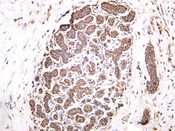
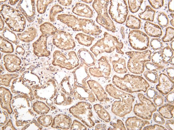
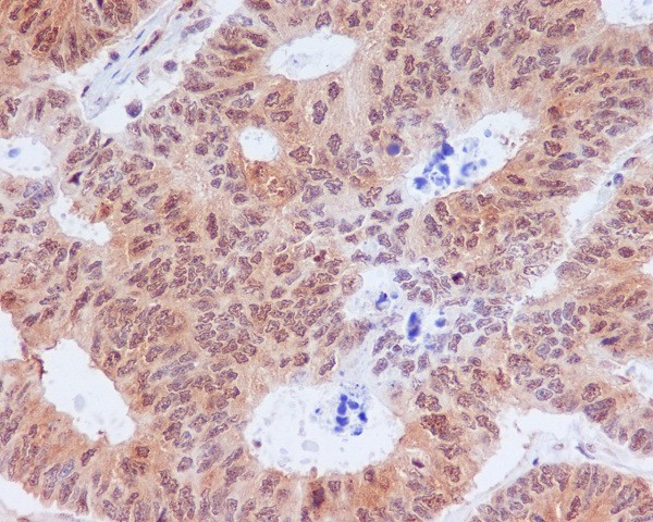
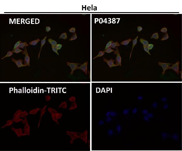
Anti-EIF2S1 IHC images show human breast, kidney, and colon cancer, plus rat kidney (catalog IHC captions: M04387-6, P04387); P04387 also has an IF image (catalog IF caption).
M04387-6 lists IHC and IF for Human, Mouse, and Rat, with IHC images of human breast, human kidney, and rat kidney (catalog: M04387-6 applications, reactivity, IHC captions). P04387 lists IHC and ICC/IF for Human, Mouse, and Rat, with a paraffin-section IHC image of human colon cancer and an IF image (catalog: P04387 applications, reactivity, IHC and IF captions).
Which to pick: For total eIF2α tissue IHC, choose rabbit monoclonal M04387-6; its IHC captions show human breast and kidney and rat kidney, but do not report processing or fixative (catalog: M04387-6 title and IHC captions). For phospho-Ser51 IHC in paraffin sections or for ICC/IF, choose rabbit monoclonal P04387; its human colon cancer IHC caption specifies paraffin embedding but does not report the fixative, and it has an IF image (catalog: P04387 title, applications, IHC and IF captions). For cross-species planning, both list Human, Mouse, and Rat reactivity; the pictured IHC species are Human and Rat for M04387-6 and Human for P04387 (catalog: both reactivity lists and IHC captions).